. 2007 Jun 27;27(26):6965-71.
doi: 10.1523/JNEUROSCI.0693-07.2007.

A bovine prion acquires an epidemic bovine spongiform encephalopathy strain-like phenotype on interspecies transmission

Affiliations

A bovine prion acquires an epidemic bovine spongiform encephalopathy strain-like phenotype on interspecies transmission

Vincent Béringue et al. J Neurosci. .

Abstract

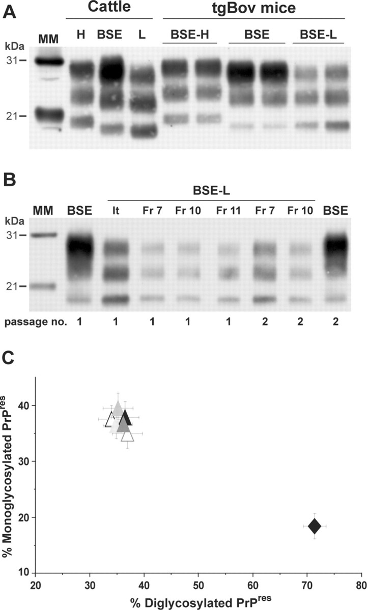
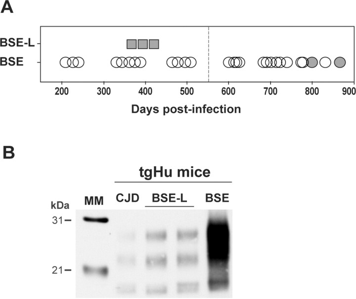
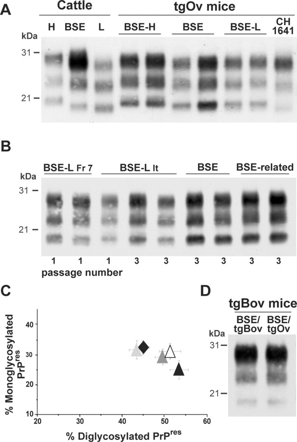
Implementation in Europe of large-scale testing to detect bovine spongiform encephalopathy (BSE)-infected cattle and prevent the transmission of this prion disease to humans has recently led to the discovery of novel types of bovine prions. We characterized atypical isolates called BSE L-type by analyzing their molecular and neuropathological properties during transmission to several mouse lines transgenic for the prion protein (PrP). Unexpectedly, such isolates acquired strain features closely similar to those of BSE-type agents when propagated in mice expressing ovine PrP, although they retained phenotypic traits distinct from BSE in other lines, including bovine PrP mice. These findings further underline the relationship between the crossing of species barrier and prion strain diversification, and, although the origin of the epidemic BSE agent has only been speculative until now, they provide new insight into the nature of the events that could have led to the appearance of this agent.


Figures

Figure 1.
Figure 1.
Brain PrPres in bovine PrP transgenic mice infected with BSE-L or BSE agents. A Western blot analysis of proteinase K-digested brain homogenates from diseased mice is presented. A, The distinct PrPres profiles of BSE H-type, BSE-L, and BSE agents are essentially preserved during cattle-to-mice transmission (2 mouse brains shown for each agent). B, The Italian (It) and French (Fr) BSE-L isolates have undistinguishable PrPres profiles that are conserved on second passage. C, Ratio of diglycosylated and monoglycosylated PrPres species (data plotted as means ± SEM). The BSE-L isolates are represented by triangles from different colors (open, first passage; filled, second passage) and BSE by a black diamond. MM, Molecular markers.
Figure 2.
Figure 2.
Brain PrPres in human PrP transgenic mice infected with BSE-L or BSE agents. Western blots of proteinase K-digested brain homogenates are shown. A, Mice were killed healthy (up to 550 d; vertical dotted line) or in extremis after primary inoculation with either BSE-L (square; Fr-7 isolate, ongoing experiment) or BSE agent (circle; data from 3 isolates). Brains positive for PrPres in Western blot are visualized by filled symbols. B, PrPres profile in BSE-L (2 brains) and BSE-infected mice. The profile produced by human CJD (low type) is shown for comparison.
Figure 3.
Figure 3.
Brain PrPres in ovine PrP transgenic mice infected with BSE-L or BSE agents. Western blots of proteinase K-digested brain homogenates are shown. The profiles in duplicate or triplicate are from different mice. A, Primary transmission to tgOv mice. The PrPres profile of BSE-L is similar to that observed with tgOv-passed BSE and differs from that in mice inoculated with BSE H-type or sheep scrapie (e.g., CH1641 strain). B, The BSE-like profile of BSE-L PrPres is maintained on subpassaging in tgOv mice. BSE-related agents are sheep BSE (left) and vCJD (right). C, Ratio of diglycoform and monoglycoform PrPres (data plotted as means ± SEM). BSE-L passages are visualized by triangles [Italian (It) first passage, black; second and third passage, dark and light gray; French (Fr-7) first passage; white] and tgOv-passed BSE (third passage) by a black diamond. D, PrPres in tgBov mice inoculated with BSE serially passed in either tgBov or tgOv mice.
Figure 4.
Figure 4.
Regional distribution of PrPres and vacuolation in the brain of bovine PrP transgenic mice infected with BSE-L or BSE agents. A–D, Histoblots of representative coronal sections at the level of the hippocampus are shown. Note the different distribution and nature of PrP deposits between BSE (A) and BSE-L [B, Italian (It); C, French (Fr); D, French second passage]. E, F, Lesion profiles in the brain of tgBov mice infected with the BSE-L (E, open circles, Italian; filled circles, French isolates) or BSE (F, open circles, cattle BSE; open squares, goat BSE) agent. The intensity of vacuolation was scored as means ± SEM in standard gray (G1–G9) and white (W1–W3) matter areas (see Materials and Methods). The BSE-L agent induced a severe vacuolation in several regions of the gray matter compared with BSE.
Figure 5.
Figure 5.
Regional distribution of PrPres and vacuolation in the brain of ovine PrP transgenic mice infected with BSE-L or BSE-type agents. A–D, Histoblots of representative coronal sections of mouse brains at the level of the hippocampus are shown. The patterns produced by cattle BSE (A, third pass) and BSE-L (B, second pass; C, third pass) are similar. The habenular (H), laterodorsal (L), geniculate (G), and submedius (S) nuclei of the thalamus are more particularly involved. The pattern produced by the CH1641 sheep scrapie strain is shown for comparison (D, second pass). E–J, Histoblots showing the PrP deposits in mice (third passage) infected with vCJD (E), cattle BSE (G), sheep BSE (I), or BSE-L (F, H, J) in the septum (E, F), hippocampus/thalamus (G, H), and midbrain at the level of the inferior colliculus (I, J). Note the similar PrPres regional distribution between the agents, except in the fasciculus retroflexus (arrowhead). K–N, Lesion profiles in tgOv mice infected with BSE-L or BSE agents. Mean ± SEM scores reflecting the vacuolation intensity on primary (open symbols) and subsequent (filled symbols) passages are shown for BSE-L (K, solid line, Italian; broken line, French), BSE-H (L), cattle BSE (M), sheep BSE (N, circles) and vCJD (N, squares). Standard brain areas are as in Figure 4.

References

    1. Aguzzi A. Prion diseases of humans and farm animals: epidemiology, genetics, and pathogenesis. J Neurochem. 2006;97:1726–1739. - PubMed
    1. Asante EA, Linehan JM, Desbruslais M, Joiner S, Gowland I, Wood AL, Welch J, Hill AF, Lloyd SE, Wadsworth JD, Collinge J. BSE prions propagate as either variant CJD-like or sporadic CJD-like prion strains in transgenic mice expressing human prion protein. EMBO J. 2002;21:6358–6366. - PMC - PubMed
    1. Baron T, Biacabe AG. Origin of bovine spongiform encephalopathy. Lancet. 2006;367:297–298. author reply 298–299. - PubMed
    1. Baron TG, Biacabe AG, Bencsik A, Langeveld JP. Transmission of new bovine prion to mice. Emerg Infect Dis. 2006;12:1125–1128. - PMC - PubMed
    1. Bartz JC, Bessen RA, McKenzie D, Marsh RF, Aiken JM. Adaptation and selection of prion protein strain conformations following interspecies transmission of transmissible mink encephalopathy. J Virol. 2000;74:5542–5547. - PMC - PubMed

Publication types