. 2007 Jun 27;27(26):7028-40.
doi: 10.1523/JNEUROSCI.1624-07.2007.

Late-stage neuronal progenitors in the retina are radial Müller glia that function as retinal stem cells

Affiliations

Late-stage neuronal progenitors in the retina are radial Müller glia that function as retinal stem cells

Rebecca L Bernardos et al. J Neurosci. .

Abstract

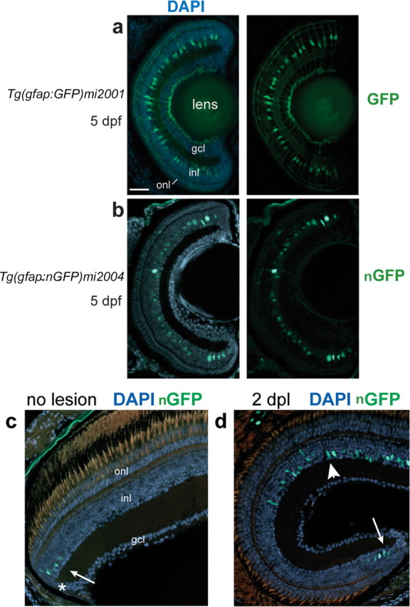
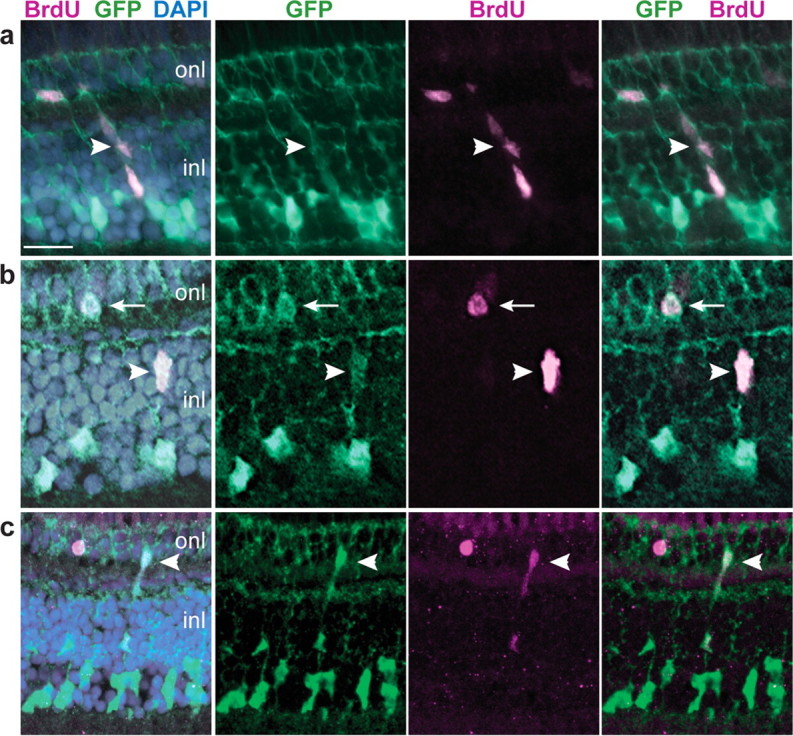
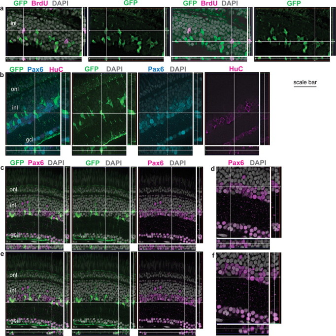
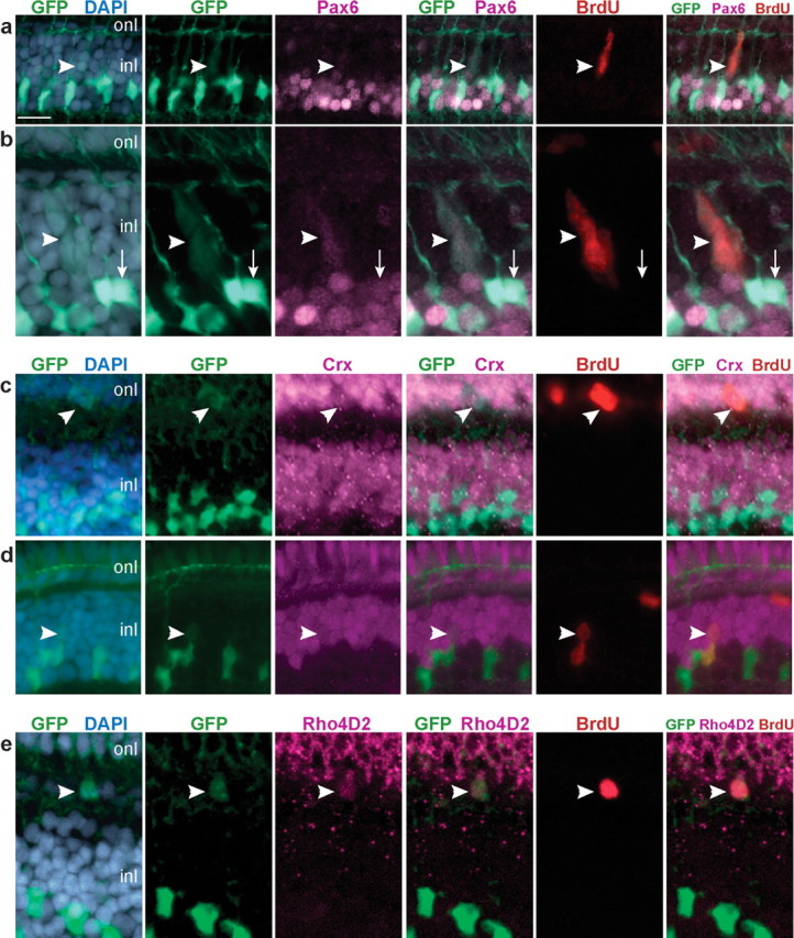
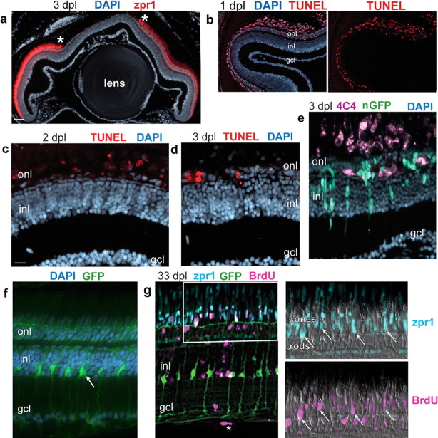
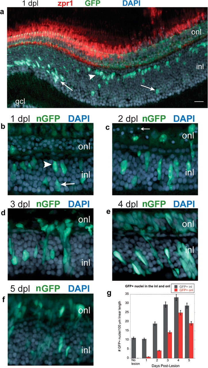
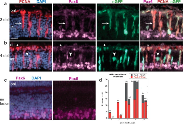
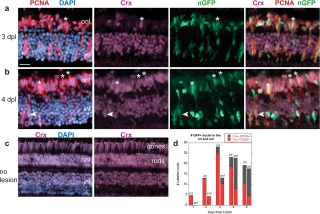
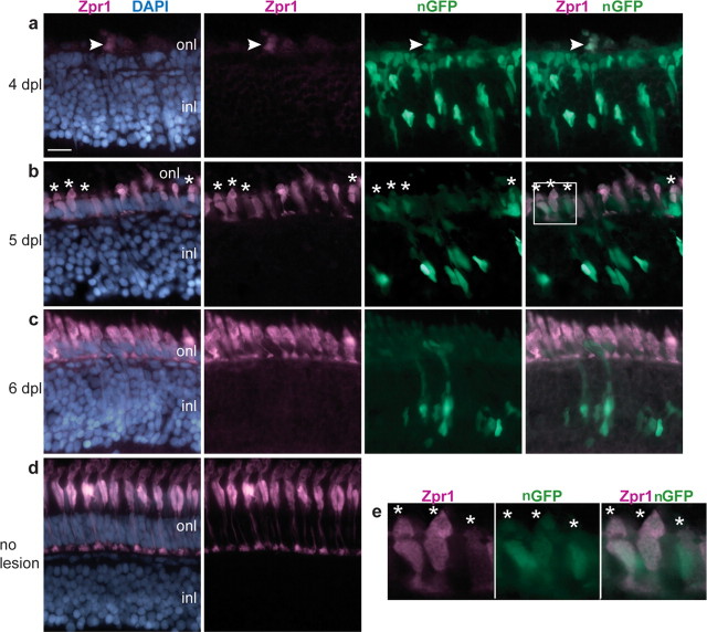
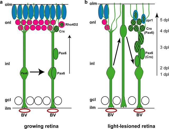
Neuronal progenitors in the mammalian brain derive from radial glia or specialized astrocytes. In developing neural retina, radial glia-like Müller cells are generated late in neurogenesis and are not considered to be neuronal progenitors, but they do proliferate after injury and can express neuronal markers, suggesting a latent neurogenic capacity. To examine the neurogenic capacity of retinal glial cells, we used lineage tracing in transgenic zebrafish with a glial-specific promoter (gfap, for glial fibrillary acid protein) driving green fluorescent protein in differentiated Müller glia. We found that all Müller glia in the zebrafish retina express low levels of the multipotent progenitor marker Pax6 (paired box gene 6), and they proliferate at a low frequency in the intact, uninjured retina. Müller glia-derived progenitors express Crx (cone rod homeobox) and are late retinal progenitors that generate the rod photoreceptor lineage in the postembryonic retina. These Müller glia-derived progenitors also remain competent to produce earlier neuronal lineages, in that they respond to loss of cone photoreceptors by specifically regenerating the missing neurons. We conclude that zebrafish Müller glia function as multipotent retinal stem cells that generate retinal neurons by homeostatic and regenerative developmental mechanisms.


Figures

Figure 1.
Figure 1.
GFP+ Müller glia in Tg(gfap:GFP)mi2001 and Tg(gfap:nGFP)mi2004 transgenic zebrafish. a–d, Retinal cryosections from larval (a, b) or adult (c, d) transgenic zebrafish counterstained with DAPI (blue). a, In the Tg(gfap:GFP)mi2001 and mi2002 cytoplasmic GFP lines, Müller glia cell bodies in the inner nuclear layer (inl) extend radial processes toward the outer nuclear layer (onl) and ganglion cell layer (gcl). b, In the Tg(gfap:nGFP)mi2004 line, reporter expression is restricted to nuclei of Müller glia in this young larval fish, at 5 dpf. The GFP expression in this preparation is visualized by immunofluorescence with an anti-GFP antibody. c, In adult Tg(gfap:nGFP)mi2004 fish, endogenous nuclear-targeted GFP expression is detected only in immature Müller glia nuclei (arrow) near the CMZ (*). d, In Tg(gfap:nGFP)mi2004 fish at 2 d after destruction of photoreceptors by exposure to high-intensity light (days postlesion), endogenous nuclear-targeted GFP expression is upregulated in Müller glia within the lesioned area (arrowhead) similar to immature Müller glia in newly generated retina at the ciliary margin (arrow). Scale bar: (in a) a, b, 40 μm; c, d, 30 μm.
Figure 2.
Figure 2.
GFP+ Müller glia proliferate in the differentiated, growing retina. a–c, Retinal cryosections from juvenile Tg(gfap:GFP)mi2001 zebrafish labeled with BrdU (magenta) and the nuclear stain DAPI (blue). Representative images from two independent experiments, six fish per experiment, 7 d continuous exposure to BrdU. a, A radial chain of GFP+, BrdU+ cells (arrowhead) extends through the inner nuclear layer (inl) into the outer nuclear layer (onl). b, A single GFP+, BrdU+ cell in the INL (arrowhead) near a GFP+, BrdU+ cell in the ONL (arrow). c, Another example of a chain of GFP+, BrdU+ cells that spans INL and ONL (arrowhead). Scale bar: (in a) a, b, 15 μm; c, 20 μm.
Figure 3.
Figure 3.
GFP+ Müller glia are colabeled with BrdU and Pax6 but not HuC. Retinal cryosections from juvenile Tg(gfap:GFP)mi2001 (a) and mi2002 (b–e) transgenic zebrafish. Reconstruction of optical slices (Z-stack) reconstructed and displayed in “cut view” through the thickness of the section in orthogonal planes. Sections were counterstained with DAPI (gray). a, This fish was exposed to BrdU for 7 d, and both of the BrdU+ nuclei (magenta) in the inner nuclear layer (inl) of this image are weakly GFP+ (green). b, This GFP+ cell (at the intersection of the crosshairs) is weakly immunoreactive for Pax6 (cyan) but not the neuronal marker HuC (magenta). As expected, many of the Pax6+, GFP neurons (amacrine cells in the INL) and retinal ganglion cells in the ganglion cell layer (gcl) are double labeled with HuC. c–f, The crosshairs show two GFP+ (green) Müller glia with characteristic polygonal nuclei that express Pax6, shown at higher magnification in d and f, respectively. Scale bar: a, b, 30 μm; c, e, 40 μm; d, f, 27 μm. onl, Outer nuclear layer.
Figure 4.
Figure 4.
GFP+ progenitors express neuronal and photoreceptor markers. a–e, Retinal cryosections from juvenile Tg(gfap:GFP)mi2001 zebrafish labeled with 7 d continuous exposure to BrdU (red). Counterstained for nuclei with DAPI (blue) and Pax6, Crx, or Rho4D2 (magenta). a, b, BrdU+, Pax6+ neural progenitors in the inner nuclear layer (inl; arrowheads) are weakly labeled with GFP (green). b, Nonproliferating (BrdU) Müller glia are strongly labeled with GFP and weakly immunoreactive for Pax6 (arrow). c, A GFP+, BrdU+ neural progenitor in the outer nuclear layer (onl) is immunoreactive for Crx (arrowhead). d, A pair of BrdU+ nuclei (arrowhead) in which the upper cell is Crx+ and weakly GFP+, and the lower cell is Crx, strongly GFP+, and has a polygonal-shaped nucleus characteristic of Müller glia. e, A GFP+, BrdU+ cell in the ONL is immunoreactive for rod opsin (Rho4D2; arrowhead). Scale bar: (in a) a, c, 15 μm; b, e, 10 μm; d, 12 μm.
Figure 5.
Figure 5.
Exposure to intense light destroys photoreceptors, which subsequently regenerate. Retinal cryosections from adult Tg(gfap:GFP)mi2001 or Tg(gfap:nGFP)mi2004 fish exposed for 30 min to intense light. a, At 3 d after a 30 min exposure to intense light, double cones labeled by zpr1 (red) are absent from central retina; the borders of the lesion are denoted by asterisks. Counterstained with DAPI (blue). b, TUNEL-labeled (red) apoptotic cells are restricted to the outer nuclear layer (onl) at 1 dpl. c, d, Fewer TUNEL-labeled cells (red) are in the ONL at 2 and 3 dpl. e, In the light-lesioned retina from a Tg(gfap:nGFP)mi2004 fish at 3 dpl, phagocytic microglia are immunolabeled with the microglial marker 4C4 (magenta). f, In the undamaged retina of an adult Tg(gfap:GFP)mi2001 fish, Müller glia (green) cell bodies and nuclei (arrow) are confined to the inner part of the inner nuclear layer (inl) and extend radial processes through the ONL and ganglion cell layer (gcl). g, Regenerated retina from a Tg(gfap:GFP)mi2002 zebrafish at 33 dpl that was subjected to continuous exposure to BrdU (magenta) for 72 h, from 2 through 4 dpl. BrdU+ nuclei (magenta) are throughout the retina, primarily in the ONL but some in the inner retina including an endothelial cell in a vitreal blood vessel (*). The boxed area is shown at higher magnification in the panels to the right. Double cones are immunoreactive with zpr1 (cyan) and several have BrdU+ nuclei (arrows), indicating that they were regenerated after the lesion. Some rod photoreceptors, whose nuclei are on the inner side of the ONL, are also BrdU labeled. Scale bar: a, 50 μm; b, 40 μm; c–e, 15 μm; f and the first panel in g, 10 μm.
Figure 6.
Figure 6.
Müller glia proliferate after photoreceptor loss. Retinal cryosections from adult Tg(gfap:GFP)mi2001 or Tg(gfap:nGFP)mi2004 zebrafish exposed for 30 min to intense light. Sections are counterstained with DAPI (blue). a, Light-lesioned retinas from Tg(gfap:GFP)mi2001 zebrafish at 1 dpl immunolabeled with the double-cone-specific zpr1 monoclonal antibody (red). Most of the GFP+ Müller glia nuclei (green) within the region of the lesion have migrated from their normal position in the inner nuclear layer (inl; arrows) to the outer portion of the INL (arrowhead). b–f, Light-lesioned retinas from Tg(gfap:nGFP)mi2004 zebrafish immunolabeled with anti-GFP (green). At 1 dpl (b) and 2 dpl (c), GFP+ Müller nuclei are scattered throughout the INL, some have elongated, and a few have crossed into the outer nuclear layer (onl). Pyknotic photoreceptor nuclei are in the ONL (arrow). d, Radially elongated clusters of GFP+ nuclei are in the INL and ONL at 3 dpl and at 4 dpl (e). f, At 5 dpl, the number of GFP+ nuclei begins to decrease in the INL and ONL. g, The number of GFP+ nuclei in the INL (gray) and ONL (red) per 100 μm linear length of retina is plotted as a function of days postlesion. Scale bar: (in a) a, 20 μm; c–e, 15 μm. gcl, ganglion cell layer.
Figure 7.
Figure 7.
Pax6 is expressed in Müller-derived neuronal progenitors in the regenerating retina. a–c, Cryosections of adult zebrafish retinas labeled with Pax6 (magenta), PCNA (red), GFP (green), and DAPI (blue). a, b, Light-lesioned retinas from Tg(gfap:nGFP)mi2004 fish. At 3 and 4 dpl, almost all GFP+ nuclei in the inner nuclear layer (inl) are PCNA+ and weakly Pax6+ (arrow). B, One nucleus is GFP+, Pax6+ but PCNA (b; arrowhead). GFP+, Pax6+, PCNA+ cells are also in the outer nuclear layer (onl) (*). c, In unlesioned retinas, expression of Pax6 is primarily restricted to the inner portion of the INL. d, The number of GFP+, PCNA+ nuclei per 100 μm linear length of retina in the INL and ONL that are Pax6+ (gray) or Pax6 (red) is plotted as a function of days postlesion. Scale bar: (in a) a–c, 15 μm.
Figure 8.
Figure 8.
Crx is expressed in Müller-derived photoreceptor progenitors in the regenerating retina. a–c, Cryosections of adult zebrafish retinas labeled with Crx (magenta), PCNA (red), GFP (green), and DAPI (blue). a, b, Light-lesioned Tg(gfap:nGFP)mi2004 retinas. A, At 3 dpl, many GFP+, PCNA+ nuclei are present in the outer nuclear layer (onl), but few are Crx+ (*). b, At 4 dpl, many GFP+, Crx+, PCNA+ nuclei are present in the ONL (*) and a few in the inner nuclear layer (inl; arrowhead). c, An uninjured retina showing the normal expression pattern of Crx in the outer half of the INL and in the entire ONL, both rod and cone nuclei. d, The number of GFP+, PCNA+ nuclei per 100 μm linear length of retina in the INL and ONL that are Crx+ (gray) or Crx (red) is plotted as a function of days postlesion. Scale bar: (in a) a–c, 15 μm.
Figure 9.
Figure 9.
Regenerated cone photoreceptors are GFP+. a–d, Cryosections of adult zebrafish retinas labeled with zpr1 (magenta), GFP (green), and DAPI (blue). a–c, Light-lesioned retinas from Tg(gfap:nGFP)mi2004 fish. a, At 4 dpl, a few cells with weak zpr1 expression are in the outer nuclear layer (onl) in the lesioned area. A GFP+ cell is colabeled with zpr1+ (arrowhead). b, By 5 dpl, many zpr1+ immature regenerating cones are present in the ONL, and several are GFP+ (*). c, At 6 dpl, zpr1+ regenerating cones are more differentiated and are no longer labeled with GFP. d, Differentiated double cones labeled with zpr1 in an unlesioned retina. e, Magnified view of the white-boxed area in b shows colocalization of zpr1 and GFP in immature, regenerating cones (*). Scale bar: (in a) a–d, 15 μm. inl, Inner nuclear layer.
Figure 10.
Figure 10.
Model of photoreceptor production by Müller glia. a, b, Cone photoreceptor nuclei (blue), rod photoreceptor nuclei (magenta), Müller glia and their progeny (green), and blood vessels (BV; red ovals). Additional cells in the inner nuclear layer (inl) have been omitted for simplicity. The radial processes of Müller glia contact blood vessels (BV) at the inner limiting membrane (ilm) and reach the outer limiting membrane (olm). Müller glia nuclei are weakly immunoreactive for Pax6. a, Müller glia divide to produce Pax6+ neural progenitors in the INL that migrate along the radial glial process to the outer nuclear layer (onl). Pax6 is downregulated in the migrating, proliferating progenitors, and Crx is upregulated. The Crx+ progenitors withdraw from the cell cycle and express rod opsin (Rho4D2 immunoreactivity). b, After light lesions that destroy photoreceptors, all Müller glia reenter the cell cycle, and their nuclei migrate toward the outer limiting membrane. By 3 dpl, many Müller-derived, progenitors in the INL express Pax6, and, by 4 dpl, progenitors in the ONL express Crx. By 5 dpl regenerated, differentiating cones appear in the ONL and begin to express zpr1, a specific marker of double cones. gcl, Ganglion cell layer.

References

    1. Adolf B, Chapouton P, Lam CS, Topp S, Tannhauser B, Strahle U, Götz M, Bally-Cuif L. Conserved and acquired features of adult neurogenesis in the zebrafish telencephalon. Dev Biol. 2006;295:278–293. - PubMed
    1. Alvarez-Buylla A, Lim DA. For the long run: maintaining germinal niches in the adult brain. Neuron. 2004;41:683–686. - PubMed
    1. Anthony TE, Klein C, Fishell G, Heintz N. Radial glia serve as neuronal progenitors in all regions of the central nervous system. Neuron. 2004;41:881–890. - PubMed
    1. Barthel LK, Raymond PA. Improved method for obtaining 3-micron cryosections for immunocytochemistry. J Histochem Cytochem. 1990;38:1383–1388. - PubMed
    1. Bernardos RL, Raymond PA. GFAP transgenic zebrafish. Gene Expr Patterns. 2006;6:1007–1013. - PubMed

Publication types