. 2007 Jun 27;27(26):7070-80.
doi: 10.1523/JNEUROSCI.1866-07.2007.

Dual regulation of TRPV1 by phosphoinositides

Affiliations

Dual regulation of TRPV1 by phosphoinositides

Viktor Lukacs et al. J Neurosci. .

Abstract

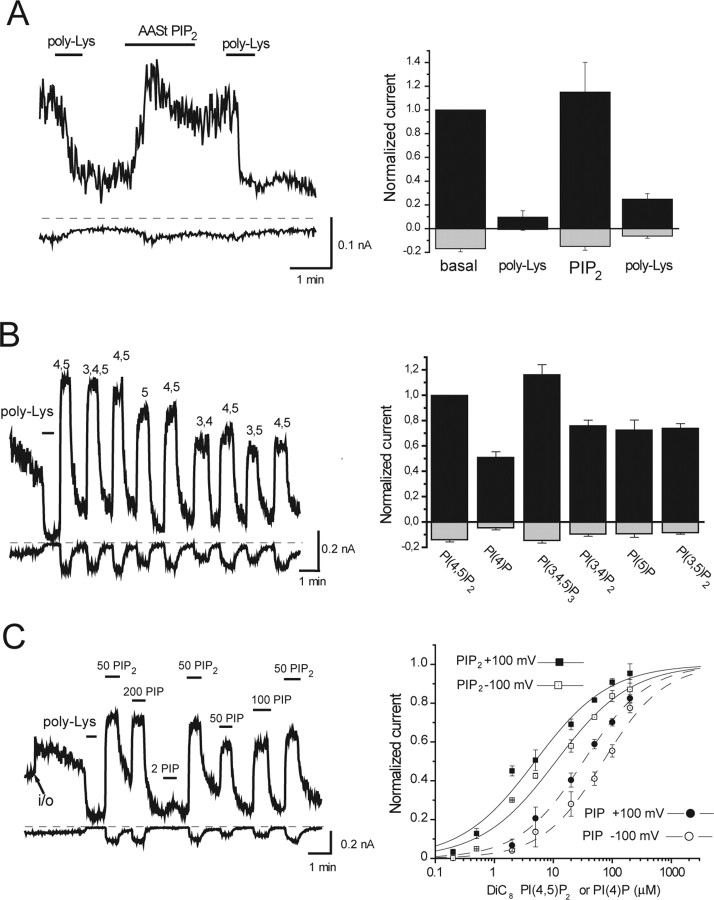
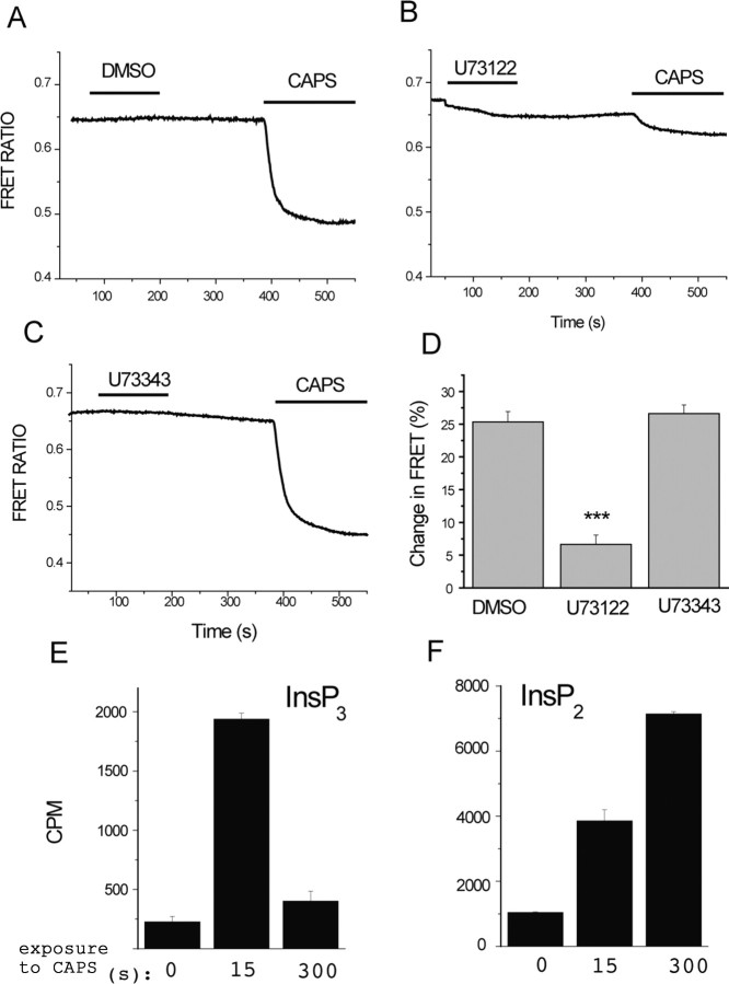
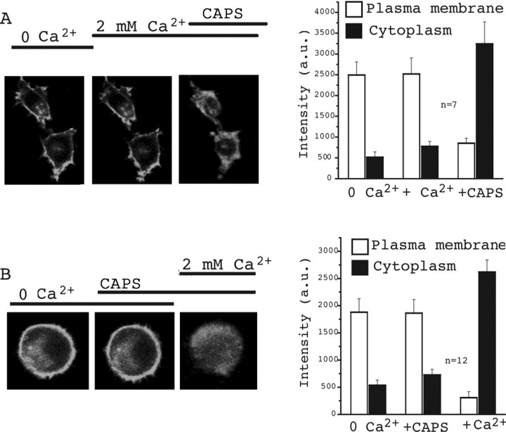
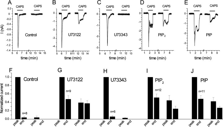
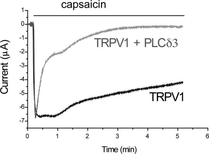
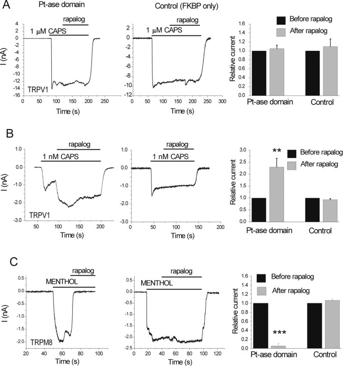
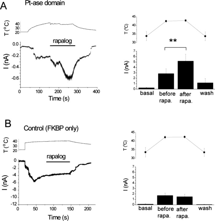
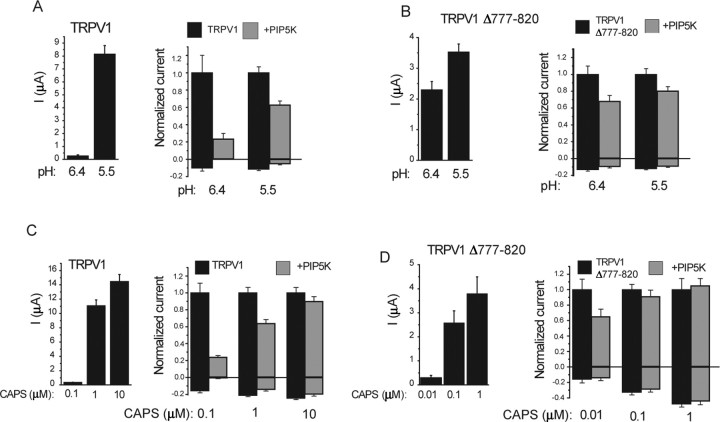
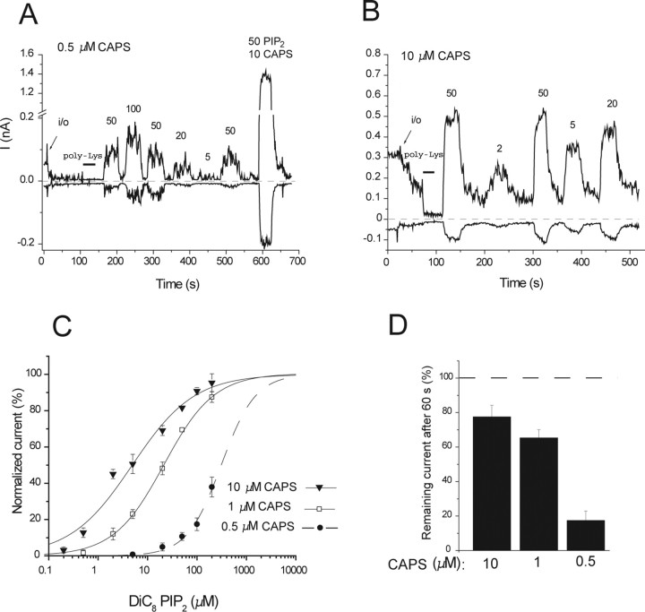
The membrane phospholipid phosphatidylinositol 4,5-bisphosphate [PtdIns(4,5)P2 or PIP2] regulates many ion channels. There are conflicting reports on the effect of PtdIns(4,5)P2 on transient receptor potential vanilloid 1 (TRPV1) channels. We show that in excised patches PtdIns(4,5)P2 and other phosphoinositides activate and the PIP2 scavenger poly-Lys inhibits TRPV1. TRPV1 currents undergo desensitization on exposure to high concentrations of capsaicin in the presence of extracellular Ca2+. We show that in the presence of extracellular Ca2+, capsaicin activates phospholipase C (PLC) in TRPV1-expressing cells, inducing depletion of both PtdIns(4,5)P2 and its precursor PtdIns(4)P (PIP). The PLC inhibitor U73122 and dialysis of PtdIns(4,5)P2 or PtdIns(4)P through the patch pipette inhibited desensitization of TRPV1, indicating that Ca2+-induced activation of PLC contributes to desensitization of TRPV1 by depletion of PtdIns(4,5)P2 and PtdIns(4)P. Selective conversion of PtdIns(4,5)P2 to PtdIns(4)P by a rapamycin-inducible PIP2 5-phosphatase did not inhibit TRPV1 at high capsaicin concentrations, suggesting a significant role for PtdIns(4)P in maintaining channel activity. Currents induced by low concentrations of capsaicin and moderate heat, however, were potentiated by conversion of PtdIns(4,5)P2 to PtdIns(4)P. Increasing PtdIns(4,5)P2 levels by coexpressing phosphatidylinositol-4-phosphate 5-kinase inhibited TRPV1 at low but not at saturating capsaicin concentrations. These data show that at low capsaicin concentrations and other moderate stimuli, PtdIns(4,5)P2 partially inhibits TRPV1 in a cellular context, but this effect is likely to be indirect, because it is not detectable in excised patches. We conclude that phosphoinositides have both inhibitory and activating effects on TRPV1, resulting in complex and distinct regulation at various stimulation levels.


Figures

Figure 1.
Figure 1.
Phosphoinositides activate TRPV1 channels. Currents were measured in large membrane patches excised from oocytes expressing TRPV1 using the ramp protocol described in Materials and Methods. Representative traces show currents at +100 mV (top trace) and −100 mV (bottom trace); dashed lines show zero current. Phosphoinositides were applied directly to the intracellular surface of the patch in the presence of 1 μm capsaicin (A, B) and 10 μm capsaicin (C) in the patch pipette. A, Effect of the long-chain AASt PtdIns(4,5)P2 (5 μm) and poly-l-lysine (1 and 30 μg/ml) for the first and second applications, respectively (n = 4). B, The effects of various phosphoinositides. The left panel is a representative trace, and the right panel shows statistics for +100 and −100 mV [n = 7–12, except PtdIns(5)P2, which is n = 3] for the effects of different phosphoinositides compared with PtdIns(4,5)P2. All phosphoinositides were short acyl-chain (DiC8) applied at 50 μm. C, Dose–response measurement with diC8 PIP2 and PIP. poly-Lys, Poly-l-lysine.
Figure 2.
Figure 2.
Capsaicin activates PLC in TRPV1-expressing cells. A–D, Fluorescence was measured in HEK 293 cells expressing TRPV1 and the CFP- and YFP-tagged PLCδ1 PH domains, as described in Materials and Methods. Cells were pretreated with DMSO (A), 3 μm U73122 (B), or 3 μm U73343 (C) for 2 min, and then 1 μm capsaicin (CAPS) was applied, as indicated by the horizontal lines. The extracellular medium contained 1 mm Ca2+. The change in FRET ratio (D) was measured by dividing the value 2 min after the application of capsaicin with the FRET ratio value before the application of capsaicin to result Q, and then 100 − (100 × Q) was plotted (n = 14–17). ***p < 0.005. E, F, HEK cells expressing TRPV1 were incubated with 3H-inositol, as described in Materials and Methods, and stimulated with 2 μm capsaicin for the time periods indicated. Inositol phosphates were extracted and separated on HPLC, and the radioactivity was plotted for IP3 (InsP3; E) and IP2 (InsP2; F).
Figure 3.
Figure 3.
Capsaicin (CAPS) induces PtdIns(4)P depletion in the presence of extracellular Ca2+. HEK cells were transfected with TRPV1 and the GFP-tagged OSH2–tandem PH domain that is targeted to the plasma membrane through binding to PtdIns(4)P. PtdIns(4)P depletion was assessed by the translocation of the PH-OSH2-GFP from the plasma membrane to the cytoplasm. A, The images show fluorescence of two representative cells in control (0 Ca2+), 2 min after the addition of 2 mm Ca2+, and 2 min after application of 1 μm capsaicin. The right panel shows summary of the data. An area was selected on the plasma membrane (more intense GFP) as well as in the cytoplasm (less intense), and the average pixel intensity in the area of selection was represented in arbitrary units (a.u.). B is the same as A, but the order of application of capsaicin and Ca2+ is reversed.
Figure 4.
Figure 4.
Desensitization of TRPV1 currents is inhibited by the PLC blocker U73122 and by PtdIns(4,5)P2 and PtdIns(4)P. Whole-cell patch-clamp measurements were performed at −60 mV on HEK cells expressing TRPV1, as described in Materials and Methods. A–E, Representative measurements. F–J, Statistics. A–C, F–H, Cells were preincubated with vehicle (A, F), 3 μm U73122 (B, G), and 3 μm U73343 (C, H), and 1 min capsaicin pulses were applied in the presence of 1 mm extracellular Ca2+. D, E, I, J, The pipette solution was supplemented with 100 μm diC8 PtdIns(4,5)P2 (D, I) or 100 μm DiC8 PtdIns(4)P (E, J). The first capsaicin pulse was applied in all experiments 5–10 min after the establishment of the whole-cell configuration. The data were normalized to the first peak current, and the current amplitudes were plotted for the end of the first capsaicin pulse, peak current of the second capsaicin pulse, and the end of the second capsaicin pulse (n = 6–12). CAPS, Capsaicin.
Figure 5.
Figure 5.
Coexpression of PLCδ3 accelerates desensitization of TRPV1 currents in oocytes. TEVC measurements were performed in Xenopus oocytes at a −60 mV holding potential, as described in Materials and Methods. Traces representing desensitization kinetics of oocytes expressing TRPV1 and those coexpressing PLCδ3 and the channel are shown; oocytes were stimulated with 5 μm capsaicin, in the presence of 2 mm calcium. See statistics in Results.
Figure 6.
Figure 6.
Translocation of the PIP2 5-phosphatase to the plasma membrane activates TRPV1 at low but not at high capsaicin (CAPS) concentrations. HEK cells were transfected with TRPV1, and the plasma membrane targeted CFP-tagged FRB (T2098L) and either the RFP-tagged FKBP12 (control) or FKBP12 fused to the phosphatase domain of the PIP2 5-phosphatase (Pt-ase domain) (Varnai et al., 2006). Measurements were performed at −60 mV in the whole-cell configuration, in nominally Ca2+-free solution, to avoid desensitization. Translocation of the Pt-ase domain was induced with 500 nm rapalog. A, B, Responses of cells to 1 μm (A) or 1 nm (B) capsaicin. The right panel in A shows the summary for n = 8–9 for 1 μm capsaicin. Current values 60 s after the application of rapalog were divided by the current level before the application of rapalog and plotted. B, Right, Summary for n = 16–18 for 1 nm capsaicin. Current values 30 s after the application of rapalog were divided by the current level before the application of rapalog and plotted. C, inhibition of currents induced by 500 μm menthol in cells expressing TRPM8 and the rapalog-inducible phosphatase (left) or the control construct FKBP only (middle).
Figure 7.
Figure 7.
Conversion of PtdIns(4,5)P2 to PtdIns(4)P increases currents stimulated by moderately high temperatures. HEK cells were transfected with TRPV1 and the CFP-tagged FRB (T2098L) and either the RFP-tagged FKBP12 (control) or the FKBP12 fused to the phosphatase domain of the PIP2 5-phosphatase (Pt-ase domain). Measurements were performed at −60 mV in the whole-cell configuration. A, The effect of rapalog in the FKBP–phosphatase domain-expressing cells. Left, Representative trace for currents (I; bottom) and temperature (T; top) recorded in the same experiment. The horizontal bar shows the application of 500 nm rapalog. Right, Summary of the data (n = 9). Current and temperature values were measured just before the current started increasing (basal), before the application of rapalog (before rapa.), 20–30 s after the application of rapalog (after rapa.), and at the end of the decreasing phase of the temperature protocol after the washout of rapalog (wash). B, Similar experiments in control cells (n = 9).
Figure 8.
Figure 8.
The effect of coexpression of PIP5K on TRPV1 currents in Xenopus oocytes. TRPV1 currents were measured with TEVC, as described in Materials and Methods. Currents at +100 and −100 mV are shown. Left panels show average current amplitudes, and right panels show currents normalized to current amplitudes elicited by the actual stimulus without PIP5K coexpression. A, B, The effect of PIP5K on low pH (6.5 and 5.5)-induced currents is shown for the wild type (A) and the Δ777–820 mutant (B) of TRPV1. C, D, The effect of PIP5K on capsaicin (CAPS)-induced currents is shown for the wild type (C) and the Δ777–820 mutant (D) of TRPV1.
Figure 9.
Figure 9.
High capsaicin (CAPS) concentrations increase the apparent affinity of TRPV1 for PtdIns(4,5)P2. A, B, Representative traces in the presence of 0.5 μm capsaicin (A) and 10 μm capsaicin (B) for dose responses with diC8 PtdIns(4,5)P2. C, Hill fits of the PtdIns(4,5)P2 dose responses at various capsaicin concentrations (+100 mV). D, Statistics of the current rundown in the presence of various capsaicin concentrations (n = 8, 44, and 11 for 10, 1, and 0.5 μm capsaicin, respectively). In the presence of 1 and 10 μm capsaicin, there was an immediate change in current after excision, which was quite variable: sometimes an increase, sometimes a decrease or no change; therefore, the values given are normalized to the current immediately after excision. In the presence of 0.5 μm capsaicin in many patches, there was a fast increase, but it disappeared so rapidly that its amplitude was not always possible to determine; thus, the values were normalized to the cell-attached level.

Similar articles

Cited by

References

    1. Balla A, Tuymetova G, Tsiomenko A, Varnai P, Balla T. A plasma membrane pool of phosphatidylinositol 4-phosphate is generated by phosphatidylinositol 4-kinase type-III alpha: studies with the PH domains of the oxysterol binding protein and FAPP1. Mol Biol Cell. 2005;16:1282–1295. - PMC - PubMed
    1. Balla T. Pharmacology of phosphoinositides, regulators of multiple cellular functions. Curr Pharm Des. 2001;7:475–507. - PubMed
    1. Baukrowitz T, Schulte U, Oliver D, Herlitze S, Krauter T, Tucker SJ, Ruppersberg JP, Fakler B. PIP2 and PIP as determinants for ATP inhibition of KATP channels. Science. 1998:1141–1144. - PubMed
    1. Bhave G, Zhu W, Wang H, Brasier DJ, Oxford GS, Gereau RW. cAMP-dependent protein kinase regulates desensitization of the capsaicin receptor (VR1) by direct phosphorylation. Neuron. 2002;35:721–731. - PubMed
    1. Caterina MJ, Julius D. The vanilloid receptor: a molecular gateway to the pain pathway. Annu Rev Neurosci. 2001;24:487–517. - PubMed

Publication types

MeSH terms