. 2007 Sep;18(9):3607-19.
doi: 10.1091/mbc.e07-02-0124. Epub 2007 Jun 27.

LEKTI fragments specifically inhibit KLK5, KLK7, and KLK14 and control desquamation through a pH-dependent interaction

Affiliations

LEKTI fragments specifically inhibit KLK5, KLK7, and KLK14 and control desquamation through a pH-dependent interaction

Celine Deraison et al. Mol Biol Cell. 2007 Sep.

Abstract

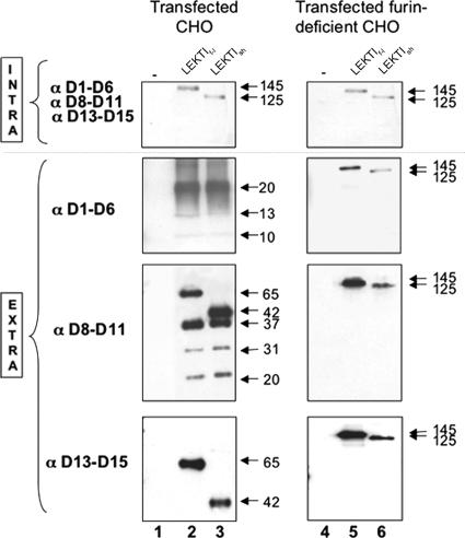
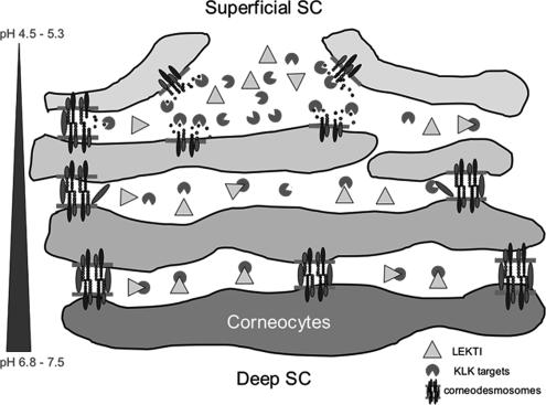
LEKTI is a 15-domain serine proteinase inhibitor whose defective expression underlies the severe autosomal recessive ichthyosiform skin disease, Netherton syndrome. Here, we show that LEKTI is produced as a precursor rapidly cleaved by furin, generating a variety of single or multidomain LEKTI fragments secreted in cultured keratinocytes and in the epidermis. The identity of these biological fragments (D1, D5, D6, D8-D11, and D9-D15) was inferred from biochemical analysis, using a panel of LEKTI antibodies. The functional inhibitory capacity of each fragment was tested on a panel of serine proteases. All LEKTI fragments, except D1, showed specific and differential inhibition of human kallikreins 5, 7, and 14. The strongest inhibition was observed with D8-D11, toward KLK5. Kinetics analysis revealed that this interaction is rapid and irreversible, reflecting an extremely tight binding complex. We demonstrated that pH variations govern this interaction, leading to the release of active KLK5 from the complex at acidic pH. These results identify KLK5, a key actor of the desquamation process, as the major target of LEKTI. They disclose a new mechanism of skin homeostasis by which the epidermal pH gradient allows precisely regulated KLK5 activity and corneodesmosomal cleavage in the most superficial layers of the stratum corneum.


Figures

Figure 1.
Figure 1.
LEKTI proteolytic fragments in the epidermis, normal human keratinocytes, and transfected CHO - Model of LEKTI proteolysis. Protein extracts from human foreskin epidermis (lane 1), cultured normal human keratinocytes (lane 2), or transfected CHO cells (lanes 3–5) were analyzed by Western-blot using three anti-LEKTI antibodies indicated on the left. CHO cells were transiently transfected with the empty pEF-DEST51 vector (−, lane 3); pEF-DEST51-SPINK5f-l (LEKTIf-l, lane 4); or pEF-DEST51-SPINK5sh (LEKTIsh, lane 5). Intracellular and extracellular fractions of cultured cells were analyzed. The molecular weights of the different bands obtained are indicated in kDa. In intracellular fractions of NHK and transfected CHO, precursors of 145 and 125 kDa are observed with each antibody. Conversely, in the epidermis and in extracellular fractions of NHK and CHO, only proteolytic fragments of LEKTI are visualized. For each antibody used, the molecular weight of these proteolytic LEKTI fragments is indicated. Note the absence of LEKTI precursors detected in human epidermis. Right, schematic representation of LEKTI processing in human epidermis and NHK. A schematic representation of LEKTI processing in human epidermis and NHK is proposed according to the results obtained with the different antibodies used. The two LEKTI precursors (145 and 125 kDa) are processed into several physiological LEKTI fragments. The identity of some LEKTI fragments was proposed according to several parameters, including the antibody used, the molecular weight of the unglycosylated forms (see Figure 3), and the existence of LEKTI fragments already published.
Figure 2.
Figure 2.
LEKTI processing involves a furin-dependant mechanism. Intracellular and extracellular extracts of CHO cells (lanes 1–3) and furin-deficient CHO cells (lanes 4–6) transiently transfected with the empty pEF-DEST51 vector (−, lanes 1 and 4); pEF-DEST51-SPINK5f-l (LEKTIf-l, lanes 2 and 5); or pEFDEST51-SPINK5sh (LEKTIsh, lanes 3 and 6) were analyzed by Western-blotting using anti-LEKTI antibodies indicated on the left. In both CHO cells and furin-deficient CHO cells, the 145- and 125-kDa LEKTI precursors are detected with each of the three anti-LEKTI antibodies in the intracellular fraction. However, in the extracellular fraction, LEKTI proteolytic fragments are visualized in CHO cells, whereas only LEKTI precursors are detected in furin-deficient CHO cells, with each of the antibody.
Figure 3.
Figure 3.
N- and O-glycosylation status of LEKTI proteolytic fragments. (A) Schematic representation of predicted N- and O-glycosylation sites on the full length (LEKTIf-l) and the short length (LEKTIsh) LEKTI precursors. N-glycosylation prediction involves two sites: one in the D8 domain, the other one in the D12 domain. Five O-glycosylation sites are predicted in the D15 domain. (B) Proteins from NHK conditioned medium were incubated in the absence (−) or presence (+) of PNGase F. After PNGase F treatment, the 31- and 37-kDa fragments recognized by the αD8–D11 antibody migrate at 29 and 35-kDa, respectively, whereas the 20-kDa band remains unchanged. Using the αD13–D15 antibody, C-terminal fragments of 42 and 65 kDa migrate at 38 and 61 kDa, respectively, when N-deglycosylated. (C) To assess O-deglycosylation, extracellular extracts of NHK were incubated at 37°C in the absence (−) or presence (+) of neuraminidase (1 h) and O-glycosidase (3 h). This treatment had no effect on the 42-kDa bands, but reduced the 65-kDa C-terminal fragment to a 61-kDa fragment.
Figure 4.
Figure 4.
Inhibition properties of LEKTI domains toward epidermal kallikreins. Proteinases were incubated with increased concentrations of inhibitors before addition of substrates (described in Table 1). The curves represent the percentage of resulting proteolytic activity according to the ratio [inhibitor]/[enzyme]. Inhibition constants Ki were calculated as described in Materials and Methods. As illustrated by the slope of the curves, the D8–D11 LEKTI fragment is the most potent inhibitor of KLK5, KLK7, and KLK14. In contrast, D6 has no inhibitory property on KLK14, and neither has D9–D15 on KLK7.
Figure 5.
Figure 5.
Surface Plasmon Resonance analysis of target proteinase binding to LEKTI fragments. LEKTI domains were immobilized onto a sensorchip. Proteinase solutions were injected over the sensorchip at concentrations ranging from 1.25 to 100 nM. Sensorgrams reflect binding during the association phase followed by the dissociation phase at the end of the injection. Representative sensorgrams show dose-dependent interactions of KLK5, KLK7 and KLK14 on D5, D6, D8–D11, and D9-D15 LEKTI domains. Each curve represents the specific interaction between the considered LEKTI fragment and proteinase. Raw binding data were analyzed using the BIAevaluation 4.0.1 software and fitted to obtain kinetics parameters. The kinetics constants ka (association constant), kd (dissociation constant), and KD (affinity constant) are indicated on each graph. ▾, stop of proteinase injection and replacement with buffer; RU, resonance units; Diff. Resp., differential response.
Figure 6.
Figure 6.
Effects of pH on the interaction between D8–D11 LEKTI fragment and target kallikreins, KLK5 and KLK7. (A) D8–D11 LEKTI domains were immobilized onto a sensorchip, over which KLK5 was injected at 2.5 nM in HBS-EP buffer at pH 7.5. At the end of the injection, this buffer was replaced by HBS-EP adjusted at various pH conditions varying from pH 7.5 to pH 4.5. The curves show D8–D11/KLK5 dissociation in response to pH changes. kd values are indicated on the graph. (B and C) D8–D11 LEKTI domains were immobilized onto a sensorchip, over which 2.5 nM KLK5 (B) or 20 nM KLK7 (C) was injected in HBS-EP buffer at various pH values. Each curve represents the specific interaction between D8–D11 LEKTI fragment and the proteinase for each pH condition. The kinetic constants ka (association constant) and kd (dissociation constant) are indicated on the right of the sensorgrams. τ, stop of proteinase injection and replacement with buffer; RU, resonance units; Norm. Diff. Resp., normalized differential response.
Figure 7.
Figure 7.
In situ zymography analysis. Protease activities were detected on skin cryosections from WT and Spink5−/− mice. The ability of D8–D11 LEKTI fragment to reduce these proteolytic activities was assessed on KO cryosections. (A) In WT epidermis, total protease activity detected by the degradation of the BODIPY FL casein substrate is mainly found in the SC. (B) In the epidermis of Spink5−/− mice, the caseinolytic activity is increased in the SC. (C) This activity is decreased in the presence of D8–D11 LEKTI fragment. (D and E) Trypsin-like activity detected by cleavage of the synthetic peptide Boc-Val-ProArg-AMC is increased in the SC of KO epidermis in comparison with normal epidermis. (F) Addition of D8–D11 LEKTI fragment decreases trypsin-like activity on KO frozen sections. (G and H) Incubation of frozen skin sections with the Suc-Leu-Leu-Val-Tyr-AMC peptide solution reveals that chymotrypsin-like activity is also markedly enhanced in the stratum corneum of KO epidermis, compared with WT. (I) These activities are decreased in the presence of D8–D11 LEKTI fragment. The color gradient represents the intensity values of the fluorescence signals ranging from dark to white. Bar, 50 μm.
Figure 8.
Figure 8.
LEKTI D8–D11 inhibits native KLK5 and KLK7 activities in Spink5−/− mouse epidermal extracts. Epidermal extracts from 2 WT and 2 Spink5−/− animals were analyzed by casein gel zymography at pH 8 to detect proteolytic activity. Hyperactivity of KLK5, KLK7, and an unknown 28-kDa proteinase is observed in KO animals. Preincubation of the gel with D8–D11 LEKTI domain (5 μM) abolishes KLK5 activity and decreases KLK7 activity, and has little effect on the 28-kDa proteinase.
Figure 9.
Figure 9.
Model of desquamation: pH controls KLK activities by regulating their interaction with LEKTI. In the deep SC, neutral pH allows a strong interaction between LEKTI and its KLK targets in the corneocyte interstices, thus preventing corneodesmosomes cleavage. As the pH acidifies along the SC, LEKTI, and KLK5 dissociate, allowing proteinase to progressively degrade its corneodesmosomal targets. In the most superficial layers of SC, pH is low enough to ensure a strong dissociation between LEKTI and its KLK targets. The release of KLK inhibition, together with other proteinase activities, lead to complete degradation of corneodesmosomal components, resulting in the detachment of the most superficial corneocytes.

Similar articles

Cited by

References

    1. Ahmed A., Kandola P., Ziada G., Parenteau N. Purification and partial amino acid sequence of proteins from human epidermal keratinocyte conditioned medium. J. Protein Chem. 2001;20:273–278. - PubMed
    1. Bitoun E., et al. LEKTI proteolytic processing in human primary keratinocytes, tissue distribution and defective expression in Netherton syndrome. Hum. Mol. Genet. 2003;12:2417–2430. - PubMed
    1. Brattsand M., Stefansson K., Lundh C., Haasum Y., Egelrud T. A proteolytic cascade of kallikreins in the stratum corneum. J. Invest. Dermatol. 2005;124:198–203. - PubMed
    1. Caubet C., Jonca N., Brattsand M., Guerrin M., Bernard D., Schmidt R., Egelrud T., Simon M., Serre G. Degradation of corneodesmosome proteins by two serine proteases of the kallikrein family, SCTE/KLK5/hK5 and SCCE/KLK7/hK7. J. Invest. Dermatol. 2004;122:1235–1244. - PubMed
    1. Chavanas S., et al. Mutations in SPINK5, encoding a serine protease inhibitor, cause netherton syndrome. Nat. Genet. 2000;25:141–142. - PubMed

Publication types

MeSH terms