. 2007 Sep;18(9):3568-81.
doi: 10.1091/mbc.e07-02-0112. Epub 2007 Jun 27.

Geranylgeranyl diphosphate synthase in fission yeast is a heteromer of farnesyl diphosphate synthase (FPS), Fps1, and an FPS-like protein, Spo9, essential for sporulation

Affiliations

Geranylgeranyl diphosphate synthase in fission yeast is a heteromer of farnesyl diphosphate synthase (FPS), Fps1, and an FPS-like protein, Spo9, essential for sporulation

Yanfang Ye et al. Mol Biol Cell. 2007 Sep.

Abstract

Both farnesyl diphosphate synthase (FPS) and geranylgeranyl diphosphate synthase (GGPS) are key enzymes in the synthesis of various isoprenoid-containing compounds and proteins. Here, we describe two novel Schizosaccharomyces pombe genes, fps1(+) and spo9(+), whose products are similar to FPS in primary structure, but whose functions differ from one another. Fps1 is essential for vegetative growth, whereas, a spo9 null mutant exhibits temperature-sensitive growth. Expression of fps1(+), but not spo9(+), suppresses the lethality of a Saccharomyces cerevisiae FPS-deficient mutant and also restores ubiquinone synthesis in an Escherichia coli ispA mutant, which lacks FPS activity, indicating that S. pombe Fps1 in fact functions as an FPS. In contrast to a typical FPS gene, no apparent GGPS homologues have been found in the S. pombe genome. Interestingly, although neither fps1(+) nor spo9(+) expression alone in E. coli confers clear GGPS activity, coexpression of both genes induces such activity. Moreover, the GGPS activity is significantly reduced in the spo9 mutant. In addition, the spo9 mutation perturbs the membrane association of a geranylgeranylated protein, but not that of a farnesylated protein. Yeast two-hybrid and coimmunoprecipitation analyses indicate that Fps1 and Spo9 physically interact. Thus, neither Fps1 nor Spo9 alone functions as a GGPS, but the two proteins together form a complex with GGPS activity. Because spo9 was originally identified as a sporulation-deficient mutant, we show here that expansion of the forespore membrane is severely inhibited in spo9Delta cells. Electron microscopy revealed significant accumulation membrane vesicles in spo9Delta cells. We suggest that lack of GGPS activity in a spo9 mutant results in impaired protein prenylation in certain proteins responsible for secretory function, thereby inhibiting forespore membrane formation.


Figures
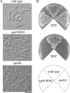
Figure 1.
Figure 1.
spo9 mutant exhibits a sporulation deficiency and temperature-sensitive growth. (A) Wild type (TN8), spo9-B261 (YF2), and spo9Δ (YF16) were incubated at 28°C on sporulation medium (MEA) for 2 d. Bar, 10 μm. (B) The same strain as shown in A was streaked on complete medium (YEA) and incubated at 25 or 36°C for 3 d.
Figure 2.
Figure 2.
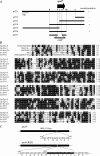
Structure of the spo9+ gene. (A) Restriction map, subcloning, and construction of null mutants. The arrow indicates the region and direction of the spo9+ ORF, which encodes a protein composed of 351 amino acid residues. All the subclones were derived from pYF1. Complementation by each subclone: +, complemented; −, did not complement. Restriction enzyme sites: A, AvrII; B, BamHI; Bg, BglII; H, HindIII; S, StuI; and X, XbaI. (B) Comparison of the amino acid sequences two of S. pombe FPS-like proteins, Spo9 (CAA19054), and Fps1 (CAB11097) and other members of the FPS family. SpSpo9, S. pombe Spo9; SpFps1, S. pombe Fps1; ScErg20, S. cerevisiae FPS (CAA89462); AtFPS, Arabidopsis thaliana FPS2 (AAB07247); and HsFPS, Homo sapiens FPS (P14324). Identical amino acid residues are shown in white against black; similar residues are shaded. (C) Determination of spo9-B261 mutation. Duplicated nucleotide region is shown in white against black.
Figure 3.
Figure 3.
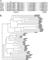
(A) Comparison of the amino acid sequences of conserved regions of Fps1 and Spo9 and other members of FPSs and GGPSs. ScBts1, S. cerevisiae GGPS, Bts1 (Q12051); AtGGPS, Arabidopsis thaliana GGPS6 (BAA23157); and HsGGPS, Homo sapiens GGPS1 (AAH05252). FARM and SARM are boxed. The GQ motif is shown in bold. The arrow indicates the fifth amino acid before the FARM motif, which is important for determination of the final product. (B) A phylogenetic tree of GGPSs and FPSs from various organisms calculated by the neighbor joining method. Fps1 and Spo9 are marked by asterisks. Abbreviations: Ag, Abies grandis; At, A. thaliana; Bm, Bombyx mori; Bt, Bos taurus; Cp, Claviceps purpurea; Dj, Dendroctonus jeffreyi; Dm, Drosophila melanogaster; Ec, E. coli; Gf, Gibberella fujikuroi; Gg, Gallus gallus; Hb, Hevea brasiliensis; Hs, Homo sapiens; Kg, Kitasatospora griseola; Kl, Kluyveromyces lactis; La, Lupinus albus; Lc, Lactarius chrysorrheus; Ld, Leishmania donovani; Mm, Mus musculus; Mp, Myzus persicae; Mt, Methanothermobacter thermautotrophicus; Nc, Neurospora crassa; Pa, Parthenium argentatum; Rn, Rattus norvegicus; Sa, Sulfolobus acidocaldarius; Sar, Streptomyces argenteolus; Sc, S. cerevisiae; Sm, Sphaceloma manihoticola; Sp, S. pombe; Tb, Trypanosoma brucei; Tg, Toxoplasma gondii; Tt, Thermus thermophilus; and Zm, Zea mays.
Figure 4.
Figure 4.
Transcription of spo9+ and fps1+ genes. Wild type (MKW5) precultured in growth medium (MM+N) was incubated in liquid sporulation medium (MM−N) at 28°C. After the shift, samples were harvested at the indicated times and subjected to Northern blotting. Top, spo9+ mRNA; middle, fps1+ mRNA; bottom, rRNA stained with ethidium bromide.
Figure 5.
Figure 5.
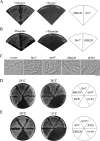
(A) S. cerevisiae strains YF79 [erg20Δ harboring pYEL2(ERG20)] or YF80 [erg20Δ harboring pYEL2(fps1)] was incubated on YP-galactose (induced) or YPD medium (repressed) at 30°C for 2 d. (B) S. pombe strains YF77 [fps1Δ harboring pREP81(fps1)] and YF78 [fps1Δ harboring pREP81(ERG20)] were incubated on MM medium with (repressed) or without thiamine (induced) at 25°C for 3 d. (C) Overexpression of various genes in spo9Δ. S. pombe strain YF16 (spo9Δ) was transformed with either empty pREP1, pREP1(fps1), pREP1(spo9), pREP1(ERG20), or pREP1(BTS1). The Leu+ transformants were incubated on sporulation medium (MEA) at 28°C for 2 d. Bar, 10 μm. (D) The same transformants as shown in C were streaked on YEA medium and incubated at 25 or 36°C for 3 d. (E) Overexpression of various genes in S. cerevisiae bts1Δ. S. cerevisiae strain SFNY368 (bts1Δ) was transformed with empty pYEL2, pYEL2(BTS1), pYEL2(spo9), pYEL2(fps1), or pYEL2(ERG20). The Leu+ transformants were incubated on YP-galactose medium at 30°C for 3 d and at 15°C for 9 d.
Figure 6.
Figure 6.
fps1+, but not spo9+, complements an E. coli ispA deletion mutant. Ubiquinone was extracted from E. coli wild-type strain DH5α and SF7 (ΔispA) harboring pGEX-KG, pGEX(fps1) or pGEX(spo9). UQ6 was included as an internal standard. The amount of UQ-8 plus UQ-7 was estimated to be 64, 9.2, 24, and 8.6 ng per A600 for growth in DH5α (wild type), SF7 harboring pGEX-KG, pGEX(fps1), and pGEX(spo9), respectively.
Figure 7.
Figure 7.
Coexpression of Spo9 and Fps1 is required for GGPS activity. E. coli DH10B carrying pACCAR25ΔcrtE was transformed with pMAL-c, pMAL(BTS1), pMAL(spo9), pMAL(fps1), pMAL (spo9+fps1), pMAL(spo9-R109Q+fps1), pMAL(spo9+fps1-R104Q), or pMAL(spo9-R109Q+fps1-R104Q); plated on LB medium supplemented with ampicillin and chloramphenicol; and incubated at 28°C for 3 d.
Figure 8.
Figure 8.
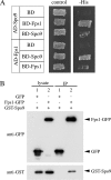
Spo9 interacts with Fps1. (A) Yeast two-hybrid analysis. Plasmids expressing the respective Gal4 activation domain (AD) and Gal4 DNA-binding domain (BD) fusions were tested for two-hybrid interaction. (B) Coimmunoprecipitation of Spo9 and Fps1. Cell extracts were prepared from vegetative cells expressing tagged proteins, GFP and GST-Spo9 or Fps1-GFP and GST-Spo9, and subjected to immunoprecipitation with anti-GFP antibody. Precipitates were analyzed by Western blotting by using anti-GFP, or anti-GST antibody.
Figure 9.
Figure 9.
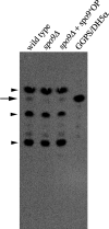
GGPS activity is significantly reduced in spo9 mutant. GGPS was assayed using [1-14C]IPP and FPP as substrates with crude extracts from wild-type (TN8), spo9Δ (YF16), and the spo9Δ harboring spo9+ (YF81) cells and E. coli DH5α harboring pBH expressing human GGPS1. The reaction products were hydrolyzed by acid phosphatase, and the resulting alcohols were analyzed by reversed phase TLC. An arrow indicates the product of GGOH. Other than GGOH, there are three intense spots; the uppermost and the lowest spots are farnesol and decaprenol, respectively, and a middle one is presumably ergosterol. These spots are indicated by arrowheads.
Figure 10.
Figure 10.
spo9 mutation causes mislocalization of Ypt7 but not Rhb1. Wild-type cells (YF51) carrying GFP-Ypt7 and spo9Δ cells (YF54) were ruptured, and then they were subjected to differential centrifugation to fractionate into a P100 membrane fraction and an S100 supernatant. Each fraction was resolved by SDS-polyacrylamide gel electrophoresis and subjected to Western blotting using either anti-GFP, anti-Rhb1, or anti-Spo14 (control) antibody.
Figure 11.
Figure 11.
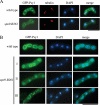
Aberrant assembly of the forespore membrane in spo9 mutant. (A) Assembly of the forespore membrane during metaphase II. Wild type (YN68) and spo9-B261 (YF5) were cultured in SSL-N to induce meiosis at 28°C for 8 h. Fixed cells were doubly stained with anti-α-tubulin and DAPI. (B) Classification of terminal phenotypes of the forespore membrane in spo9 zygotes. Strain, culture conditions and staining procedures are the same as described in Figure 11A. Type I, forespore membranes engulfed each nucleus but prespores were remarkably small. Type II, four aggregates of GFP-Psy1 were formed close to nuclei. Type III, forespore membrane formation arrested. Bars, 10 μm.
Figure 12.
Figure 12.
Fine structures of spo9Δ asci. Mature spores in wild type (A) and anucleated spore-like bodies in spo9Δ mutant (B–D). N, SPB, and FSM denote the nucleus, spindle pole body, and forespore membrane, respectively. Note that many membrane vesicles (indicated by arrows) and Golgi-like structures (indicated by arrowheads) are present in the cytoplasm of spo9Δ. Bar, 0.5 μm.

Similar articles

Cited by

References

    1. Blanchard L., Karst F. Characterization of a lysine-to-glutamic acid mutation in a conservative sequence of farnesyl diphosphate synthase from Saccharomyces cerevisiae. Gene. 1993;125:185–189. - PubMed
    1. Bresch C., Muller G., Egel R. Genes involved in meiosis and sporulation of a yeast. Mol. Gen. Genet. 1968;102:301–306. - PubMed
    1. Burke C. C., Wildung M. R., Croteau R. Geranyl diphosphate synthase: cloning, expression, and characterization of this prenyltransferase as a heterodimer. Proc. Natl. Acad. Sci. USA. 1999;96:13062–13067. - PMC - PubMed
    1. Casey P. J. Biochemistry of protein prenylation. J. Lipid Res. 1992;33:1731–1740. - PubMed
    1. Casey P. J. Lipid modifications of G proteins. Curr. Opin. Cell Biol. 1994;6:219–225. - PubMed

Publication types

MeSH terms

Substances