. 2007 Oct 1;110(7):2390-8.
doi: 10.1182/blood-2006-11-057273. Epub 2007 Jun 27.

FANCJ (BACH1) helicase forms DNA damage inducible foci with replication protein A and interacts physically and functionally with the single-stranded DNA-binding protein

Affiliations

FANCJ (BACH1) helicase forms DNA damage inducible foci with replication protein A and interacts physically and functionally with the single-stranded DNA-binding protein

Rigu Gupta et al. Blood. .

Abstract

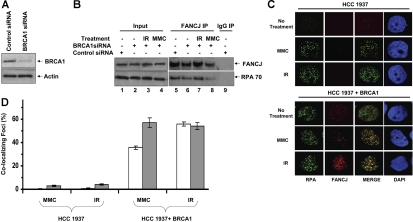
The BRCA1 associated C-terminal helicase (BACH1, designated FANCJ) is implicated in the chromosomal instability genetic disorder Fanconi anemia (FA) and hereditary breast cancer. A critical role of FANCJ helicase may be to restart replication as a component of downstream events that occur during the repair of DNA cross-links or double-strand breaks. We investigated the potential interaction of FANCJ with replication protein A (RPA), a single-stranded DNA-binding protein implicated in both DNA replication and repair. FANCJ and RPA were shown to coimmunoprecipitate most likely through a direct interaction of FANCJ and the RPA70 subunit. Moreover, dependent on the presence of BRCA1, FANCJ colocalizes with RPA in nuclear foci after DNA damage. Our data are consistent with a model in which FANCJ associates with RPA in a DNA damage-inducible manner and through the protein interaction RPA stimulates FANCJ helicase to better unwind duplex DNA substrates. These findings identify RPA as the first regulatory partner of FANCJ. The FANCJ-RPA interaction is likely to be important for the role of the helicase to more efficiently unwind DNA repair intermediates to maintain genomic stability.


Figures

Figure 1
Figure 1
FANCJ and RPA are associated with each other in vivo and directly interact. (A) Coimmunoprecipitation of RPA with FANCJ. FANCJ antibody coprecipitates FANCJ and RPA from HeLa or FA-J–corrected cells but not from the FA-J extracts. The blot was probed with rabbit anti-FANCJ (top) and mouse anti-RPA (bottom) antibodies. (Lane 1) HeLa nuclear extract (15% of input), (lane 2) control immunoprecipitate from HeLa nuclear extract using normal rabbit IgG, (lane 3) immunoprecipitate from HeLa nuclear extract using rabbit anti-FANCJ antibody, (lane 4) FA-J whole-cell extract (WCE; 15% of input), (lane 5) immunoprecipitate from FA-J WCE using rabbit anti-FANCJ antibody, (lane 6) control precipitate from FA-J WCE using normal rabbit IgG, (lane 7) WCE from HeLa included as control for Western detection of FANCJ and RPA, (lane 8) FA-J corrected WCE (15% of input), (lane 9), immunoprecipitate from FA-J–corrected WCE using rabbit anti-FANCJ antibody, and (lane 10) control immunoprecipitate from FA-J–corrected WCE using normal rabbit IgG. (B) Coimmunoprecipitation of BRCA1 with FANCJ. FANCJ antibody coprecipitates FANCJ and BRCA1 from HeLa nuclear extract. The blot was probed with rabbit anti-FANCJ (top) and mouse anti-BRCA1 (bottom) antibodies. (Lane 1) HeLa nuclear extract (15% of input), (lane 2) immunoprecipitate from HeLa nuclear extract using rabbit anti-FANCJ antibody, and (lane 3) control immunoprecipitate from HeLa nuclear extract using normal rabbit IgG. (C) FANCJ antibody coprecipitates FANCJ and RPA from HeLa nuclear extracts in the presence of ethidium bromide or DNaseI. The blot was probed with rabbit anti-FANCJ (top) and mouse anti-RPA (bottom) antibodies. (Lane 1) HeLa nuclear extract (15% of input), (lane 2) immunoprecipitate from HeLa nuclear extract using rabbit anti-FANCJ antibody, (lanes 3 and 4) immunoprecipitate from HeLa nuclear extracts in the presence of 2 μg/mL DNaseI or 10 μg/mL ethidium bromide using rabbit anti-FANCJ antibody, and (lane 5) control immunoprecipitate from HeLa nuclear extract using normal rabbit IgG. (D,E) FANCJ and RPA form a complex by direct physical interaction. (D) RPA (96 nM heterotrimer, □) or ESSB (96 nM homotetramer, ■) was coated onto the ELISA plate. After blocking with 3% BSA, the wells were incubated with increasing concentrations of purified recombinant FANCJ protein (0-150 nM) for 60 minutes at 37°C. Wells were aspirated and washed 3 times, and bound FANCJ-WT protein was detected by ELISA with a rabbit polyclonal antibody against FANCJ. (E) Same as described for panel D except 2 μg/mL DNase I or 10 μg/mL ethidium bromide (EtBr) were incubated with RPA (96 nM) and FANCJ (77 nM) during the binding step in the corresponding wells. The values represent the mean of 3 independent experiments performed in duplicate with standard deviation (SD) indicated by error bars. (F) FANCJ and RPA interact by the 70-kDa subunit of RPA. Purified RPA, ESSB, and BSA (as indicated above the lanes) were subjected to SDS–polyacrylamide gel electrophoresis on 3 identical gels. The proteins bound to membranes were stained with Ponceau S or transferred to nitrocellulose membrane and incubated with either purified FANCJ (+FANCJ) or no protein (−FANCJ). Western blotting with anti-FANCJ antibody was then used to detect the presence of FANCJ on each membrane. The positions of the 70-, 32-, and 14-kDa subunits of RPA are indicated by asterisks. The positions of the molecular mass standards running parallel are shown on the left.
Figure 2
Figure 2
DNA damage-inducible colocalization of FANCJ helicase and RPA. (A) HeLa cells were treated with the DNA-damaging agents MMC (500 ng/mL) or hydroxyurea (2 mM) or exposed to 10 Gy IR as described in “Immunofluorescence cellular localization studies” After fixation and permeabilization, cells were incubated with anti-RPA (green) and anti-FANCJ (red) antibodies. After treatment with MMC, hydroxyurea, or IR, RPA localizes in nuclear foci that coincide with FANCJ foci as shown in the overlapped images. The yellow color results from the overlapping of the red and green foci in the merged images. In control untreated cells, RPA and FANCJ staining is diffuse. (B) Quantitation of FANCJ-RPA colocalizing foci as described in panel A. Percentages of RPA foci colocalizing with FANCJ are represented □, and percentages of FANCJ foci colocalizing with RPA are represented (▩). Experimental data are the mean of at least 3 independent experiments with standard deviations indicated by error bars. (C) FA-J vector and FA-J corrected cells were treated with the indicated DNA-damaging agents and processed for immunofluorescence as described above.
Figure 3
Figure 3
Coimmunoprecipitation of FANCJ and RPA after DNA damage. Nuclear extracts were prepared from HeLa cells that were untreated or exposed to IR (10 Gy) or MMC (500 ng/mL) and immunoprecipitated with anti-FANCJ or anti-RPA antibody as indicated. The blot was probed with rabbit anti-FANCJ (top) and mouse anti-RPA70 or anti-RPA32 (bottom) antibodies.
Figure 4
Figure 4
FANCJ-RPA interaction is intact in FA mutant cells coimmunoprecipitation of FANCJ and RPA in FA-A and FA-D2 cells. Coimmunoprecipitation of RPA with FANCJ in FA-A vector and FA-A corrected cells (A) or FA-D2 vector and FA-D2–corrected cells (B) with the use of FANCJ antibody. The blot was probed with rabbit anti-FANCJ (top) and mouse anti-RPA (bottom) antibodies. Input represents 15% of WCE input for coimmunoprecipitation experiments. Control immunoprecipitate from WCE of FA-A–corrected or FA-D2–corrected cells with the use of normal rabbit IgG is shown in lane 5.
Figure 5
Figure 5
Effect of BRCA1 deficiency on the DNA damage-inducible association of FANCJ and RPA in BRCA1-deficient cells. (A) siRNA depletion of BRCA1. WCE of HeLa cells transfected with control or BRCA1 siRNA were probed using an antibody against BRCA1. Actin serves as a loading control. (B) WCEs were prepared from control siRNA or BRCA1 siRNA HeLa cells that were untreated or exposed to IR (10 Gy) or MMC (500 ng/mL) as indicated and immunoprecipitated with FANCJ antibody. The blot was probed with rabbit anti-FANCJ (top) and mouse anti-RPA70 (bottom) antibodies. (C) HCC 1937 cells form RPA but not FANCJ DNA damage-inducible foci. BRCA1 mutant HCC 1937 or reconstituted cells were treated with the DNA-damaging agent MMC (500 ng/mL) or exposed to 10 Gy IR as described in “Immunofluorescence cellular localization studies.” After fixation and permeabilization, cells were incubated with anti-RPA (green) and anti-FANCJ (red) antibodies. (D) Quantitation of FANCJ-RPA colocalizing foci as described in panel C. Percentages of RPA foci colocalizing with FANCJ are represented (□). Percentages of FANCJ foci colocalizing with RPA are represented (▩). Experimental data are the mean of at least 3 independent experiments with SD indicated by error bars.
Figure 6
Figure 6
Limited unwinding reaction catalyzed by FANCJ is stimulated by RPA. (A) FANCJ helicase catalyzes a limited unwinding reaction. Helicase assays were as described, using the indicated concentrations of FANCJ-WT or FANCJ-M299I and forked duplex DNA substrates of 22 bp and 47 bp. Incubation was at 30°C for 15 minutes. Reaction products were analyzed by nondenaturing gel electrophoresis. Quantitation of results from helicase assays with standard deviations indicated by error bars. FANCJ-WT, 22 bp, ▲; FANCJ-M299I, 22 bp, △; FANCJ-WT, 47 bp, ■; FANCJ-M299I, 47 bp, □. Percentage of displacement is expressed as a function of FANCJ-WT or FANCJ-M299I protein concentration. (B-D) Stimulation of FANCJ-WT helicase activity by RPA. FANCJ-WT protein (4.8 nM) was incubated with the 47-bp forked duplex in the presence of the indicated concentrations of RPA heterotrimer (B) or ESSB homotetramer (C) under standard helicase reaction conditions. Incubation was at 30°C for 60 minutes. (D) Quantitation of results from helicase assays with SD indicated by error bars. Percentage of displacement is expressed as a function of single-stranded DNA-binding protein concentration (RPA heterotrimer or ESSB homotetramer). (Inset) Quantitation of results with percentage of displacement expressed as a function of the ratio (SSB binding unit)/(DNA-binding unit).
Figure 7
Figure 7
Kinetic analyses of DNA unwinding of the 47-bp forked duplex DNA substrate by FANCJ-WT or FANCJ polymorphic variants in the presence of RPA or ESSB. (A) FANCJ-WT (4.8 nM) or its associated variants (FANCJ-M299I or FANCJ-P47A) was incubated with the 47-bp forked duplex in the absence or presence of 24 nM RPA heterotrimer under standard helicase reaction conditions. Incubation was at 30°C for the indicated times. Quantitation of results from helicase assays with SD are indicated by error bars. FANCJ-WT (△); FANCJ-WT + RPA (▲); FANCJ-M299I (○); FANCJ-M299I + RPA (●); FANCJ-P47A (□); FANCJ-P47A + RPA (■).(B) Coimmunoprecipitation of FANCJ (FANCJ) sequence variants with RPA. FA-J cells were transiently transfected with plasmid DNA encoding Myc-tagged FANCJ-WT, FANCJ-M299I, or FANCJ-P47A, and WCEs were used for coimmunoprecipitation experiments using FANCJ antibody. The blot was probed with rabbit anti-FANCJ (top) and mouse anti-RPA (bottom) antibodies. Input represents 15% of WCE input for coimmunoprecipitation experiments. (C) RPA directly binds to FANCJ variants. RPA (96 nM heterotrimer) was coated onto the ELISA plate. After blocking with 3% BSA, the wells were incubated with increasing concentrations of purified recombinant FANCJ-M299I or FANCJ-P47A protein (0-150 nM) for 60 minutes at 37°C. Wells were aspirated and washed 3 times, and bound FANCJ protein was detected by ELISA using a rabbit polyclonal antibody against FANCJ.

References

    1. Kennedy RD, D'Andrea AD. The Fanconi anemia/BRCA pathway: new faces in the crowd. Genes Dev. 2005;19:2925–2940. - PubMed
    1. Mirchandani KD, D'Andrea AD. The Fanconi anemia/BRCA pathway: a coordinator of cross-link repair. Exp Cell Res. 2006;312:2647–2653. - PubMed
    1. Meijer GA. The 13th Fanconi anemia gene identified: FA. Cell Oncol. 2007;29:181–182. - PMC - PubMed
    1. Levitus M, Waisfisz Q, Godthelp BC, et al. The DNA helicase BRIP1 is defective in Fanconi anemia complementation group J. Nat Genet. 2005;37:934–935. - PubMed
    1. Levran O, Attwooll C, Henry RT, et al. The BRCA1-interacting helicase BRIP1 is deficient in Fanconi anemia. Nat Genet. 2005;37:931–933. - PubMed

Publication types

MeSH terms

Substances