. 2007 Aug 31;282(35):25376-84.
doi: 10.1074/jbc.M704698200. Epub 2007 Jun 27.

Mosquito heparan sulfate and its potential role in malaria infection and transmission

Affiliations

Mosquito heparan sulfate and its potential role in malaria infection and transmission

Photini Sinnis et al. J Biol Chem. .

Abstract

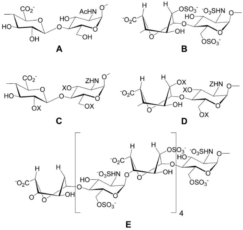
Heparan sulfate has been isolated for the first time from the mosquito Anopheles stephensi, a known vector for Plasmodium parasites, the causative agents of malaria. Chondroitin sulfate, but not dermatan sulfate or hyaluronan, was also present in the mosquito. The glycosaminoglycans were isolated, from salivary glands and midguts of the mosquito in quantities sufficient for disaccharide microanalysis. Both of these organs are invaded at different stages of the Plasmodium life cycle. Mosquito heparan sulfate was found to contain the critical trisulfated disaccharide sequence, -->4)beta-D-GlcNS6S(1-->4)-alpha-L-IdoA2S(1-->, that is commonly found in human liver heparan sulfate, which serves as the receptor for apolipoprotein E and is also believed to be responsible for binding to the circumsporozoite protein found on the surface of the Plasmodium sporozoite. The heparan sulfate isolated from the whole mosquito binds to circumsporozoite protein, suggesting a role within the mosquito for infection and transmission of the Plasmodium parasite.


Figures

FIGURE 1
FIGURE 1. The life cycle of Plasmodium in the mammalian host (A) and the mosquito (B)
A, sporozoites are injected during blood feeding by infected anophelene mosquitoes, go to the liver, and invade hepatocytes, where they divide, rupture from the hepatocyte, and enter erythrocytes. The released merozoites invade erythrocytes, mature, divide, rupture from the cell, and enter new erythrocytes. The asexual erythrocytic cycle gives rise to the clinical symptoms associated with malaria. Some merozoites differentiate to gametocytes, which are infective for mosquitoes. This image is reprinted with permission from Ref. . B, mosquitoes become infected with Plasmodium when they ingest gametocytes (1) during blood feeding. In the midgut (MG) of the mosquito, these cells differentiate (2), fertilization occurs, and the resultant zygote differentiates into an ookinete (3), which traverses the midgut wall (4) and comes to rest extracellularly between the basal lamina and the midgut (5). The ookinete then differentiates into an oocyst, and sporozoites develop (6), and when mature, they emerge into the hemocoel (HC) of the mosquito and invade salivary glands (SG) (7). When the mosquito takes another blood meal, these salivary gland sporozoites are injected into the mammalian host (8), thus continuing the cycle. This image is reprinted with permission from Ref. .
FIGURE 2
FIGURE 2. Structure of heparan sulfate glycosaminoglycans
A, unsulfated disaccharide sequence prominent in heparan sulfate. B, fully sulfated disaccharide sequence prominent in heparin. C, structural variants of glucuronic acid containing disaccharide sequences found in heparan sulfate and heparin (X = SO3, H). D, structural variants of iduronic acid containing disaccharide sequences found in heparan sulfate and heparin (X = SO3, H; Z = SO3, COCH3, H). E, proposed apoE and CSP binding domain.
FIGURE 3
FIGURE 3. Reversed-phase ion pairing-HPLC determination of chondroitin lyase-derived glycosaminoglycan disaccharides
A, female mosquitoes; B, salivary glands; C, midguts; D, malpighian tubules and ovaries. Peak 1, ΔDi-0S; peak 2, ΔDi-4S. Other conditions were as described under “Experimental Procedures.”
FIGURE 4
FIGURE 4. RPIP-HPLC determination of heparin lyase-derived glycosaminoglycan disaccharides
A, female mosquitoes; B, salivary glands; C, midguts; D, malpighian tubules and ovaries. Peak 1, ΔUA-GlcNAc; peak 2, ΔUA-GlcNS; peak 3, ΔUA-GlcNAc6S; peak 4, ΔUA-GlcNS6S; peak 5, ΔUA2S-GlcNS; peak 6, ΔUA2S-GlcNS6S. Other conditions were as described under “Experimental Procedures.”
FIGURE 5
FIGURE 5. Surface plasmon resonance studies on whole mosquito heparan sulfate binding to CSP
Shown are sensorgrams at heparan sulfate (solid lines) concentrations of (beginning from the bottom solid curve) 1, 5, 10, 20, and 50 μM. The start of injection begins at 0 s and ends at 180 s (the arrow indicates stop of injection). The dashed curve represents interaction between CSP and 10 μM porcine liver HS.

References

    1. Frevert U, Sinnis P, Cerami C, Shreffler W, Takacs B, Nussenzweig V. J Exp Med. 1993;177:1287–1298. - PMC - PubMed
    1. Pinzon-Ortiz C, Friedman J, Esko J, Sinnis P. J Biol Chem. 2001;276:26784–26791. - PMC - PubMed
    1. Lyon M, Dakin JA, Gallagher JT. J Biol Chem. 1994;269:11208–11215. - PubMed
    1. Hernáiz MJ, Yang HO, Gunay NS, Linhardt RJ. Carbohydr Polym. 2002;48:153–160.
    1. Vongchan P, Warda M, Toyoda H, Toida T, Marks RM, Linhardt RJ. Biochim Biophys Acta. 2005;1721:1–8. - PubMed

Publication types

MeSH terms

LinkOut - more resources