. 2007 Aug 24;371(4):971-88.
doi: 10.1016/j.jmb.2007.05.075. Epub 2007 Jun 2.

Protein-protein Förster resonance energy transfer analysis of nucleosome core particles containing H2A and H2A.Z

Affiliations

Protein-protein Förster resonance energy transfer analysis of nucleosome core particles containing H2A and H2A.Z

Duane A Hoch et al. J Mol Biol. .

Abstract

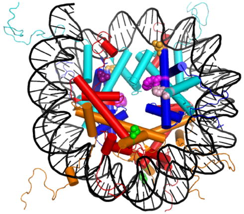
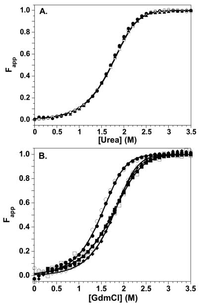
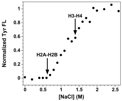
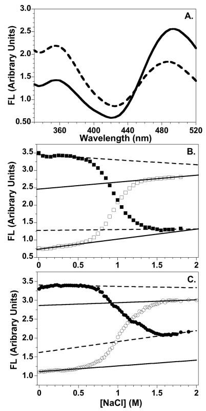
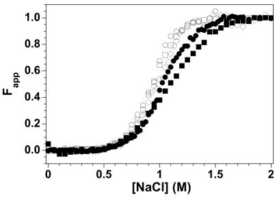
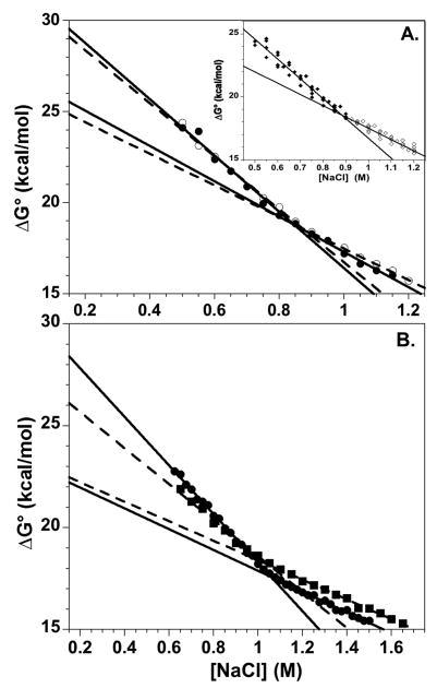
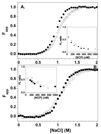
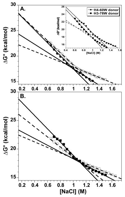
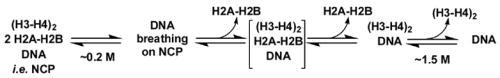
A protein-protein Förster resonance energy transfer (FRET) system, employing probes at multiple positions, was designed to specifically monitor the dissociation of the H2A-H2B dimer from the nucleosome core particle (NCP). Tryptophan donors and Cys-AEDANS acceptors were chosen because, compared to previous NCP FRET fluorophores, they: (1) are smaller and less hydrophobic, which should minimize perturbations of histone and NCP structure; and (2) have an R0 of 20 A, which is much less than the dimensions of the NCP (approximately 50 A width and approximately 100 A diameter). Equilibrium protein unfolding titrations indicate that the donor and acceptor moieties have minimal effects on the stability of the H2A-H2B dimer and (H3-H4)2 tetramer. NCPs containing the various FRET pairs were reconstituted with the 601 DNA positioning element. Equilibrium NaCl-induced dissociation of the modified NCPs showed that the 601 sequence stabilized the NCP to dimer dissociation relative to weaker positioning sequences. This finding implies a significant role for the H2A-H2B dimers in determining the DNA sequence dependence of NCP stability. The free energy of dissociation determined from reversible and well-defined sigmoidal transitions revealed two distinct phases reflecting the dissociation of individual H2A-H2B dimers, confirming cooperativity as suggested previously; these data allow quantitative description of the cooperativity. The FRET system was then used to study the effects of the histone variant H2A.Z on NCP stability; previous studies have reported both destabilizing and stabilizing effects. H2A.Z FRET NCP dissociation transitions suggest a slight increase in stability but a significant increase in cooperativity of the dimer dissociations. Thus, the utility of this protein-protein FRET system to monitor the effects of histone variants on NCP dynamics has been demonstrated, and the system appears equally well-suited for dissection of the kinetic processes of dimer association and dissociation from the NCP.


Figures

Figure 1
Figure 1
The structure of the NCP (1kx5.pdb) showing the sites mutated to introduce FRET fluorophores (Table 1). Histones are colored: H2A, red; H2B, orange; H3, cyan; and H4, blue. Mutations: H2A Leu-108 (dark purple); H2B Ser-109 (green); H3 Phe-78 (pink); H4 Leu-49 (orange), and H4 Val-60 (light purple). This figure was rendered with Pymol (Delano Scientific, LLC, San Carlos, CA).
Figure 2
Figure 2
Equilibrium stability of the WT and engineered histones. CD data, open symbols; FL data, closed symbols. Equilibrium titration data were collected from 0 to 4 M denaturant, but the transition region is expanded for clarity. A) Urea-induced denaturation of H2A-H2B dimers at 10 μM monomer. H2A-108Cys-AEDANS/H2B, circles; H2A/H2B-109Cys-AEDANS, triangles. The transition for WT H2A-H2B is represented by the solid line. B) GdmCl-induced denaturation of the H3-H4 oligomers at 4 μM monomer in 1 M TMAO. H3-78W/H4, circles; H3/H4-49W, diamonds; H3/H4-60W, squares. The solid lines represent global fits of the entire data sets for the histone mutants. The WT H3-H4 transition is represented by the dashed line. Conditions: 200 mM KCl, 0.1 mM EDTA, 20 mM KPi pH 7.2, 25°C.
Figure 3
Figure 3
NaCl-induced dissociation of NCPs reconstituted with the 601 DNA sequence and recombinant WT histones monitored by Tyr FL. Arrows indicate the beginning of the transitions corresponding to the dissociation the H2A-H2B dimers and H3-H4 tetramer. Conditions: 250 nM NCP, 0.1 mM EDTA, 0.1 mM β-ME, 20 mM Tris-Cl pH 7.6, 25°C.
Figure 4
Figure 4
NCPs reconstituted with 149 bp 601 DNA electrophoresed on 5% native acrylamide gels and stained with EtBr. A. NCPs with major H2A. From left to right: 100 bp ladder; H4-60W/H2A-108C-AEDANS; H4-60W/H2B-109C-AEDANS; H4-49W/H2A-108C-AEDANS; H4-49W/H2B-109C-AEDANS; H3-78W/H2A-108C-AEDANS; H3-78W/H2B-109-C-AEDANS; 149 bp 601 DNA. B. NCPs with H2A.Z. From left to right: 100 bp ladder; H3-78W/H2B-109C-AEDANS; H4-60W/H2B-109C-AEDANS.
Figure 5
Figure 5
Representative FL data for the salt-induced dissociation of the FRET NCPs. A. FL emission spectra of the H4-49W/H2A-108Cys-AEDANS FRET NCP at 0 M (solid line) and 1.5 M NaCl concentrations (dashed line). B and C. Salt dependence of the FL intensities for the H4-60W/H2A-108Cys-AEDANS and the H3-78W/H2B-109Cys-AEDANS NCPs, respectively. Trp donor FL at 350 nm: □ and ○; Cys-AEDANS FL at 490 nm: ■ and ●. Baselines for intact NCPs and the dimer-dissociated complex are indicated by solid and dashed lines, respectively. Conditions: 250 nM NCP; 20 mM Tris-Cl pH 7.6, 0.1 mM EDTA, 0.1 mM β-ME, 25°C.
Figure 6
Figure 6
Fapp normalization of the NaCl-dependence of the Trp FL data for the FRET NCPs. Open symbols: NCPs with the H2A-108Cys-AEDANS acceptor and the H3-78W (○), H4-49W (◇), and H4-60W (□) donors. Closed symbols: NCPs with the H2B-109Cys-AEDANS acceptor and the H3-W78 (●) and H4-W60 (■) donors. Conditions are given in the legend of Figure 5.
Figure 7
Figure 7
The salt dependence of the free energy of dissociation for the H2A-H2B dimers from the NCP, showing extrapolations to physiological ionic strengths. A) H3-78W/H2A-108Cys-AEDANS NCP. Values were calculated from FL intensities at 350 nm (●, solid lines) and 490 nm (○, dashed lines) and were fit to two lines which intersect near the transition midpoint (Figure 6). Inset: overlay of data from the three H2A-108Cys-AEDANS NCPs; the lines represent the average ΔG°(H2O) and m values given in the text. B. H2B-109Cys-AEDANS NCPs with H3-78W (●, solid lines) or H4-60W (■, dashed lines). Conditions are given in legend of Figure 5.
Figure 8
Figure 8
Salt-induced equilibrium dissociation transitions for H2B-109Cys-AEDANS FRET NCPs with H2A (open symbols) and H2A.Z (solid symbols) normalized to Fapp values. A) H3-78W donor; B) H4-60W donor. The insets show the normalized ratios of the 350 nm and 490 nm FL as a function of NCP concentration at 1 M NaCl. Buffer conditions are given in the legend of Figure 5.
Figure 9
Figure 9
Comparison of the salt dependence of the free energy of dissociation of the H2A-H2B dimers from H2A.Z and H2A NCPs, utilizing the H2B-109Cys-AEDANS acceptor. Linear extrapolations are shown to physiological ionic strengths. NCPs with the major H2A: open symbols, dashed lines; NCPs with the H2A.Z variant: solid symbols, solid lines. A) H3-78W donor. B) H4-60W donor. Inset panel A) Comparison of the two H2A.Z NCPs with H3-78W donor (circles) and H4-60W donor (squares). Conditions are given in legend of Figure 5.
Scheme 2
Scheme 2
Salt-induced NCP unfolding

References

    1. Workman JL, Kingston RE. Alteration of nucleosome structure as a mechanism of transcriptional regulation. Ann Rev Biochem. 1998;67:545–79. - PubMed
    1. Khorasanizadeh S. The nucleosome: from genomic organization to genomic regulation. Cell. 2004;116:259–72. - PubMed
    1. Luger K. Dynamic nucleosomes. Chromosome Res. 2006;14:5–16. - PubMed
    1. Wolffe AP. Chromatin remodeling: why it is important in cancer. Oncogene. 2001;20:2988–90. - PubMed
    1. Huang C, Sloan EA, Boerkoel CF. Chromatin remodeling and human disease. Curr Opin Genet Dev. 2003;13:246–52. - PubMed

Publication types

LinkOut - more resources