. 2007 Sep;14(18):1309-18.
doi: 10.1038/sj.gt.3302981. Epub 2007 Jun 28.

Modulating mtDNA heteroplasmy by mitochondria-targeted restriction endonucleases in a 'differential multiple cleavage-site' model

Affiliations

Modulating mtDNA heteroplasmy by mitochondria-targeted restriction endonucleases in a 'differential multiple cleavage-site' model

S R Bacman et al. Gene Ther. 2007 Sep.

Abstract

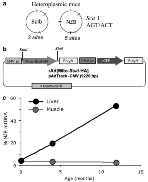
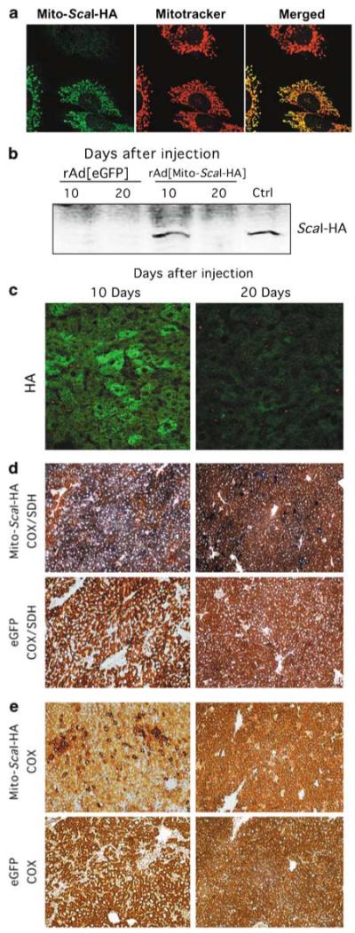
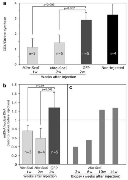
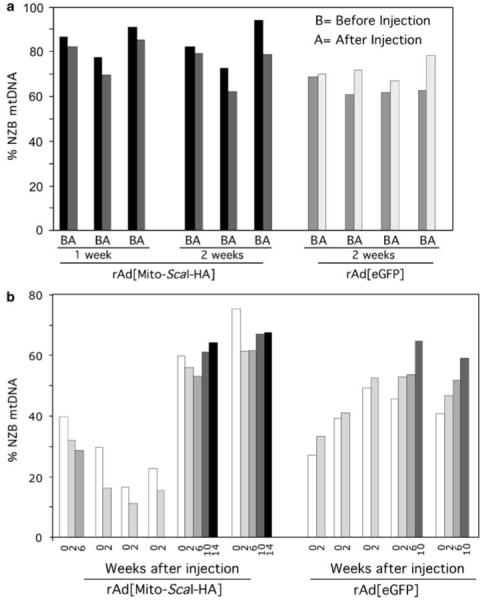
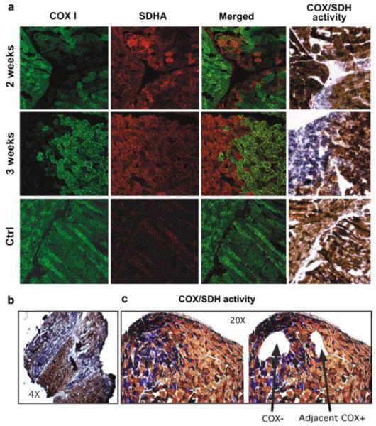
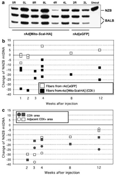
The ability to manipulate mitochondrial DNA (mtDNA) heteroplasmy would provide a powerful tool to treat mitochondrial diseases. Recent studies showed that mitochondria-targeted restriction endonucleases can modify mtDNA heteroplasmy in a predictable and efficient manner if it recognizes a single site in the mutant mtDNA. However, the applicability of such model is limited to mutations that create a novel cleavage site, not present in the wild-type mtDNA. We attempted to extend this approach to a 'differential multiple cleavage site' model, where an mtDNA mutation creates an extra restriction site to the ones normally present in the wild-type mtDNA. Taking advantage of a heteroplasmic mouse model harboring two haplotypes of mtDNA (NZB/BALB) and using adenovirus as a gene vector, we delivered a mitochondria-targeted Scal restriction endonuclease to different mouse tissues. Scal recognizes five sites in the NZB mtDNA but only three in BALB mtDNA. Our results showed that changes in mtDNA heteroplasmy were obtained by the expression of mitochondria-targeted ScaI in both liver, after intravenous injection, and in skeletal muscle, after intramuscular injection. Although mtDNA depletion was an undesirable side effect, our data suggest that under a regulated expression system, mtDNA depletion could be minimized and restriction endonucleases recognizing multiple sites could have a potential for therapeutic use.


Figures

Figure 1
Figure 1
Production and characterization of the tools and reagents used to test mitochondria-targeted restriction endonucleases. (a) ScaI restriction endonuclease can digest both BALB and NZB mtDNA, recognizing three sites in the BALB mtDNA and five sites in the NZB mtDNA. ScaI was chosen because it resembles a human disease-related mutation model, namely the creation of an extra ApaI restriction endonuclease site in MELAS syndrome mtDNA, in addition to sites present in the wild-type human mtDNA. (b) The recombinant rAd[Mito-ScaI-HA] contained a mitochondrial targeting sequence (cytochrome oxidase subunit 8 (Cox8)), a gene coding for Streptomyces caespitosus ScaI restriction endonuclease synthesized in vitro and an HA tag for immunological detection. The construct was cloned in a recombinant adenovirus vector also encoding the enhanced green fluorescence protein (eGFP) under the control of cytomegalovirus promoter (CMV). (c) The mouse model utilized (NZB/BALB mtDNA) shows an age-related shift in mtDNA heteroplasmy in liver, where the NZB haplotype increases with age. On the other hand, mtDNA heteroplasmy in tissues such as muscle remains unchanged. mtDNA, mitochondrial DNA; MELAS, mitochondrial encephalopathy, lactic acidosis and stroke-like episodes; NZB, New Zealand Black.
Figure 2
Figure 2
Strong expression of rAd[Mito-ScaI-HA] in liver leads to COX deficiency. (a) The mitochondrial localization of ScaI was confirmed by confocal microscopy of mouse cell line (hepatocyte-derived) infected with rAd[Mito-ScaI-HA]. Colocalization of Mito-ScaI-HA and Mitotracker was evident in transduced cells. (b) Steady-state levels of Mito-ScaI-HA were detected by western blot (anti-HA) of liver samples after injection. Strong protein expression was observed in a liver sample transduced with rAd[Mito-ScaI-HA] 10 days after jugular injection. No HA expression was detected at 20 days after rAd[Mito-ScaI-HA] or after rAd[eGFP] injection. Cultured cells transduced with rAd[Mito-ScaI-HA] were used as positive controls. (c) The same pattern of expression was observed by immunocytochemistry analysis with an anti-HA antibody (green). COX (e) and COX/SDH (d) activities in liver samples at 10 or 20 days after injection. COX-negative regions with increased SDH staining were observed in rAd[Mito-ScaI-HA]-transduced liver 10 days after injection. These areas were reduced 20 days after systemic gene delivery. rAd[eGFP]-injected mice showed no change in COX or COX/SDH staining (d and e). COX, cytochrome c oxidase; eGFP, enhanced green fluorescence protein; HA, hemag-glutinin; SDH, succinate dehydrogenase.
Figure 3
Figure 3
Transduction of rAd[Mito-ScaI-HA] in liver leads to a partial COX deficiency and mtDNA depletion. (a) Enzymatic activity measured spectrophotometrically in liver homogenates of rAd-injected mice. Specific COX activity is expressed as ratios to citrate synthase activity. rAd[Mito-ScaI-HA]-infected liver samples showed decreased COX activity when compared to rAd[eGFP] samples or uninjected controls. (b) mtDNA levels were analyzed by Southern blot. rAd[Mito-ScaI-HA]-infected samples from old-mice group showed decreased ratios of mtDNA/nuclear DNA when compared to the respective pre-injection biopsy samples. (c) Serial liver biopsies were analyzed for mtDNA content in 4-month-old mice. Each bar represents the liver biopsy performed in the same animal analyzed at different times after rAd[Mito-ScaI-HA] delivery. Error bars represent standard deviation. Significance was assessed by the Student’s t-test (individual comparisons to the GFP-treated group). COX, cytochrome c oxidase; eGFP, enhanced green fluorescence protein; mtDNA, mitochondrial DNA.
Figure 4
Figure 4
Transduction of rAd[Mito-ScaI-HA] in liver leads to a shift in mtDNA heteroplasmy. (a) The percentage of NZB mtDNA genotype was quantified by the last-cycle hot PCR/RFLP analysis. NZB mtDNA was decreased in 1-year-old mice 1 or 2 weeks after injection of rAd[Mito-ScaI-HA], and increased as expected after rAd[eGFP] administration. Bars represent the percentage of NZB mtDNA in liver samples before and after adenovirus injection. The decrease of NZB mtDNA in the rAd[Mito-ScaI-HA]-injected liver was 7.2±2.4% (1 week, n = 3) and 11.5±7.1% (2 weeks, n = 3). In contrast, the rAd[eGFP]-injected livers showed an increase in NZB mtDNA (13.5±10.3%, n = 4). The difference in heteroplasmy shift between the 2 weeks injected animals (rAd[Mito-ScaI-HA] vs rAd[eGFP]) was significant (P 0.015). (b) Serial liver biopsies were performed after rAd[Mito-ScaI-HA] or rAd[eGFP] injection. Each cluster of bars represents data from one animal, which was analyzed for different periods. Each bar corresponds to liver biopsies analyzed at different times (1-14 weeks) after adenoviral injection. eGFP, enhanced green fluorescence protein; mtDNA, mitochondrial DNA; NZB, New Zealand Black.
Figure 5
Figure 5
Expression of mitochondrial ScaI in muscle leads to COX deficiency in injected areas. (a) Immunostaining of muscle samples at different time points after focal injection with rAd[Mito-ScaI-HA] with MT-CO1-Alexa-conjugated monoclonal antibody shows a decrease in CO1. Mitochondrial proliferation can be inferred from the increased staining with the SDHA antibody. CO1-deficient areas also showed decreased COX activity with increased SDH staining (fourth column). (b) Muscle sections stained for the COX/SDH activity showed a pattern of COX-deficient areas (at the sites of focal injections) that coexists with normal COX areas (as shown with different magnifications). (c) Laser capture microscope microdissected areas of COX-negative (COX-) areas and COX-positive (COX+) areas adjacent to the deficient areas were collected for analyses. COX, cytochrome c oxidase; SDH, succinate dehydrogenase.
Figure 6
Figure 6
Shift in mtDNA heteroplasmy after rAd[Mito-ScaI-HA] transduction in muscle. (a) Representative PAGE analysis showing the percentage of NZB mtDNA of injected samples (R = right) after DNA extraction and last-cycle hot PCR-RFLP procedures. Each sample was a pool of muscle fiber segments (10-20) from individual mice. Samples 5, 8 and 4 right (R) from rAd[Mito-ScaI-HA]-injected muscle at 1 week after virus administration showed a decrease in the relative ratio of NZB mtDNA, when compared to the uninjected leg (L = left). Sample 2R (leg injected with rAd[eGFP]) did not show a shift in mtDNA heteroplasmy. ‘Uncut’, PCR fragment non-digested. NZB or BALB indicates each of the two mtDNA haplotypes after PCR/RFLP analysis. The original percentage of NZB mtDNA ranged from 25 to 40% of the total mtDNA. (b) Differences in the mtDNA haplotype in LCM-microdissected muscle samples. Each square represents the difference in the %NZB haplotype between injected and uninjected hind limbs of single rAd[Mito-ScaI-HA] (black squares)- or rAd[eGFP] (white squares)-injected mice. (c) Differences in the proportion of NZB mtDNA between injected and uninjected femoris COX-positive (COX+) fibers located adjacent to COX-negative (COX-) fibers were microdissected and %NZB mtDNA quantified. Both COX-areas (gray squares or circles) and adjacent COX+ areas (corresponding white squares or circles) showed decreased NZB mtDNA at all time points. COX, cytochrome c oxidase; LCM, laser capture microscope; mtDNA, mitochondrial DNA; NZB, New Zealand Black.

References

    1. Schaefer AM, Taylor RW, Turnbull DM, Chinnery PF. The epidemiology of mitochondrial disorders - past, present and future. Biochim Biophys Acta. 2004;1659:115–120. - PubMed
    1. Chinnery PF, Johnson MA, Wardell TM, Singh-Kler R, Hayes C, Brown DT, et al. The epidemiology of pathogenic mitochondrial DNA mutations. Ann Neurol. 2000;48:188–193. - PubMed
    1. Solignac M, Monnerot M, Mounolou JC. Mitochondrial DNA heteroplasmy in Drosophila mauritiana. Proc Natl Acad Sci USA. 1983;80:6942–6946. - PMC - PubMed
    1. Chomyn A, Martinuzzi A, Yoneda M, Daga A, Hurko O, Johns D, et al. MELAS mutation in mtDNA binding site for transcription termination factor causes defects in protein synthesis and in respiration but no change in levels of upstream and downstream mature transcripts. Proc Natl Acad Sci USA. 1992;89:4221–4225. - PMC - PubMed
    1. Hanna MG, Nelson IP, Morgan-Hughes JA, Harding AE. Impaired mitochondrial translation in human myoblasts har-bouring the mitochondrial DNA tRNA lysine 8344 A→G (MERRF) mutation: relationship to proportion of mutant mitochondrial DNA. J Neurol Sci. 1995;130:154–160. - PubMed

Publication types

MeSH terms