. 2007 Nov 1;69(2):270-84.
doi: 10.1002/prot.21471.

Mass spectrometry analysis of HIV-1 Vif reveals an increase in ordered structure upon oligomerization in regions necessary for viral infectivity

Affiliations

Mass spectrometry analysis of HIV-1 Vif reveals an increase in ordered structure upon oligomerization in regions necessary for viral infectivity

Jared R Auclair et al. Proteins. .

Abstract

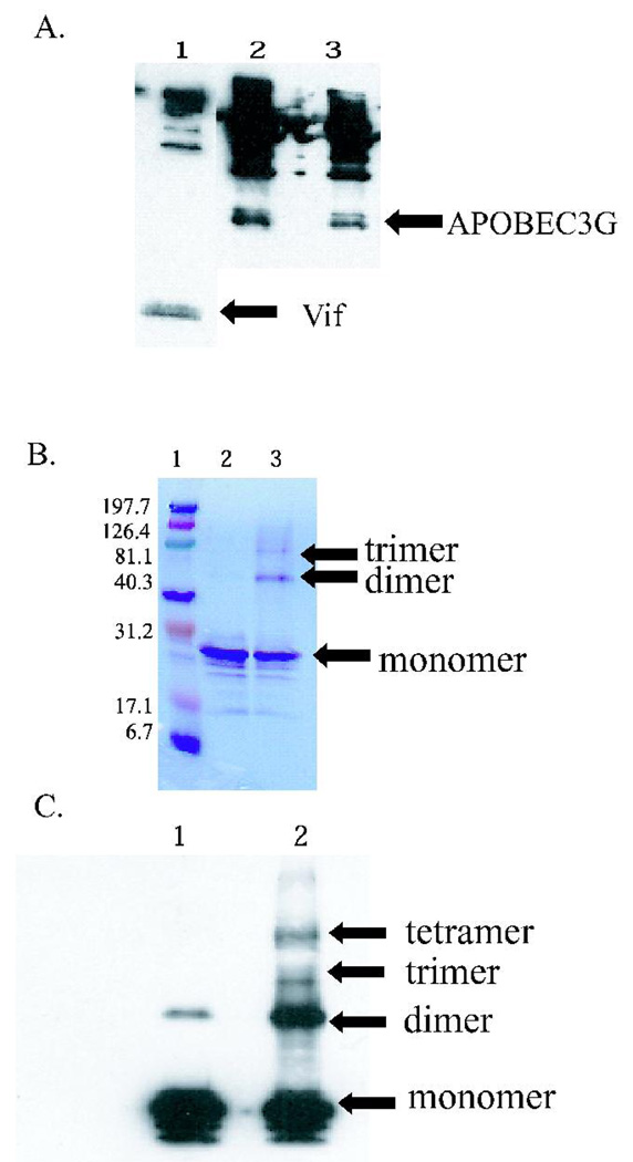
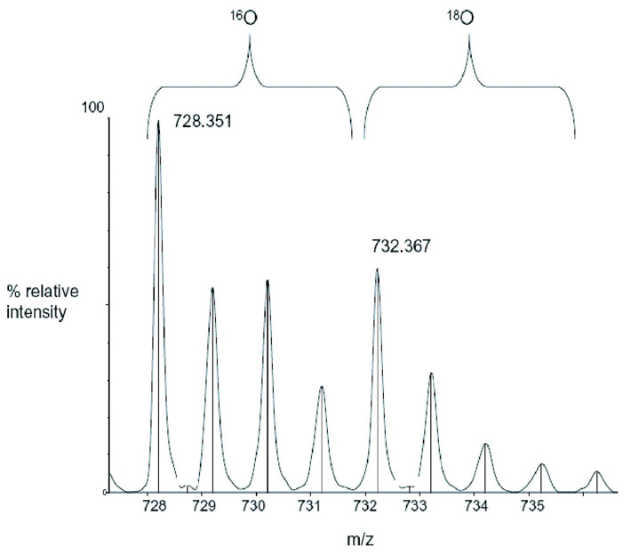
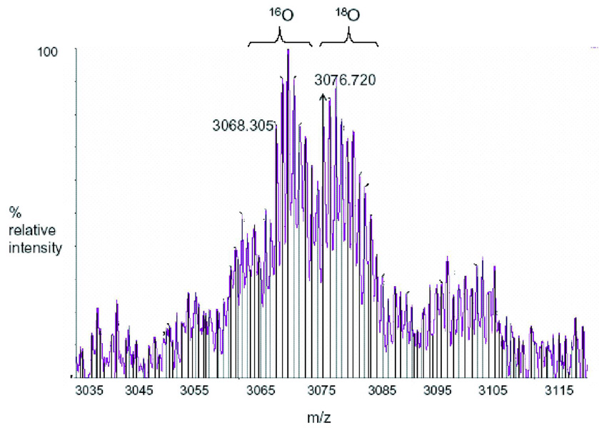
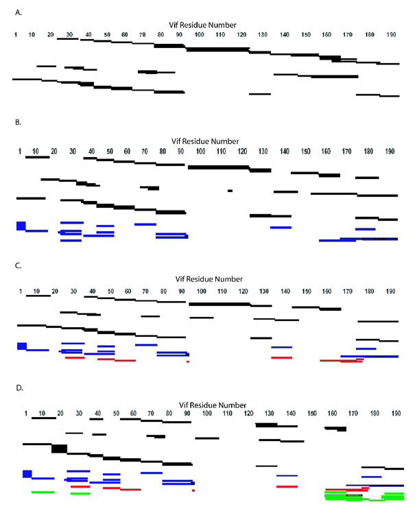
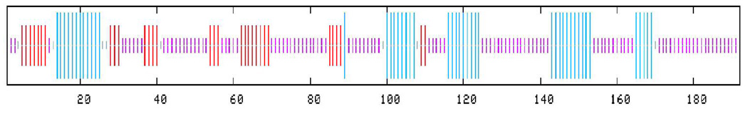
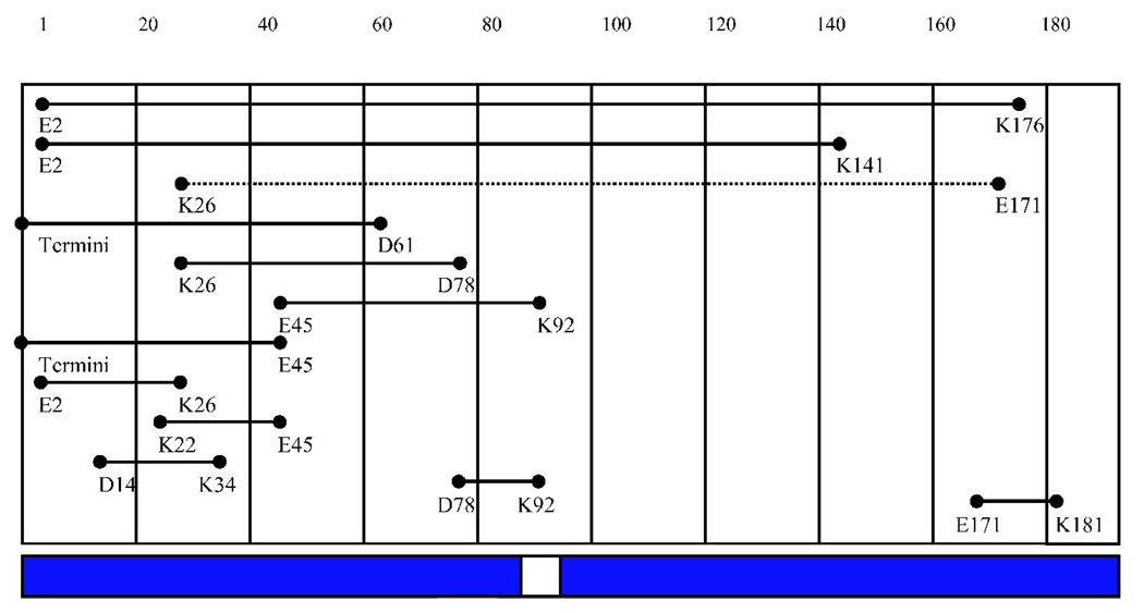
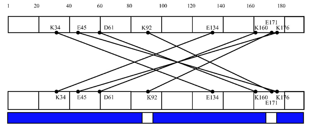
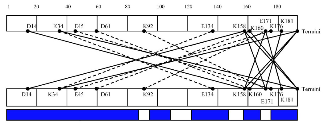
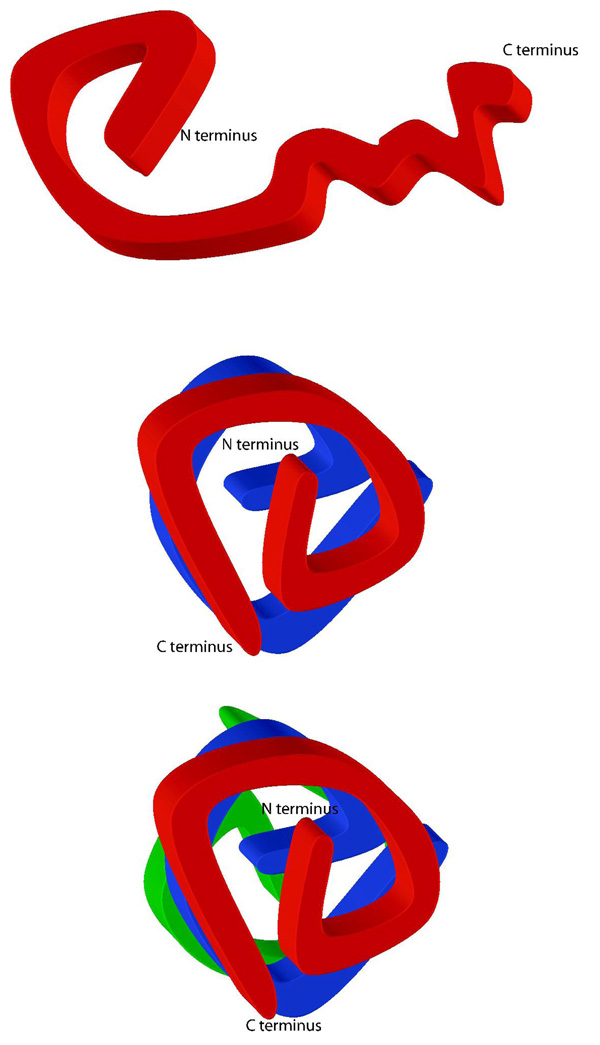
HIV-1 Vif, an accessory protein in the viral genome, performs an important role in viral pathogenesis by facilitating the degradation of APOBEC3G, an endogenous cellular inhibitor of HIV-1 replication. In this study, intrinsically disordered regions are predicted in HIV-1 Vif using sequence-based algorithms. Intrinsic disorder may explain why traditional structure determination of HIV-1 Vif has been elusive, making structure-based drug design impossible. To characterize HIV-1 Vif's structural topology and to map the domains involved in oligomerization we used chemical cross-linking, proteolysis, and mass spectrometry. Cross-linking showed evidence of monomer, dimer, and trimer species via denaturing gel analysis and an additional tetramer via western blot analysis. We identified 47 unique linear peptides and 24 (13 intramolecular; 11 intermolecular) noncontiguous, cross-linked peptides, among the noncross-linked monomer, cross-linked monomer, cross-linked dimer, and cross-linked trimer samples. Almost complete peptide coverage of the N-terminus is observed in all samples analyzed, however reduced peptide coverage in the C-terminal region is observed in the dimer and trimer samples. These differences in peptide coverage or "protections" between dimer and trimer indicate specific differences in packing between the two oligomeric forms. Intramolecular cross-links within the monomer suggest that the N-terminus is likely folded into a compact domain, while the C-terminus remains intrinsically disordered. Upon oligomerization, as evidenced by the intermolecular cross-links, the C-terminus of one Vif protein becomes ordered by wrapping back on the N-terminal domain of another. In addition, the majority of the intramolecular cross-links map to regions that have been previously reported to be necessary for viral infectivity. Thus, this data suggests HIV-1 Vif is in a dynamic equilibrium between the various oligomers potentially allowing it to interact with other binding partners.


Figures

Figure 1
Figure 1. HIV-1 Vif is functional and can form higher order oligomers
(A) Co-immunoprecipitation of HIV-1 Vif with APOBEC3G. Vif (APOBEC3G) was immobilized on Protein A affinity beads, incubated overnight with APOBEC3G (Vif), and eluted via boiling. Lane 1: Immobilized APOBEC3G interacts with HIV-1 Vif. Lane 2: Immobilized HIV-1 Vif interacts with baculovirus-expressed APOBEC3G. Lane 3: Immobilized HIV-1 Vif interacts with E. coli-expressed APOBEC3G. The higher molecular weight bands observed are the result of heavy chains, light chains, and Protein A background routinely observed in immunoprecipitations. (B) SDS PAGE analysis of HIV-1 Vif cross-links. Lane 1: Molecular weight markers (kDa). Lane 2: Noncross-linked HIV-1 Vif control that is predominately monomer (23 kDa). Lane 3: EDC cross-linked HIV-1 Vif, with evidence for a dimer (46 kDa) and trimer (69 kDa). (C) Western blot of HIV-1 Vif cross-links. Lane 1: A noncross-linked HIV-1 Vif control that is predominantly monomer, with a small amount of dimer. Lane 2: EDC cross-linked HIV-1 Vif, with evidence for dimer, trimer and tetramer forms.
Figure 2
Figure 2. Analysis of HIV-1 Vif using reflectron MALDI-TOF-MS
The mass spectrometry data presented here represents an example of the spectra of a peptide (Figure 2B) and a cross-link (Figure 2C) in Tables 1 and 2, respectively. m/z represents the mass-to-charge ratio of the ion and in the case of a singly charged ion represents the peptides MH+ (molecular weight). (A) Predicted regions of intrinsic disorder for HIV-1 Vif using PONDR®, Predictors of Natural Disordered Regions ,. The four colors indicate four separate predictor algorithms: blue VL3, red VL-XT, yellow XL1-XT, and green CaN-XT. (B) An unlabelled peptide at m/z 728.351 and its labeled (18O) counterpart 4Da larger at m/z 732.367. This peptide corresponds to residues 37–41 in the protein. (C) An unlabelled intermolecular cross-linked peptide at m/z 3068.305 and its labeled (18O) counterpart 8Da larger at m/z 3076.720. This region corresponds to a cross-link between peptides 51–63 and 159–173 and is only seen in the dimer and trimer. The arrow indicates the beginning of the +8 Da ion series.
Figure 3
Figure 3. Peptide Sequence Coverage Map from MALDI-TOF, LC-ion trap-MS, and LC-QTof-MS Data
Coverage maps of tryptic and chymotryptic peptides identified from MALDI-TOF, LC-ion trap-MS, and LC-QTof-MS, and tryptic cross-links identified from MALDI-TOF. Peptides and cross-links were identified via comparison of the experimental molecular weight with a list of theoretical molecular weights calculated from ProteinProspector or GPMAW as well as by a corresponding ion 4Da larger for a peptide and 8Da larger for a cross-link. (A) Noncross-linked. (B) Monomer. (C) Dimer. (D) Trimer. Identified regions are those that are cross-linked to each other and those that are protected. Black: peptides; blue: intramolecular cross-links; red: cross-links in the dimer and trimer; green: trimer cross-links.
Figure 4
Figure 4. Cross-links of HIV-1 Vif
Schematic diagram of cross-links observed in different oligomeric states of HIV-1 Vif that were analyzed by MALDI-TOF and heavy water labeling. (A) Consensus secondary structure prediction for HIV-1 Vif . Red: beta-sheet. Blue: alpha-helix. Purple: random coil (B) Cross-links observed in the monomer sample. (C) Cross-links observed in the dimer sample and shown going from both the N- to C-terminal and from the C-to N-terminal regions. (D) Cross-links observed in the trimer and shown going from both the N-to C-terminal and from the C- to N-terminal regions. Cross-linking: Dotted lines indicate that cross-links appeared in both the dimer and trimer samples. The blue bars represent peptide coverage for each oligomeric state.
Figure 5
Figure 5. Topology and Multimerization of HIV-1 Vif
(A) Intramolecular cross-links suggest that the N-terminus is folded into a compact domain and the C-terminus is less structured. The HIV-1 Vif monomer is globular in shape. (B and C) Schematic of how the HIV-1 Vif dimer and trimer may fold. The carboxyl terminus becomes more ordered upon oligomerization.

References

    1. Baraz L, Kotler M. The Vif protein of human immunodeficiency virus type 1 (HIV-1): enigmas and solutions. Current Medical Chemistry. 2004;11:221–231. - PubMed
    1. Lake J-a, Carr J, Feng F, Mundy L, et al. The role of Vif during HIV-1 infection: interaction with novel host cellular factors. Journal of Clinical Virology. 2003;26:143–152. - PubMed
    1. Navarro F, Landau NR. Recent insights into HIV-1 Vif. Current Opinion in Immunology. 2004;16:477–482. - PubMed
    1. Rose KM, Marin M, Kozak SL, Kabat D. The viral Infectivity factor (Vif) of HIV-1 unveiled. TRENDS in Molecular Medicine. 2004;10(6):291–297. - PubMed
    1. Madani N, Kabat D. Cellular and viral specificities of human immunodeficiency virus type 1 vif protein. Journal of Virology. 2000;74:5982–5987. - PMC - PubMed

Publication types

MeSH terms

LinkOut - more resources