

Newly codified glial neoplasms of the 2007 WHO Classification of Tumours of the Central Nervous System: angiocentric glioma, pilomyxoid astrocytoma and pituicytoma
- PMID: 17598825
- PMCID: PMC8095654
- DOI: 10.1111/j.1750-3639.2007.00082.x
Newly codified glial neoplasms of the 2007 WHO Classification of Tumours of the Central Nervous System: angiocentric glioma, pilomyxoid astrocytoma and pituicytoma
Abstract
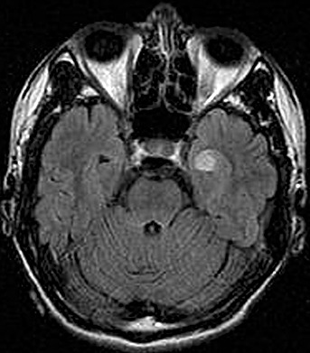
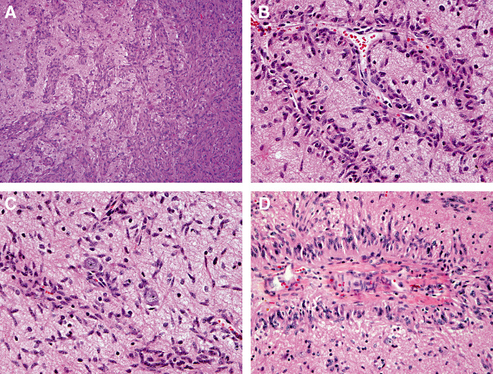
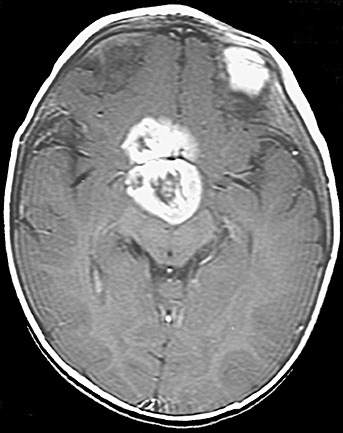
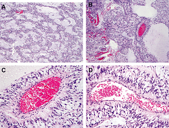
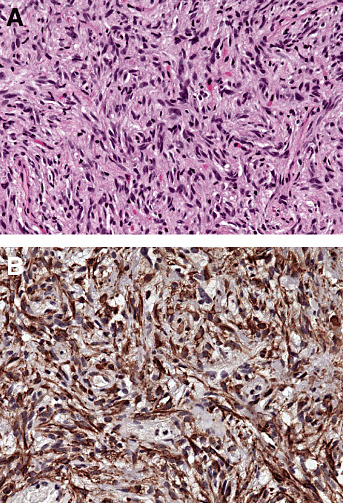
The 4(th) edition of the WHO Classification of Tumours of the Nervous System (WHO 2007) introduces changes that reflect both the recognition of new brain tumour types and a better understanding of neoplastic behavior. Three new tumours, angiocentric glioma (AG), pilomyxoid astrocytoma (PMA), and pituicytoma are added to the section on gliomas. AG is a slowly growing cerebral tumour that typically presents with seizures in children and young adults. It is characterized by monomorphous, bipolar tumour cells with a striking perivascular growth pattern. Although the 'cell of origin' of AG is not clear, ultrastructural evidence points to an ependymal derivation. Typically, AG can be cured by total resection, and is designated WHO grade I. PMA is a solid, circumscribed tumour occurring mainly in the hypothalamic region of young children. It is composed of a monomorphous population of bipolar tumour cells within a rich myxoid background, with a conspicuous anglocentric arrangement. While PMA is considered a more aggressive variant of pilocytic astrocytoma, this relationship awaits further clarification. The PMA has been designated WHO grade II. The pituicytoma, involves the posterior pituitary and/or its stalk and affects adults. It is solid in architecture, composed of spindle cells and presumably derived from pituicytes. Pituicytomas are indolent tumours, and are designated WHO grade I.
Figures
References
-
- Benveniste RJ, Purohit D, Byun H (2006) Pituicytoma presenting with spontaneous hemorrhage. Pituitary 9:53–58. - PubMed
-
- Brat DJ, Scheithauer BW, Staugaitis SM, Holtzman RN, Morgello S, Burger PC (2000) Pituicytoma: a distinctive low‐grade glioma of the neurohypophysis. Am J Surg Pathol 24:362–368. - PubMed
-
- Burger PC, Jouvet A, Preusser M, Hans VH, Rosenblum MK, Lellouch‐Tubiana A (2007) Angiocentric glioma. In: World Health Organization Classification of Tumors of the Central Nervous System. Louis DN, Ohgaki H, Wiestler OD, Cavenee WK (eds), IARC: Lyon. In press.
-
- Cenacchi G, Giovenali P, Castrioto C, Giangaspero F (2001) Pituicytoma: ultrastructural evidence of a possible origin from folliculo‐stellate cells of the adenohypophysis. Ultrastruct Pathol 25:309–312. - PubMed
MeSH terms
LinkOut - more resources
Full Text Sources
Medical
