. 2007 Jul 25;26(14):3308-21.
doi: 10.1038/sj.emboj.7601778. Epub 2007 Jun 28.

PtdIns(4,5)P-restricted plasma membrane localization of FAN is involved in TNF-induced actin reorganization

Affiliations

PtdIns(4,5)P-restricted plasma membrane localization of FAN is involved in TNF-induced actin reorganization

Dirk Haubert et al. EMBO J. .

Abstract

The WD-repeat protein factor associated with nSMase activity (FAN) is a member of the family of TNF receptor adaptor proteins that are coupled to specific signaling cascades. However, the precise functional involvement of FAN in specific cellular TNF responses remain unclear. Here, we report the involvement of FAN in TNF-induced actin reorganization and filopodia formation mediated by activation of Cdc42. The pleckstrin-homology (PH) domain of FAN specifically binds to phosphatidylinositol-4,5-bisphosphate (PtdIns(4,5)P), which targets FAN to the plasma membrane. Site-specific mutagenesis revealed that the ability of FAN to mediate filopodia formation was blunted either by the destruction of the PtdIns(4,5)P binding motif, or by the disruption of intramolecular interactions between the PH domain and the adjacent beige and Chediak-Higashi (BEACH) domain. Furthermore, FAN was shown to interact with the actin cytoskeleton in TNF-stimulated cells via direct filamentous actin (F-actin) binding. The results of this study suggest that PH-mediated plasma membrane targeting of FAN is critically involved in TNF-induced Cdc42 activation and cytoskeleton reorganization.


Figures
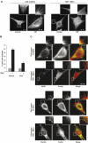
Figure 1
Figure 1
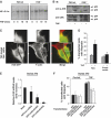
Impaired filopodia formation in FAN −/− fibroblasts after TNF stimulation. (A) TNF-induced filopodia formation in FAN wild-type (wt) MEFs. FAN wt and FAN−/− MEFs were stimulated with TNF for 10 min or left untreated and stained for F-actin using AlexaFluor568-conjugated phalloidin. Insets are enlargements of the boxed area. Scale bar, 20 μm. (B) Cells in panel A were quantified for filopodia formation. Cells were scored positive when presenting at least five filopodia. For each experiment, >100 cells were evaluated, and values are represented as mean±s.d. of at least three independent experiments. (C) TNF-induced filopodia contain filopodial markers. FAN wt and FAN−/− MEFs were stimulated with TNF for 10 min and stained for F-actin using AlexaFluor568-conjugated phalloidin (red) and VASP or paxillin using specific antibodies following incubation with AlexaFluor488-conjugated secondary antibodies (green). Scale bar, 20 μm.
Figure 2
Figure 2
FAN deficiency specifically affects TNF-induced filopodia formation. (A) FAN deficiency does not affect TNF-induced NF-κB activation. A total of 5 × 10⁶ FAN wt or FAN−/− MEFs were stimulated with TNF for the indicated times, and analyzed for NF-κB binding activity. (B) FAN deficiency does not affect TNF-induced JNK activation. A total of 105 FAN wt or FAN−/− MEFs were stimulated with TNF for 15 min, and total cell lysates were used for immunoblotting (IB) and probed for phosphorylated and total JNK using specific antibodies. (C) Overexpression of FAN restores TNF-induced filopodia formation in FAN−/− MEFs. FAN−/− MEFs were transfected with pEGFP-FAN (green), stimulated with TNF for 10 min and stained for F-actin using AlexaFluor546-conjugated phalloidin (red). Scale bar, 20 μm. (D) Quantitation of FAN−/− MEFs transfected with pEGFP-FAN or pEGFP empty vector bearing filopodia. For each experiment, >100 transfected cells were evaluated, and values are represented as mean±s.d. of at least three independent experiments. (E) siRNA-mediated downregulation of FAN. Swiss 3T3 fibroblasts were mock transfected or transfected with siRNAs against FAN (FAN-siRNA1, FAN-siRNA2) or scrambled siRNA (scr-siRNA) as control, and analyzed for FAN mRNA levels using LightCylcler PCR. Data are represented as mean±s.d. of triplicates of two independent experiments. (F) siRNA-mediated FAN knockdown abrogates TNF-induced filopodia formation. Swiss 3T3 fibroblasts were transfected with FAN-siRNA1, FAN-siRNA2 or scr-siRNA, together with pEGFP empty vector. GFP fluorescence was used to visualize transfected cells, and >100 transfected cells were evaluated for filopodia formation as described in panel D.
Figure 3
Figure 3
FAN mediates TNF-induced filopodia formation through Cdc42 activation. (A) TNF-induced filopodia formation is blocked by overexpression of dominant-negative Cdc42 (Cdc42N17). FAN wt MEFs were transfected with an myc-tagged dominant-negative Cdc42 (Cdc42N17-myc), treated with TNF for 10 min and stained for F-actin using AlexaFluor568-conjugated phalloidin (red) and anti-myc (green) to visualize filopodia and myc-tagged Cdc42N17, respectively. (B) Quantitation of FAN wt MEFs bearing filopodia transfected with Cdc42N17 or GFP and stimulated with TNF. For each experiment, >100 transfected cells were evaluated, and values are represented as mean±s.d. of at least three independent experiments. (C) TNF-induced activation of Cdc42. A total of 4 × 10⁶ FAN wt or FAN−/− MEFs were stimulated with TNF for 5 min or left untreated. Activated GTP-bound Cdc42 was precipitated from total lysates and detected on Western blot using a Cdc42-specific antibody. An aliquot of the total lysate used for precipitation was analyzed for total Cdc42 content in cell lysates. (D) Cdc42 activation after TNF stimulation was quantified at indicated times by Western blotting using AlphaEasy FC software (Alpha Innotech, San Leandro, USA). Data are shown as mean±s.d. of three independent experiments. (E) Activation of Cdc42 by PDGF and bradykinin. FAN wt and FAN−/− MEFs were stimulated with PDGF or bradykinin for 10 min or left untreated. Activated GTP-bound Cdc42 was detected as in panel C and quantified as in panel D. (F) Constitutively active Cdc42 overcomes the defective TNF-induced filopodia formation in FAN−/− MEFs. FAN wt and FAN−/− MEFs were transfected with an myc-tagged constitutively active Cdc42 (Cdc42L61-myc) and stained for F-actin (red) and myc-Cdc42L61 (green). Cells were cotransfected with dominant-negative RacN17 to avoid Rac activation by Cdc42 (Nobes and Hall, 1995) (see also Supplementary Figure S2B). (G) Quantitation of FAN wt and FAN−/− cells bearing filopodia transfected with Cdc42L61 or GFP as in panel B. (H) TNF-induced activation of Rac1 and RhoA. FAN wt and FAN−/− MEFs were stimulated with TNF for 10 min or left untreated, and activated GTP-bound GTPases were precipitated from total lysates and detected on Western blot using specific antibodies. Results were quantified as in panel D.
Figure 4
Figure 4
Impaired Golgi apparatus reorientation in FAN−/− MEFs. (A) Golgi reorientation in a scratch-wound test. After scratching of confluent layers of FAN wt and FAN−/− MEFs (0 h), cells were immediately treated with TNF and stained after the indicated times for the Golgi apparatus using anti-Rab6 antibody and Hoechst 33258 to visualize the nuclei. The dotted line indicates the direction of the wound. (B) Quantification of Golgi reorientation. Cells with the Golgi orientated toward the wound were scored positive at the indicated times. For each experiment, >100 cells were evaluated. Results are represented as mean±s.d. of at least three independent experiments.
Figure 5
Figure 5
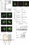
The PH domain directs FAN to the plasma membrane via specific binding to PtdIns(4,5)P. (A) A schematic representation of the FAN variants. (B) Colocalization of plasma membrane TNF-RI and FAN. COS cells were transfected with pEGFP-FAN (green) and TNF-RI lacking functional DD to avoid toxic effects (Tcherkasowa et al, 2002). Cells were stained for TNF-RI (red) without permeabilization for selective visualization of plasma-membrane TNF-RI, and analyzed by confocal microscopy. (C) PH-mediated plasma membrane localization of FAN. Confocal images of COS cells transiently expressing pEGFP-FAN or different deletion mutants of FAN. Arrows indicate membrane staining of pEGFP-FAN. (D) Quantification of membrane staining in panel C. Intensity profiles along the indicated white lines of the pictures in panel C were generated using ImageJ (Abramoff et al, 2004). (E) Expression and purification of recombinant His-tagged FAN-PH. FAN-PH and FAN-PHK199A/H212A were recombinantly expressed and affinity purified. Equal amounts were analyzed by SDS–PAGE following silver staining or immunoblot (IB) analysis with anti-His antibody. (F) Lipid overlay assay using purified FAN-PH protein. The recombinantly expressed PH domain of FAN was incubated with a nitrocellulose membrane spotted with different phospholipids. Bound protein was detected using anti-His antibody. (G) Lipid overlay assay as in panel E using purified FAN-PH and FAN-PHK199A/H212A protein. Equal amounts of protein were incubated with phospholipid-spotted membranes and detected using anti-His antibody. PI, phosphatidylinositol; PE, phosphatidylethanolamine; PtdIns(4)P, phosphatidylinositol-4-phosphate; PtdIns(3,4)P, phosphatidylinositol-3,4-phosphate; PtdIns(4,5)P, phosphatidylinositol-4,5-phosphate; PtdIns(3,4,5)P, phosphatidylinositol-3,4,5-phosphate; Ins(1,4,5)P, inositol-1,4,5-phosphate.
Figure 6
Figure 6
Plasma membrane association of FAN is indispensable for TNF-induced filopodia formation. (A) FAN−/− MEFs transfected with pEGFP-FAN or pEGFP-FAN-ΔPH (green) were stimulated with TNF for 10 min or left untreated. Cells were stained for F-actin using AlexaFluor546-conjugated phalloidin (red) to visualize filopodia. Insets are enlargements of the boxed area. Scale bar, 20 μm. (B) FAN−/− MEFs transfected with the indicated pEGFP-FAN fusion constructs were stimulated with TNF for 10 min or left untreated. Cells were stained for F-actin using AlexaFluor568-conjugated phalloidin and quantified for filopodia formation. For each experiment, >100 transfected cells were counted, and results are represented as mean±s.d. of at least three independent experiments.
Figure 7
Figure 7
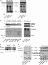
FAN interacts with the cytoskeleton. (A) GST pull-down assay after TNF treatment. HEK 293 cells were transiently transfected with DNA constructs coding for FAN-GST or GST alone and stimulated with TNF for 10 min or left untreated. Cells were lysed after 24 h and subjected to a GST pull-down assay. Precipitates and total cell lysate were immunoblotted and probed with the indicated antibodies (IB) to detect co-precipitated proteins. (B) Immunoprecipitation (IP) of VASP and actin after TNF treatment. HEK 293 cells were transiently transfected with DNA constructs coding for FAN-GST or GST alone and stimulated with TNF for 10 min or left untreated. Cells were lysed after 24 h, and IP was carried out using specific antibodies against actin, VASP or Bax. Precipitates and total cell lysate were immunoblotted and probed with the indicated antibodies. Asterisks (*) represent actin band recognized by actin-specific antibody before reprobing with anti-VASP-specific antibody. (C) F-actin sedimentation assay. FAN-GST or control GST proteins were subjected to a F-actin sedimentation assay as described in Materials and methods. Supernatants (S) and pellets (P) were analyzed by immunoblot (IB) using anti-GST and anti-β-actin antibodies. Samples without F-actin were included as binding control. (D) GST pull-down assay with cells ectopically expressing TNF-RI. HEK 293 cells were transiently cotransfected with DNA constructs coding for full-length TNF-RI, together with FAN-GST, FANΔPH-GST, FAN-PH-GST, or GST alone. After 24 h, cells were lysed and subjected to a GST pull-down assay as in panel A.

Similar articles

Cited by

References

    1. Abramoff M, Magelhaes P, Ram S (2004) Image processing with ImageJ. Biophotonics International 11: 36–42
    1. Adam D, Wiegmann K, Adam-Klages S, Ruff A, Krönke M (1996) A novel cytoplasmic domain of the p55 tumor necrosis factor receptor initiates the neutral sphingomyelinase pathway. J Biol Chem 271: 14617–14622 - PubMed
    1. Adam-Klages S, Adam D, Wiegmann K, Struve S, Kolanus W, Schneider-Mergener J, Krönke M (1996) FAN, a novel WD-repeat protein, couples the p55 TNF-receptor to neutral sphingomyelinase. Cell 86: 937–947 - PubMed
    1. Adam-Klages S, Schwandner R, Adam D, Kreder D, Bernardo K, Krönke M (1998) Distinct adapter proteins mediate acid versus neutral sphingomyelinase activation through the p55 receptor for tumor necrosis factor. J Leukoc Biol 63: 678–682 - PubMed
    1. Banno T, Gazel A, Blumenberg M (2004) Effects of tumor necrosis factor-alpha (TNF alpha) in epidermal keratinocytes revealed using global transcriptional profiling. J Biol Chem 279: 32633–32642 - PubMed

MeSH terms

Substances