. 2007 Jul 25;26(14):3346-59.
doi: 10.1038/sj.emboj.7601767. Epub 2007 Jun 28.

Functional dynamics of H3K9 methylation during meiotic prophase progression

Affiliations

Functional dynamics of H3K9 methylation during meiotic prophase progression

Makoto Tachibana et al. EMBO J. .

Abstract

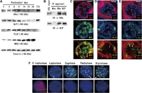
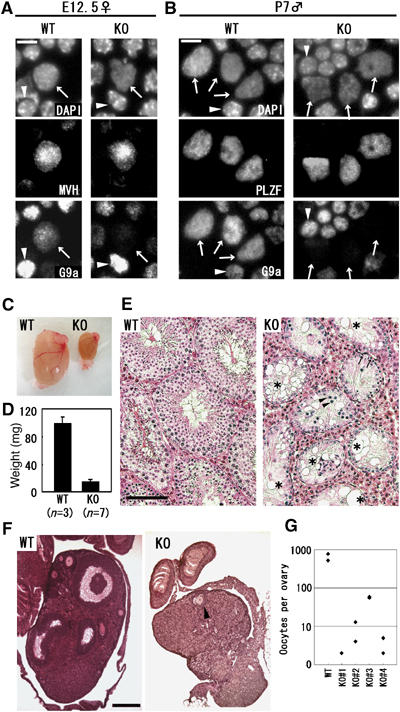
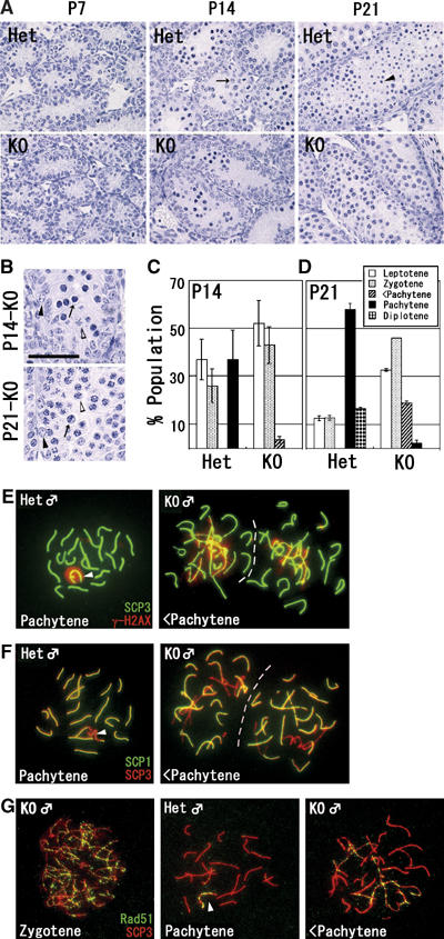
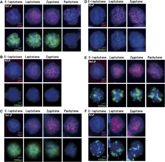
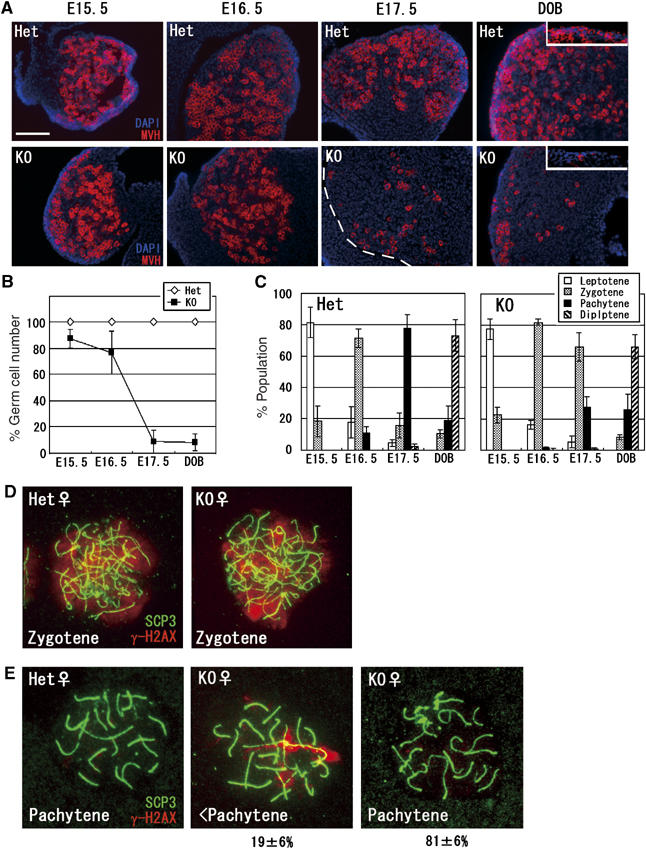
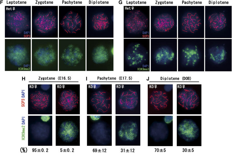
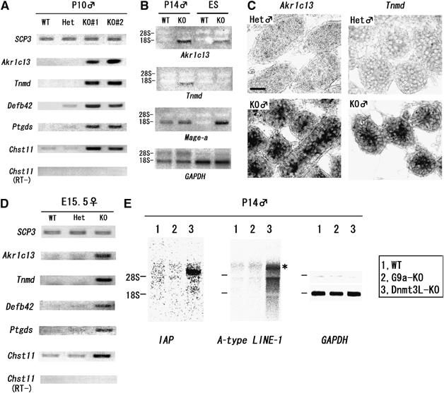
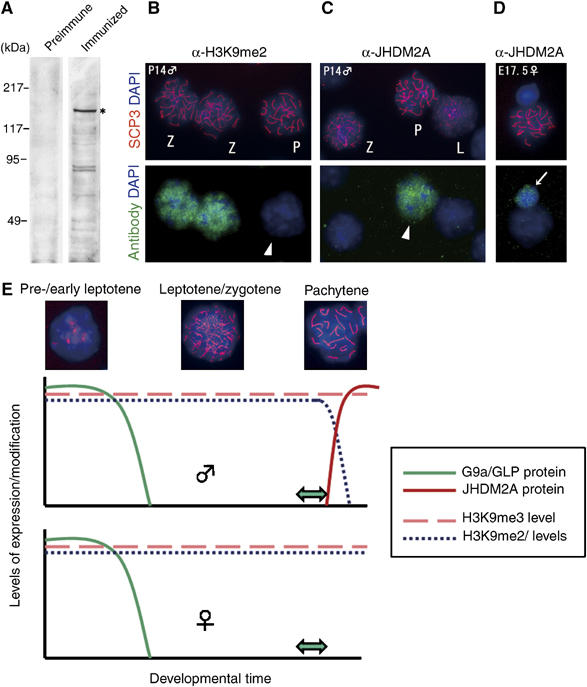
Histone H3 lysine 9 (H3K9) methylation is a crucial epigenetic mark of heterochromatin formation and transcriptional silencing. G9a is a major mammalian H3K9 methyltransferase at euchromatin and is essential for mouse embryogenesis. Here we describe the roles of G9a in germ cell development. Mutant mice in which G9a is specifically inactivated in the germ-lineage displayed sterility due to a drastic loss of mature gametes. G9a-deficient germ cells exhibited perturbation of synchronous synapsis in meiotic prophase. Importantly, mono- and di-methylation of H3K9 (H3K9me1 and 2) in G9a-deficient germ cells were significantly reduced and G9a-regulated genes were overexpressed during meiosis, suggesting that G9a-mediated epigenetic gene silencing is crucial for proper meiotic prophase progression. Finally, we show that H3K9me1 and 2 are dynamically and sex-differentially regulated during the meiotic prophase. This genetic and biochemical evidence strongly suggests that a specific set of H3K9 methyltransferase(s) and demethylase(s) coordinately regulate gametogenesis.


Figures

Figure 1
Figure 1
Temporal G9a/GLP expression in germ cell development. (A) Postnatal testicular cells prepared at various stages were subjected to immunoblot analysis. Total amounts of proteins loaded were normalized by determining tubulin content. ES cell lysate (TT2 strain) served as a positive control for G9a/GLP proteins. PLZF and mouse VASA homologue (MVH) were marker proteins for undifferentiated spermatogonia and pan-germ cells, respectively. (B) G9a/GLP complex formation was conserved in testicular cells. (C) Double-immunofluorescent staining profiles with anti-G9a/MVH on P10 testes sections. G9a protein is expressed in a subpopulation of male germ cells (arrowhead). The arrow represents a G9a-negative germ cell. (D) Double-immunofluorescent staining profiles with anti-G9a/PLZF on P10 testes sections. PLZF-positive spermatogonia are G9a positive (arrowhead). A subpopulation of G9a-positive cells is PLZF negative (arrow). (E) Double-immunofluorescent staining profiles with anti-G9a/c-Kit on P10 testes sections. G9a-positive germ cells are c-Kit positive (arrowhead). Scale bars in (C–E), 20 μm. (F) G9a expression profile during male meiosis. G9a protein is detected in only early leptotene (designated as E-leptotene) nuclei. Arrowheads indicate XY bodies.
Figure 2
Figure 2
Loss of germ cells in G9a-KO gonads. (A–B) Depletion of G9a protein in G9a-KO gonads. Enzymatically dissociated cells from E12.5 female gonads (A) and P7 testes (B) were squashed onto slide glass and stained with the indicated antibodies. Calculated efficiencies of G9a depletion are shown in Table II. Arrows and arrowheads indicate germ cells and control somatic cells, respectively. Bars, 10 μm. (C) Gross morphology of wild-type (WT) and germ cell G9a-KO (KO) testes at 3-month old age. (D) Testes weight of WT and KO mice at 3 months of age. The average weights were 98 and 15 mg, respectively. Error bars represent standard deviation. (E) Histological examination of WT and KO testes from 3-month-old mice. Paraffin-embedded sections were stained with hematoxylin and eosine. Seminiferous tubules devoid of germ cells were indicated by an asterisk. Leptotene-like spermatocytes and spermatogonia were indicated by arrowheads and arrows, respectively. Scale bars, 100 μm. (F) Histological examination of WT and KO ovaries at 2 months of age. Paraffin-embedded sections were stained with hematoxylin and eosine. The KO ovary was significantly smaller than that of WT mice. Only one oocyte was recognized in a section of KO ovary (arrowhead). Scale bar, 200 μm. (G) Gross numbers of oocytes per ovary at 2 months of age.
Figure 3
Figure 3
Developmental defects at the pachytene stage in G9a-KO male germ cells. (A) Paraffin-embedded sections (3 μm) of testes at various developmental time points (postnatal day (P) 7, and 14, and 21) were prepared and stained with hematoxylin. Pachytene spermatocytes at P14 and haploid spermatids at P21 in heterozygous testes were indicated by arrow and arrowhead, respectively. Scale bar, 50 μm. (B) Magnification of KO tubules. Apoptotic nuclei were detected both at P14 (top) and P21 (bottom) in KO tubules (arrows). Black and white arrowheads indicate Intermediate spermatogonia and early pachytene spermatocytes. Scale bar, 50 μm. (C–D) Histogram of male meiotic prophase stages at P14 (C) and P21 (D) between heterozygous and KO spread preparations. Developmental stages were classified by staining profiles with anti-SCP3/γH2AX. Bars represent the average of two samples per genotype (>150 cells were analyzed per sample). <Pachytene represents incomplete pachytene spermatocyte. (E–F) Perturbed synapsis formation in KO spermatocyte. P14 spermatocytes were stained with a combination of anti-SCP3/γH2AX (E) and anti-SCP1/SCP3 antibodies (F). Typical pachytene spermatocytes from heterozygous testes and incomplete pachytene spermatocytes (<Pachytene) from KO testes were represented. Arrowheads indicate XY bodies. (G) Immunofluorescence staining profile with anti-Rad51 antibodies in KO spermatocytes. At the pachytene stage, the signals of Rad51 protein were retained only at the XY body in heterozygous nuclei (arrowhead). In contrast, the signals were still retained on asynapsed chromosomes in KO spermatocyte (right).
Figure 4
Figure 4
The kinetics of H3K9 methylation during male meiotic prophase. (A–B) Kinetics of H3K9me2 in male meiotic prophase. Meiotic nuclear spreads were prepared from P14 testes of heterozygous (A) and KO (B) male mice, and the status of H3K9me2 was monitored in combination with anti-SCP3 antibody. (C–D) Kinetics of H3K9me1 in male meiotic prophase in nuclei of heterozygous (C) and KO spermatocytes (D). (E–F) Kinetics of H3K9me3 in male meiotic prophase in nuclei of heterozygous (E) and KO spermatocytes (F).
Figure 5ae
Figure 5ae
Developmental defects in G9a-KO female germ cells. (A) Germ cell development during meiotic prophase in KO ovaries. Frozen sections of heterozygous and KO ovaries at various developmental time points were prepared and stained with anti-MVH. Primordial oocytes were represented in the box of DOB panels. Scale bar, 100 μm. (B) Kinetics of germ cell reduction in KO ovaries. MVH-positive cells per 0.01 mm2 of ovary section were counted and the numbers of germ cells in heterozygous ovaries are normalized as 100%. Data represent the average of two samples per genotype (>100 cells were counted per ovary). (C) Developmental dynamics of female germ cells. Ovaries at various developmental stages were collected and oocytes spreads were stained with anti-SCP3/γH2AX antibodies for determining meiotic stages. Three independent ovaries were used for calculation at every embryonic stage (>50 cells were analyzed per ovary). (D–E) Perturbed synapsis formation in G9a-KO oocytes. Oocytes at the zygotene (D) and the pachytene stages (E) were prepared from E17.5 ovaries and stained with anti-SCP3/γH2AX antibodies. In the pachytene stage, γH2AX signals disappeared from heterozygous nuclei (E, left panel). In contrast, a subpopulation of KO oocytes exhibited synapsis perturbation similar to males, in which γH2AX signals were still retained along certain axial elements (E, middle panel). The other population of pachytene oocytes from KO ovaries exhibited γH2AX-negative staining profiles (E, right panel). Percentages represent population distributions (>30 cells were analyzed per ovary). (F–G) Kinetics of H3K9me2
Figure 5fj
Figure 5fj
(F) and me3 (G) in female heterozygous nuclei during the meiotic prophase. (H–J) Mosaicism of H3K9me2 staining profiles of female KO germ cells during meiotic prophase. Staining profiles of H3K9me2-positive and -negative oocytes at the zygotene stage at E16.5 (H), the pachytene stage at E17.5 (I), and the diplotene stage at DOB (J) were represented, and their population distributions are described below. The data were obtained from at least two independent KO female mice (>50 cells were analyzed per ovary).
Figure 6
Figure 6
G9a suppresses specific genes during meiotic prophase. (A) De-repression of genes in G9a-KO testes. Transcripts of Akr1c13, Tnmd, Defb42, Ptgds, and Chst11 were amplified by using cDNAs from independently prepared testes at P10 with the indicated genotype. (B) Comparison of transcriptional perturbation between G9a-KO germ cells and ES cells by Northern blot analysis. (C) RNA in situ hybridization analysis of Akr1c13 and Tnmd in P10 testes sections from heterozygous (top) and KO animals (bottom). Bar, 20 μm. (D) Several G9a-regulated genes were conserved during meiotic prophase in both sexes. cDNA was prepared from total RNA from E15.5 ovaries and amplified using specific primers as shown in (A). (E) Retrotransposons were suppressed in G9a-KO testes. Total RNAs from P14 whole testes from the indicated genotypes were blotted with specific probes as indicated. Transcripts of IAP (∼5.4 kb transcripts of IΔ1 type IAP) and LINE-1 (dominant ∼7 kb transcripts, shown as asterisk) were upregulated in Dnmt3L-KO but not in G9a-KO testes.
Figure 7
Figure 7
Strong correlation between JHDM2A expression and stage- and sex-dependent removal of H3K9 methylation. (A) Generation of specific antibodies against mouse JHDM2A. Antiserum against mouse JHDM2A (details were described in Supplementary data) was used for immunoblot analysis of whole-cell extracts of TT2-ES cells (left, preimmune serum; right, immunized serum). Expression of the JHDM2A in ES cells was confirmed by RT–PCR analysis beforehand (data not shown). Specific signals around 150 kDa (asterisk) are in accordance with the molecular weight of endogenous JHDM2A protein as described previously (Yamane et al, 2006). (B–D) Male pachytene-specific expression of JHDM2A protein. (C) Immunostaining profiles of IgG fractions from antiserum against JHDM2A on G9a-heterozygous spermatocyte spreads (P14). The same preparations were also stained with anti-H3K9me2 antibodies as a control (B). L, leptotene; Z, zygotene; P, pachytene. (D) Oocytes spreads were prepared from G9a-heterozygous ovaries (E17.5) and then stained with the anti-JHDM2A antibodies. Arrow indicates a somatic cell in which JHDM2A was expressed weakly. (E) The kinetics of H3K9 methylation in meiotic prophase. In spermatogenesis, G9a/GLP proteins were detected from spermatogonia until early leptotene spermatocytes and then rapidly extinguished. This suggests that H3K9me2/1 marks were added within this stage. Predeposited H3K9me2/1 marks were persistent until the late zygotene stage even without catalytic enzymes. However, after the completion of synapsis, the marks were rapidly and actively removed. The ‘active' removal of H3K9me2/1 marks seems to be catalyzed by JHDM2A protein at least in part, which is expressed after the completion of synapsis. As the ‘active' removal mechanism was absent in females at least until early diplotene stage, H3K9me2/1 modification was maintained throughout meiotic prophase. Double arrows indicate the meiotic catastrophic points by the loss of G9a and H3K9me2/1.

Similar articles

Cited by

References

    1. Bellve AR, Cavicchia JC, Millette CF, O'Brien DA, Bhatnagar YM, Dym M (1977) Spermatogenic cells of the prepuberal mouse. Isolation and morphological characterization. J Cell Biol 74: 68–85 - PMC - PubMed
    1. Bourc'his D, Bestor TH (2004) Meiotic catastrophe and retrotransposon reactivation in male germ cells lacking Dnmt3L. Nature 431: 96–99 - PubMed
    1. Buaas FW, Kirsh AL, Sharma M, McLean DJ, Morris JL, Griswold MD, de Rooij DG, Braun RE (2004) Plzf is required in adult male germ cells for stem cell self-renewal. Nat Genet 36: 647–652 - PubMed
    1. Cao R, Zhang Y (2004) The functions of E(Z)/EZH2-mediated methylation of lysine 27 in histone H3. Curr Opin Genet Dev 14: 155–164 - PubMed
    1. Carmell MA, Girard A, van de Kant HJ, Bourc'his D, Bestor TH, de Rooij DG, Hannon GJ (2007) MIWI2 is essential for spermatogenesis and repression of transposons in the mouse male germline. Dev Cell 12: 503–514 - PubMed

Publication types

MeSH terms