. 2007 Sep;14(9):1590-604.
doi: 10.1038/sj.cdd.4402194. Epub 2007 Jun 29.

The pyroptosome: a supramolecular assembly of ASC dimers mediating inflammatory cell death via caspase-1 activation

Affiliations

The pyroptosome: a supramolecular assembly of ASC dimers mediating inflammatory cell death via caspase-1 activation

T Fernandes-Alnemri et al. Cell Death Differ. 2007 Sep.

Abstract

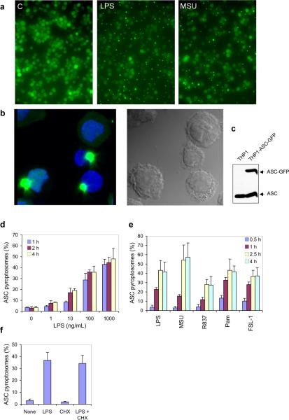
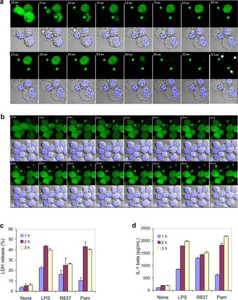
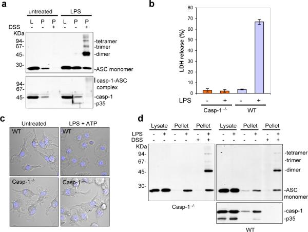
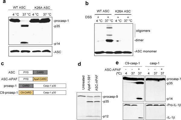
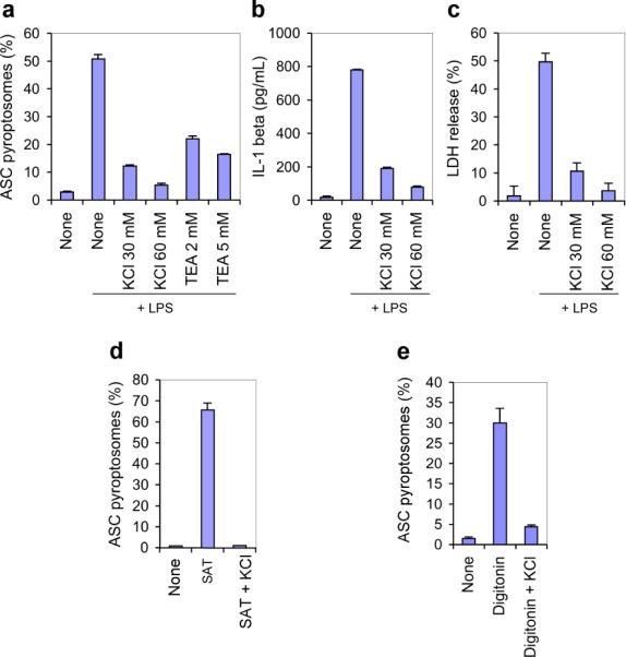
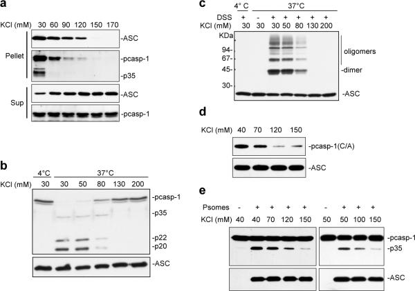
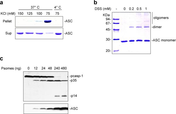
Pyroptosis is a caspase-1-dependent inflammatory form of cell death. The adapter protein ASC binds directly to caspase-1 and is critical for caspase-1 activation in response to a broad range of stimuli. To elucidate the mechanism of activation of caspase-1 by ASC and its exact role in macrophage pyroptosis, we performed time-lapse confocal bioimaging analysis on human THP-1 macrophages stably expressing an ASC-GFP fusion protein. We show that stimulation of these cells with several proinflammatory stimuli trigger the formation of a large supramolecular assembly of ASC, termed here pyroptosome. Only one distinct pyroptosome in each stimulated cell is formed, which rapidly recruits and activates caspase-1 resulting in pyroptosis and the release of the intracellular proinflammatory cytokines. The pyroptosome is largely composed of oligomerized ASC dimers. Dimerization of ASC is driven by subphysiological concentrations of potassium as in vitro incubation of purified recombinant ASC in the presence of subphysiological concentrations of potassium induces the assembly of a functional pyroptosome. Furthermore, stimulation of potassium efflux in THP-1 cells with potassium-depleting agents induces formation of the pyroptosome, while increasing potassium concentrations in the culture medium or pharmacological inhibition of this efflux inhibits its assembly. Our results establish that macrophage pyroptosis is mediated by a unique pyroptosome, distinct from the inflammasome.


Figures

Figure 1
Figure 1
Proinflammatory stimuli induce formation of ASC pyroptosome in THP-1 cells. (a) Live PMA-primed THP-1-ASC-GFP cells were left untreated (c, left micrograph) or treated with LPS (1 μg/ml) (middle micrograph) or MSU (100 μg/ml) (right micrograph) for 60 min and observed by fluorescence microscopy (× 20 magnification). (b) PMA-primed THP-1-ASC-GFP cells seeded on glass cover slips were treated with crude LPS for 60 min and then fixed and stained with DAPI. Cells were then observed and photographed by fluorescence confocal microscopy (× 63 magnification). Notice the uniformly distributed green fluorescent ASC–GFP in one cell that did not form ASC pyroptosome (top half of left micrograph) and the large perinuclear green fluorescent ASC pyroptosomes formed in three other cells (bottom half of left micrograph). The right micrograph shows a phase contrast confocal micrograph of the same cells in the left micrograph. (c) Western blot analysis of lysates from parental THP-1 or stable THP-1-ASC-GFP cells with anti-ASC antibody. Notice that the expression level of the ASC–GFP fusion protein (upper band) in the THP-1-ASC-GFP cells is comparable to the expression level of endogenous ASC (lower band). (d) THP-1-ASC-GFP cells were pretreated with zVAD-FMK for 30 min and then treated with increasing amounts of crude LPS for the indicated periods of time. The percentages of cells containing ASC pyroptosomes were calculated as described under the `Materials and Methods'. (e) THP-1-ASC-GFP cells were treated as in (d) with LPS (1.0 μg/ml), MSU (100 μg/ml), R837 (5.0 μg/ml) Pam3CSK4 (1.0 μg/ml) or FSL-1 (1.0 μg/ml) for the indicated periods of time. The percentages of cells containing ASC pyroptosomes were determined as in (d). (f) THP-1-ASC–GFP cells were preincubated with cycloheximide (10 μg/ml) for 30 min and then treated with LPS (1.0 μg/ml) for an additional 1 h. The percentages of cells containing ASC pyroptosomes were determined as in (d)
Figure 2
Figure 2
Formation of the ASC pyroptosome causes cell death in THP-1 cells. (a and b) Time-series confocal images (× 63 magnification) of crude LPS-stimulated THP-1-ASC-GFP cells in the absence (a, 1.5 min images are shown) or presence of zVAD-FMK (70 μM) (b, 1.0 min images are shown). The arrows in the last image point to ASC pyroptosomes. Time-lapse movies showing the assembly of the ASC pyroptosome in the absence or presence of zVAD-FMK can be seen in the `Supplementary Movies 1–5'. (c and d) Pyroptosome-induced pyroptotosis causes the release of intracellular LDH and IL -1. PMA-primed parental THP-1 or THP-1-ASC-GFP cells were pretreated for 1 h with ultrapure LPS to induce pro-IL–1β and then treated with crude LPS (1.0 μg/ml), R837 (10 μg/ml) or Pam3CSK4 (0.5 μg/ml) for the indicated periods of time. LDH release into the culture medium is shown as a percentage of LDH release by detergent (c). IL–1β release into the culture media was determined by ELISA (d)
Figure 3
Figure 3
Caspase-1 is the apical caspase in pyroptosis and inflammation. (a) THP-1 cells were left untreated or treated with LPS (1.0 μg/ml) for 3 h in the presence of zVAD-FMK and then lysed. The ASC pyroptosomes present in the lysates were pelleted by centrifugation at 5000 r.p.m. as described under `Materials and Methods'. Two-third of the pellets (P) was incubated with DSS for 30 min (third and sixth lanes) and the remaining one-third was left untreated (second and fifth lanes). The lysates (L) and pellets (P) were then fractionated by SDS-PAGE and Western blotted with anti-ASC (upper panel) or anti-caspase-1 (lower panel) antibodies. (b) Bone marrow macrophages from WT or caspase-1−/− mice were treated with LPS (1.0 μg/ml) for 3 h followed by ATP (4 mm) for 1 h, and the LDH release into the culture medium was determined as described under `Materials and Methods'. (c) Confocal micrographs of bone marrow macrophages from WT or caspase-1−/− mice treated with LPS plus ATP. Notice the pyroptotic phenotype in the LPS plus ATP-treated WT (top right panel), but not the caspase-1−/− (bottom right panel) cells. (d) ASC pyroptosomes were isolated from WT or caspase-1−/− bone marrow macrophages after treatment with LPS plus ATP. The ASC pyroptosomes were isolated as in (a). The lysates and pellets were then fractionated by SDS-PAGE and Western blotted with anti-ASC (upper panel) or anti-caspase-1 (lower panel) antibodies
Figure 4
Figure 4
The ASC pyroptosome is the molecular platform responsible for recruitment and activation of caspase-1. (a) ASC pyroptosomes purified from LPS-stimulated THP-1-ASC-GFP cells were incubated with flag-tagged WT or active-site mutant (C/A) procaspase-1 together with pro-IL–1β at 37°C for 20, 40, or 60 min as indicated. The reaction products were then analyzed by SDS-PAGE and Western blotting with anti-flag (upper panel) or anti-IL–1β (middle panel) antibodies. Notice that only WT caspase-1 (lanes 1–4), but not active-site mutant caspase-1 (C/A) (lanes 5–8), can be activated by ASC pyroptosomes. The asterisks indicate a non-specific band. (b) Confocal micrographs (63 magnification) of ASC–GFP pyroptosomes purified from THP-1-ASC-GFP cell lysates (upper panels), or endogenous ASC pyroptosomes from THP-1 cell lysates (lower panels). The ASC pyroptosomes were isolated in the presence of rhodamine-labeled zVAD-fmk to label the associated caspase-1 (middle red panels). Phase contrast (right panels). Notice the highly organized star-shaped structure of the ASC pyroptosome. (c) Same as in (a) but using in vitro assembled and purified ASC pyroptosomes from THP-1 lysate. (d) The purified ASC pyroptosomes shown in (b) were fractionated by SDS–PAGE and stained with Coomassie Blue. Lane 1: endogenous ASC pyroptosomes purified from THP-1 lysate. Lane 2: ASC pyroptosomes purified from THP-1-ASC-GFP lysate. Notice the presence of endogenous ASC (lane 2, middle band) in the ASC–GFP pyroptosome preparation. The ASC–GFP (lane 2, top band) migrates slightly above the 45 kDa marker. The bottom band in lanes 1 and 2 is an endogenous short isoform of ASC (ASC-S). (e) Western blots of purified in vitro assembled ASC pyroptosomes from THP-1 cell lysates (first lanes), or purified ASC pyroptosomes from LPS-stimulated THP-1-ASC-GFP cells (second lanes). The pyroptosomes were isolated in the presence of zVAD-FMK to trap the activated caspase-1 on the pyroptosomes. The blots were probed with anti-ASC (left upper panel, exposure time 15 s), anti-caspase-1 (left lower panel, exposure time 30 s) or anti-cryopyrin (right panel, exposure time 30 min) antibodies. The third lane in the cryopyrin blot is a positive cryopyrin-containing lysates control from a stable 293 cells expressing cryopyrin
Figure 5
Figure 5
The pyrin domain of ASC mediates formation of the ASC pyroptosome. (a) Lysates from stable 293T cells (10 μg/μl) expressing flag-tagged procaspase-1 and either WT or K26A mutant ASC were activated by incubation at 37°C or left at 4°C for 1 h as indicated. The lysates were then analyzed by SDS-PAGE and Western blotted with anti-flag (upper panel) or anti-ASC (lower panel) antibodies. (b) The lysates described in (a) were incubated at 4 or 37°C for 30 min with the crosslinking agent DSS as indicated. The lysates were then analyzed by SDS-PAGE and Western blotted with anti-ASC antibody. (c) A schematic illustration of the chimeric ASC-APAF which contains the CARD of Apaf-1 at its C terminus instead of its original CARD, and the C9-procaspase-1, which contains the CARD of procaspase-9 at its N terminus instead of its original CARD. (d) Bacterially produced chimeric ASC-APAF pyroptosomes or Apaf-1–591 were incubated with 35S-labeled procaspases-9 at 37°C for 1 h as indicated. The reaction products were then analyzed by SDS-PAGE and autoradiography. Notice that both ASC-APAF and Apaf-1–591 can activate procaspase-9. (e) Bacterially produced chimeric ASC-APAF pyroptosomes were incubated with flag-tagged C9-procaspase-1 chimera (left panels) or WT procaspase-1 (right panels) together with pro-IL–1β at 4 or 37°C for 1 h as indicated. The reaction products were then analyzed by SDS-PAGE and Western blotting with anti-flag (upper panels) or anti-IL–1β (lower panels) antibodies
Figure 6
Figure 6
Potassium depletion triggers formation of the ASC pyroptosome in vivo. (a) THP-1-ASC-GFP cells were treated with crude LPS (1 μg/ml) in the absence or presence of the indicated concentrations (mM) of KCl, or the potassium channel blocker TEA for 2 h. The percentages of cells containing ASC–GFP pyroptosomes were determined as in Figure 1d. (b and c) THP-1 cells were treated with crude LPS (1 μg/ml) in the absence or presence of the indicated concentrations of KCl for the indicated periods of time. LDH release into the culture medium is shown as a percentage of LDH release by detergent (c). IL–1β release into the culture media was determined by ELISA (b). (d and e) THP-1-ASC-GFP cells were treated with SAT (10 μg/ml) or digitonin (10 μg/ml) in the absence or presence of the of KCl (60 mM) as indicated and the percentages of cells containing ASC–GFP pyroptosomes were determined as in Figure 1d
Figure 7
Figure 7
Effect of potassium on pyroptosome assembly in vitro. (a) ASC pyroptosomes were assembled in vitro by incubation of THP-1 S100 extracts (10 μg/μl) at 37°C in the presence of increasing concentrations of KCl. The reaction mixtures were centrifuged at 5000 r.p.m., and the resulting pellets, which contain the assembled pyroptosomes, and the remaining supernatants were then fractionated by SDS-PAGE followed by Western blotting with anti-ASC or anti-caspase-1 antibodies. The ASC blot (first panel from the top, pellet) was exposed for 2 min. The caspase-1 blot (second panel from the top, pellet) was exposed for 3 h to detect caspase-1. The supernatant blots (third and fourth panels from the top) were exposed for 2 min. (b and c) Lysates from THP-1 cells (10 μg/μl) were incubated at 4°C or activated at 37°C in the presence of the indicated concentrations of KCl together with (b) or without (c) the crosslinking agent DSS (4 μM). The lysates were analyzed by Western blotting with anti-caspase-1 (b) or anti-ASC (c) antibodies. (d) Purified preformed ASC pyroptosomes were incubated with 293 lysates containing inactive flag-procaspase-1 mutant (C285A) in the presence of the indicated potassium concentrations at 37°C for 1 h. The ASC pyroptosomes were then pelleted by centrifugation, washed three times and then fractionated by SDS-PAGE followed by Western blotting with anti-flag (top panel) or anti-ASC (bottom panel) antibodies as indicated. (e) Purified preformed ASC pyroptosomes were incubated with 293 lysates containing WT flag-procaspase-1 (left panels) or purified WT flag-procaspase-1 (right panels) in the presence of the indicated potassium concentrations at 37°C for 1 h. The total reaction mixtures were fractionated by SDS-PAGE followed by Western blotting with anti-flag (top panel) or anti-ASC (bottom panel) antibodies as indicated
Figure 8
Figure 8
In vitro assembly of the ASC pyroptosome with purified ASC. (a) Purified recombinant ASC (10 ng/μl) was incubated at 37°C or at 4°C in the presence of increasing concentrations of KCl as indicated. The samples were centrifuged at 5000 r.p.m., and the resulting pellets (upper panel), which contain the assembled ASC pyroptosomes, and the remaining supernatants (lower panel) were then fractionated by SDS-PAGE followed by coomassie staining. (b) The assembled recombinant ASC pyroptosomes from (a) above were incubated with increasing concentrations of DSS for 30 min as indicated, and then fractionated by SDS-PAGE followed by Coomassie staining. (c) Increasing amounts of the assembled recombinant ASC pyroptosomes from (a) above were incubated with flag-procaspase-1 for 1 h at 37°C, and then fractionated by SDS-PAGE followed by Western blotting with anti-flag (top panel) or anti-ASC (bottom panel) antibodies as indicated

Comment in

Similar articles

Cited by

References

    1. Martinon F, Tschopp J. NLRs join TLRs as innate sensors of pathogens. Trends Immunol. 2005;26:447–454. - PubMed
    1. Ting JP, Kastner DL, Hoffman HM. Caterpillers, pyrin and hereditary immunological disorders. Nat Rev Immunol. 2006;6:183–195. - PubMed
    1. Delbridge LM, O'Riordan MX. Innate recognition of intracellular bacteria. Curr Opin Immunol. 2007;19:10–16. - PubMed
    1. Dinarello CA. Interleukin-1 beta, interleukin-18, and the interleukin-1 beta converting enzyme. Ann N Y Acad Sci. 1998;856:1–11. - PubMed
    1. Martinon F, Burns K, Tschopp J. The inflammasome: a molecular platform triggering activation of inflammatory caspases and processing of proI-Lbeta. Mol Cell. 2002;10:417–426. - PubMed

Publication types

MeSH terms