. 2007 Aug 24;371(4):855-72.
doi: 10.1016/j.jmb.2007.06.013. Epub 2007 Jun 9.

Transcription termination defective mutants of Rho: role of different functions of Rho in releasing RNA from the elongation complex

Affiliations

Transcription termination defective mutants of Rho: role of different functions of Rho in releasing RNA from the elongation complex

Jisha Chalissery et al. J Mol Biol. .

Abstract

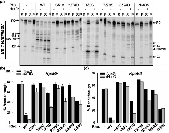
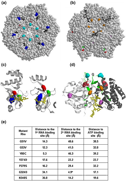
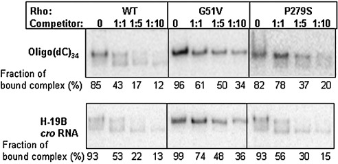
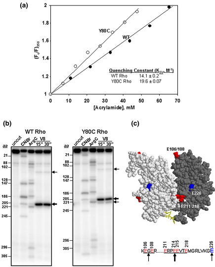
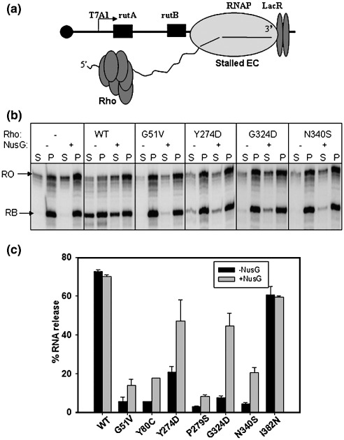
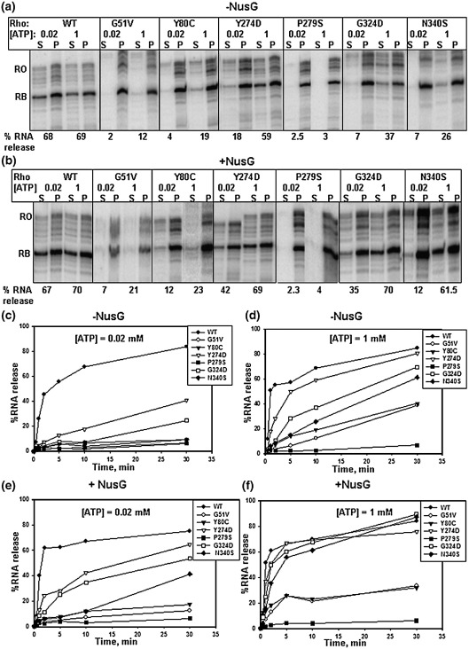
The transcription termination factor Rho of Escherichia coli is a RNA binding protein which can translocate along the RNA and unwind the RNA:DNA hybrid using the RNA-dependent ATPase activity. In order to investigate the involvement of each of these functions in releasing RNA from the elongation complex, we have isolated different termination defective mutants of Rho by random mutagenesis, characterized them for their different functions and established the structure-function correlations from the available structural data of Rho. These mutations are located within the two domains; the N-terminal RNA binding domain (G51V, G53V, and Y80C) and in the C-terminal ATP binding domain (Y274D, P279S, P279L, G324D, N340S, I382N) including the two important structural elements, the Q-loop (P279S, P279L) and R-loop (G324D). Termination defects of the mutants in primary RNA binding domain and Q-loop could not be restored under any conditions that we tested and these were also defective for most of the other functions of Rho. The termination defects of the mutants (Y274D, G324D and N340S), which were mainly defective for secondary RNA binding and likely defective for translocase activity, could be restored under relaxed in vitro conditions. We also show that a mutation in a primary RNA binding domain (Y80C) can cause a defect in ATP binding and induce distinct conformational changes in the distal C-terminal domain, and these allosteric effects are not predictable from the crystal structure. We conclude that the interactions in the primary RNA binding domain and in the Q-loop are mandatory for RNA release to occur and propose that the interactions in the primary RNA binding modulate most of the other functions of Rho allosterically. The rate of ATP hydrolysis regulates the processivity of translocation along the RNA and is directly correlated with the efficiency of RNA release. NusG improves the speed of RNA release and is not involved in any other step.


Figures

Figure 1
Figure 1
In vitro termination defects of the mutant Rho proteins. (a) Autoradiogram of the in vitro transcription termination by different Rho mutants both in the absence and presence of 200 nM NusG. Transcription was initiated from T7A1 promoter. The DNA template was immobilized on magnetic beads. Rho-dependent RNA release starts from 100 nt downstream of the transcription start site in the trp t' terminator region (as indicated). Major termination positions are marked. Half of the supernatant is denoted as S (released RNA) and the rest of the reaction mixture containing both supernatant and pellet is denoted as P (total RNA). RO denotes the run-off product. The read-through efficiency (RT) for each case was calculated from the P lanes as: RT=[RO product]/[RO product + all the products ≥ 100 nt]. (b) Measured RT for each of the mutants is shown as bar diagrams using WT RNA polymerase. Higher read through efficiencies of the Rho mutants reflect their termination defect. (c) Read through efficiencies measured in the presence of B8 RNA polymerase.
Figure 2
Figure 2
Location of the mutations on the crystal structure of Rho. In (a) and (b) the space-filled model of hexameric closed ring structure of Rho was generated from the available co-ordinates of the dimeric unit of Rho (2HT119) and the Figures were prepared using RASMOL. Positions of the mutations are indicated. The color codes are as follows. G51V, red; G53V, green; Y80C, blue; Y274D, pink; P279S/P279L, cyan; G324D, orange; N340S, violet. The position of I382N is not resolved in this structure. In (a) the view is from primary RNA binding domain and that in (b) is from secondary RNA binding domain. (c) The primary RNA binding domain of Rho generated from the co-ordinates of the structure of N-terminal domain (2A8V; left) and closed ring (2HT1; right) structures. The RNA oligonucleotide is shown in yellow and locations of G51V, G53V and Y80C are also shown as spheres with the same color code as in (a) and (b). (d) The secondary RNA binding domain is shown in the dimeric unit of the closed ring structure with the RNA (in yellow) bound at the interface of the two monomers (chains are shown in grey and black). P, Q and R loops are shown in green and the locations of Y274D, P279S/P279L, G324D and N340S are also indicated as spheres. Color codes for the mutations are same as in (a) and (b). (e) Distances of the positions of different mutations from important functional domains. The distances were calculated using the RASMOL program. For calculating the distances from primary (1°) and secondary (2°) RNA binding domains, the nearest RNA residues were considered and that from the P-loop, Cα atom of residue 184 was considered.
Figure 3
Figure 3
Stability of Rho–oligo(dC)34 complexes. Autoradiograms of native PAGE showing the amount of labeled oligo(dC)34 complexed with WT and different mutant Rho proteins survived in the presence of increasing concentrations of competitors: upper panel, unlabeled oligo(dC)34; lower panel, H-19B cro RNA. The fraction of bound complex was calculated as: [bound oligo]/[free oligo+bound oligo]. The 5 nM labeled oligo(dC)34 was used to form complex with 50 nM of WT and mutant Rho proteins.
Figure 4
Figure 4
Conformational changes in the C-terminal domain of the Y80C mutant. (a) Stern–Volmer plots for acrylamide quenching of tryptophan fluorescence of WT and Y80C Rho proteins. The quenching constant (KSV) was calculated from these plots using the equation: (F0/F)350 = 1+ KSV[Q], where Q is the concentration of the quencher, acrylamide. (b) Limited proteolysis of end-labeled WT and Y80C Rho proteins with V8 protease. Glutamic acid residues corresponding to the major cleavage products are indicated by arrows. Thickness of the arrow corresponds to the intensity of the bands. These positions were identified from the calibration curve obtained from the molecular weight markers generated with CNBr and ArgC digestions of the same end-labeled Rho. The amino acid positions from N to C-terminal are indicated to the left of the gel pictures. The thick arrow corresponds to the sensitivity towards the glutamic acids residues between 211 and 218. Individual amino acids are not resolved. Position of 106/108 and 226 are also indicated by thin arrows. (c) Locations of the V8 sensitive residues are indicated on the dimeric unit of the closed ring structure of Rho. The red spheres are the V8 sensitive patch comprising of glutamic acid residues at 211, 214, 215 and 218. Also E106/108 is also indicated as red spheres in the N-terminal domain. E226, which specifically became sensitive in the Y80C Rho is shown as a blue sphere.
Figure 5
Figure 5
Rho-mediated RNA release from the stalled elongation complex. (a) Cartoon showing the design of a stalled elongation complex using lac repressor as a roadblock. In this set-up, the EC was stalled at 161 position of the trp t' terminator. Rho was added to the reaction after the stalled complex is formed and RNA release was measured from the supernatant. (b) Autoradiogram showing the amount of released RNA in the supernatant by WT and different Rho mutants both in the absence and presence of 200 nM NusG from the stalled EC. RB and RO denote the positions of roadblocked product and run-off product, respectively. The meaning of S and P is same as described for Figure 1. (c) The RNA release efficiencies shown by bar diagrams are calculated as: [2S]/[S+P].
Figure 6
Figure 6
ATP and NusG dependence of Rho-mediated RNA release from a stalled elongation complex. Autoradiograms of the RNA release after 10 min of addition of different Rho mutants from the stalled EC (described in Figure 5) in the presence of different concentrations of ATP, both in the absence (a) and presence (b) of 200 nM NusG. (c)–(f) Curves showing the time courses of the RNA release upon addition of WT and different Rho mutants in the presence of indicated concentrations of ATP. The presence or absence of NusG in the reactions is also indicated. The RNA release efficiencies are calculated in the same way as described in Figure 5.

Similar articles

Cited by

References

    1. Wilson K.S., von Hippel P.H. Stability of Escherichia coli transcription complexes near an intrinsic terminator. J. Mol. Biol. 1994;244:36–51. - PubMed
    1. Mooney R.A., Artsimovitch I., Landick R. Information processing by RNA polymerase: recognition of regulatory signals during RNA chain elongation. J. Bacteriol. 1998;180:3265–3275. - PMC - PubMed
    1. Yarnell W.S., Roberts J.W. Mechanism of intrinsic transcription termination and antitermination. Science. 1999;284:611–615. - PubMed
    1. Richardson J.P. Transcription termination. Crit. Rev. Biochem. Mol. Biol. 1993;28:1–30. - PubMed
    1. Uptain S.M., Kane C.M., Chamberlin M.J. Basic mechanisms of transcript elongation and its regulation. Annu. Rev. Biochem. 1997;66:117–172. - PubMed

Publication types

LinkOut - more resources