. 2007 Aug 24;371(4):959-70.
doi: 10.1016/j.jmb.2007.05.056. Epub 2007 May 25.

A structural basis for regulation of actin polymerization by pectenotoxins

Affiliations

A structural basis for regulation of actin polymerization by pectenotoxins

John S Allingham et al. J Mol Biol. .

Abstract

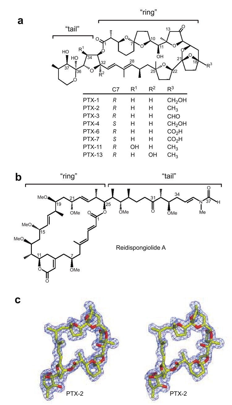
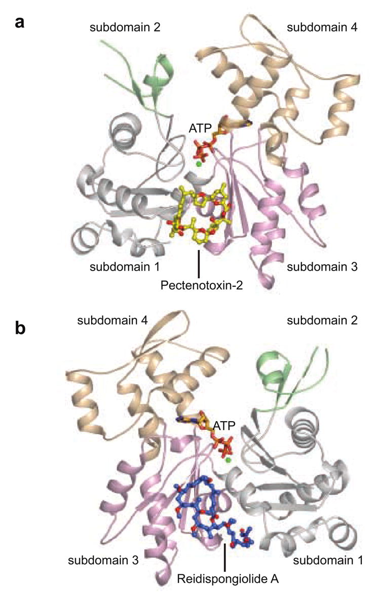
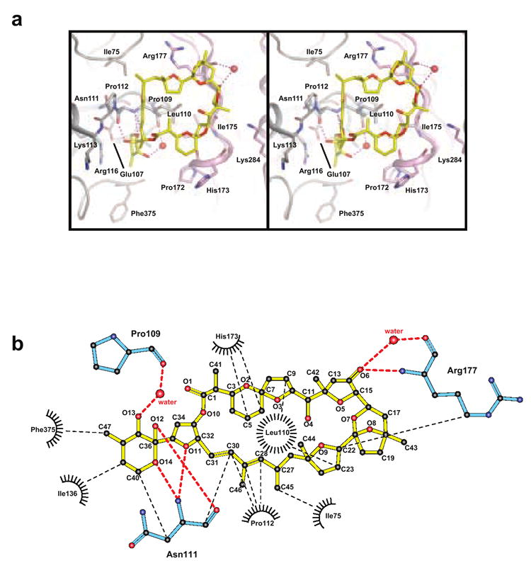
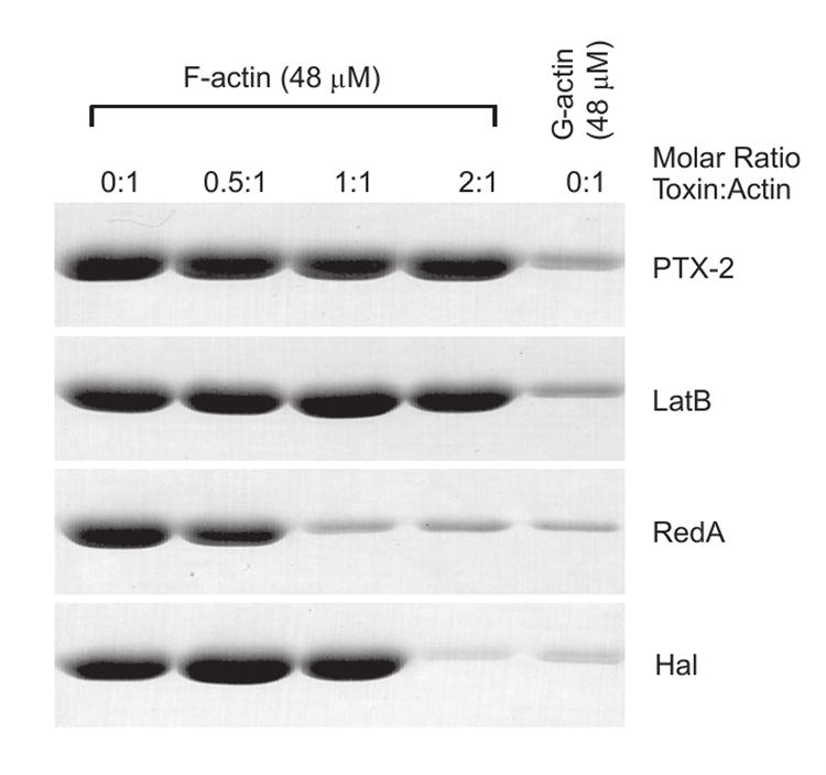
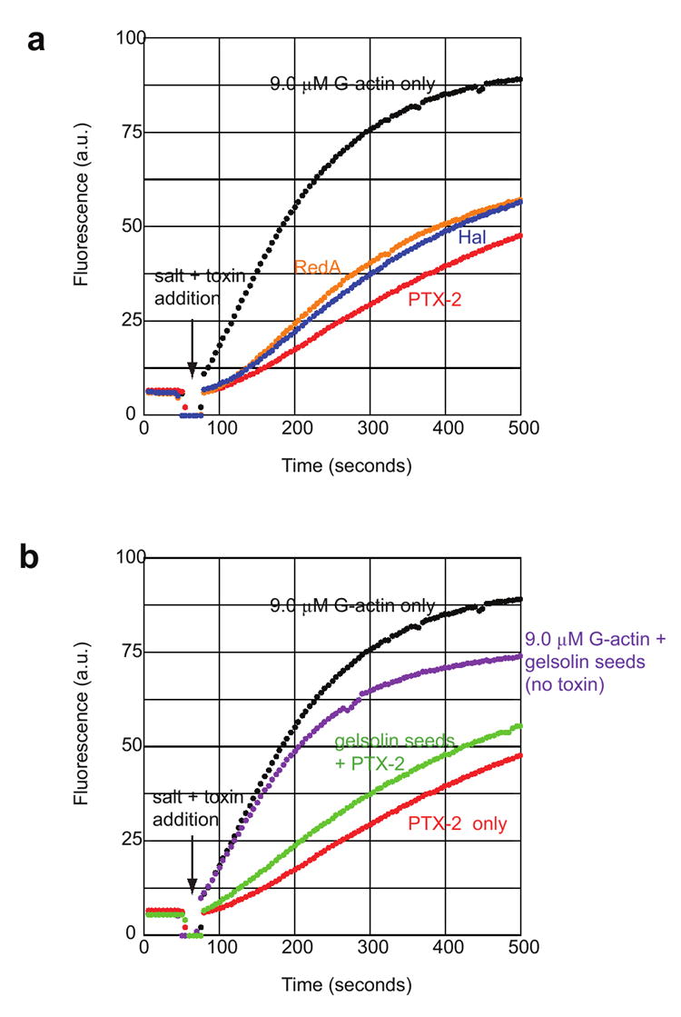
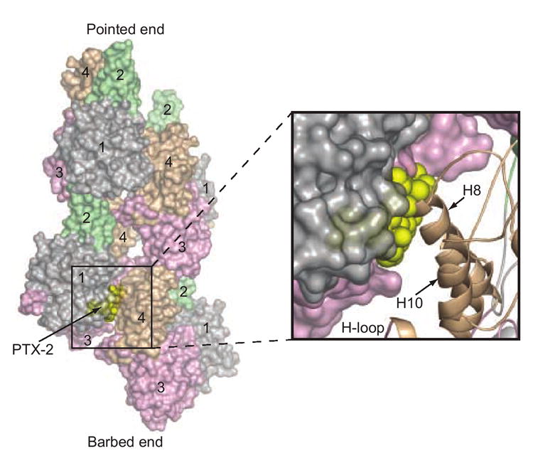
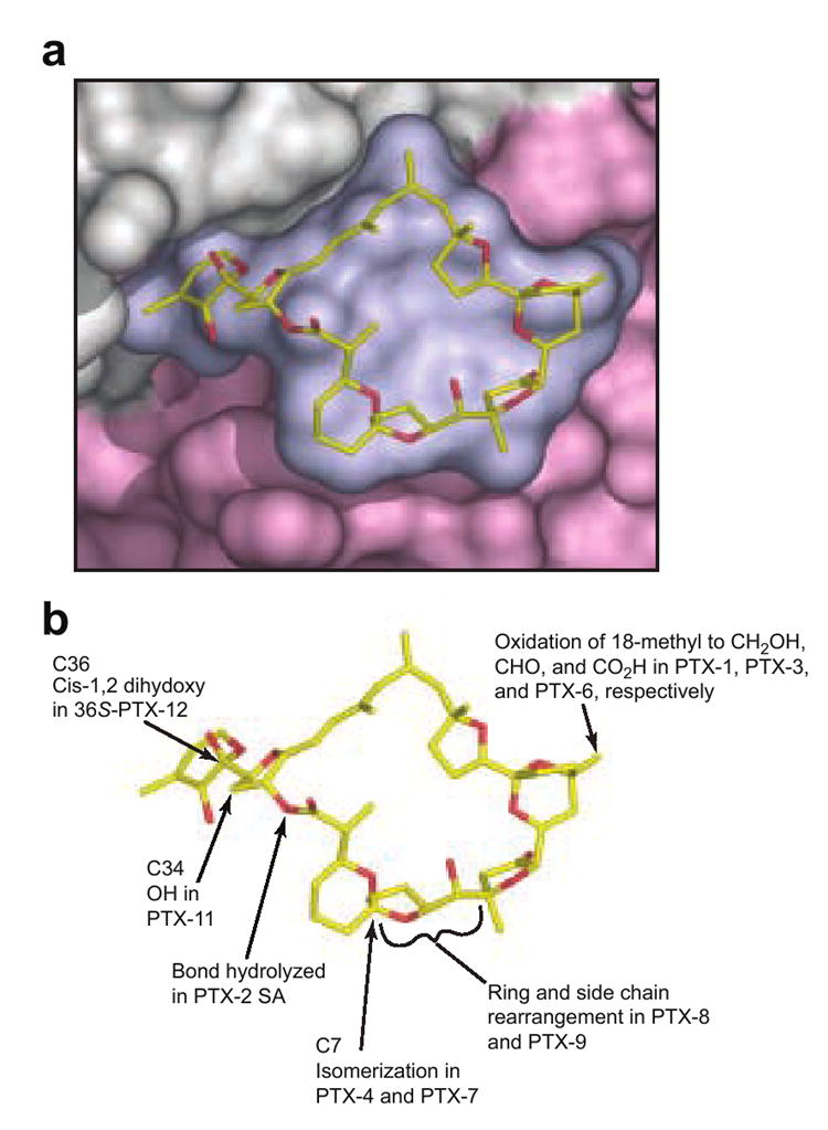
(PTXs) are polyether macrolides found in certain dinoflagellates, sponges and shellfish, and have been associated with diarrhetic shellfish poisoning. In addition to their in vivo toxicity, some PTXs are potently cytotoxic in human cancer cell lines. Recent studies have demonstrated that disruption of the actin cytoskeleton may be a key function of these compounds, although no clarification of their mechanism of action at a molecular level was available. We have obtained an X-ray crystal structure of PTX-2 bound to actin, which, in combination with analyses of the effect of PTX-2 on purified actin filament dynamics, provides a molecular explanation for its effects on actin. PTX-2 formed a 1:1 complex with actin and engaged a novel site between subdomains 1 and 3. Based on models of the actin filament, PTX binding would disrupt key lateral contacts between the PTX-bound actin monomer and the lower lateral actin monomer within the filament, thereby capping the barbed-end. The location of this binding position within the interior of the filament indicates that it may not be accessible once polymerization has occurred, a hypothesis supported by our observation that PTX-2 caused filament capping without inducing filament severing. This mode of action is unique, as other actin filament destabilizing toxins appear to exclusively disrupt longitudinal monomer contacts, allowing many of them to sever filaments in addition to capping them. Examination of the PTX-binding site on actin provides a rationalization for the structure-activity relationships observed in vivo and in vitro, and may provide a basis for predicting toxicity of PTX analogues.


Figures

Figure 1
Figure 1. Chemical structures of pectenotoxins compared to that of reidispongiolide A and the X-ray crystallographic structure PTX-2 extracted from the PTX-2–actin crystal structure
(a) The chemical scaffold of pectenotoxin is shown with eight different PTX analogues and their respective chemical modifications listed below. The overall structure has been divided into “ring” and “tail” structural components similar to other actin filament-destabilizing toxins., (b) The chemical structure of reidispongiolide A. (c) Stereo view of the actin-bound conformation of PTX-2 is shown surrounded by experimental electron density. The Fo-Fc electron density omit map was contoured at 2.5σ. Figures 1b, 2, 3, 6, and 7 were prepared with Pymol.
Figure 2
Figure 2. The PTX-2–actin complex
(a) PTX-2 (yellow) is shown as a ball-and-stick representation bound to actin between subdomains 1 and 3. (b) The structure of the reidispongiolide A–actin complex is shown for reference and is rotated 180° along the vertical axis from the orientation of the PTX-2–actin complex in (a). Reidispongiolide A is shown as blue ball-and-sticks. In this figure subdomains 1, 2, 3 and 4 of actin are colored in light gray, green, wheat, and light pink respectively. This color scheme is used throughout the figures to depict the subdomains of actin.
Figure 3
Figure 3. PTX-2–actin contacts
(a) Stereo view of specific PTX-2–actin interactions are shown. PTX-2 is in yellow and its contact residues on actin are labeled and colored according to the domains to which they belong. Polar interactions between PTX-2 and actin are shown in purple. Waters involved in hydrogen bonding between PTX-2 and actin are shown as red spheres. (b) Ligplot of the interactions between PTX-2 and actin. Van der Waals contacts are represented by black spokes radiating between interacting residues and are connected by thin dashed black lines. Water molecules are depicted as red spheres with hydrogen bonding interactions connected by thick dashed red lines. Actin residues involved in polar interactions are colored cyan. Figure produced with the program LIGPLOT.
Figure 4
Figure 4. F-actin severing activity of PTX-2
F-actin (48 μM) in F-buffer was treated with the indicated molar ratio amounts of PTX-2, latrunculin B (LatB), reidispongiolide A (RedA), or halichondramide (Hal) for 30 min and then centrifuged at 20,000 × g for 30 min. The pellet of each sample following analysis by SDS-PAGE is shown. The pellets for G-actin (48 μM) in G-buffer without the addition of toxin are shown as a control for sedimentation of non-filamentous actin within the tube.
Figure 5
Figure 5. Actin filament-capping activity of PTX-2
(a) The graph shows the change in pyrene fluorescence signal that occurs during polymerization of 9.0 μM G-actin (15% pyrenyl-G-actin) in the presence of 1 μM toxin–G-actin complexes as a function of time. A baseline signal was obtained for each actin sample before removal of the cuvette (at 50 seconds) from the fluorimeter and addition of 1 μM toxin–G-actin complex and KCl and MgCl2. Polymerization reaction profiles are labeled according to the toxin–actin complexes added (black, 9.0 μM G-actin without toxin–actin complex; blue, halichondramide; red, PTX-2; orange, reidispongiolide A). (b) 9.0 μM G-actin was pre-incubated with pre-assembled gelsolin–actin seeds added at a 1:50 gelsolin:actin ratio for five minutes prior to the addition of toxin–actin complexes and salts. Control samples of 9.0 μM G-actin only and G-actin plus gelsolin seeds are shown in black and purple, respectively. Polymerization of G-actin in the presence of 1 μM PTX-2–G-actin complex only is shown in red, while the same reaction contained gelsolin seeds is shown in green.
Figure 6
Figure 6. Superposition of PTX-2 onto the F-actin model
The G-actin-bound conformation of PTX-2 (yellow spheres) is superimposed on the model of F-actin to illustrate the mechanism by which it caps the barbed-end of the filament., Four actin subunits are shown (two for each strand) in surface representation. The pointed- and barbed-ends of this “mini-filament” are labeled. Actin subdomains 1–4 are also labeled for each subunit and are colored as in Figure 2. The binding position of PTX-2 is at the interior of the filament. The boxed region shows a magnification of the steric clash between PTX-2 and helix H8 from subdomain 4 of the lower lateral actin subunit (shown in cartoon representation).
Figure 7
Figure 7. Surface representation of the interaction of PTX-2 with actin and the location of common differences among PTX variants
(a) Closeup view of a surface representation of PTX-2 and its binding site on actin where subdomains 1 and 3 are colored in gray and light pink respectively. (b) shows the locations of the chemical differences in common PTX variants. It is immediately clear that the change from (R) configuration at C7 in PTX-2 to the (S) configuration in PTX-4 and -7 can be expected to induce a large change in the shape of the macrolactone ring.

References

    1. Draisci R, Lucentini L, Mascioni A. Pectenotoxins and yessotoxins: chemistry, toxicology, pharmacology, and analysis. In: Botana LM, editor. Seafood and Freshwater Toxins: Pharmacology, Physiology, and Detection. Marcel Dekker, Inc.; New York: 2000. pp. 289–324.
    1. Yasumoto T, Murata M, Oshima Y, Matsumoto GK, Clardy J. Diarrhetic shellfish poisoning; ACS Symposium Series, 262; New York: ACS; 1984.
    1. Yasumoto T, Murata M, Oshima Y, Sano M, Matsumoto GK, Clardy J. Diarrhetic shellfish toxins. Tetrahedron. 1985;41:1019–1025.
    1. Miles CO, Wilkins AL, Samdal IA, Sandvik M, Petersen D, Quilliam MA, Naustvoll LJ, Rundberget T, Torgersen T, Hovgaard P, Jensen DJ, Cooney JM. A novel pectenotoxin, PTX-12, in Dinophysis spp. and shellfish from Norway. Chem Res Toxicol. 2004;17:1423–1433. - PubMed
    1. Yasumoto T, Murata M, Lee J-S, Torigoe K. Polyether toxins produced by dinoflagellates. In: Natori S, Hashimoto K, Ueno Y, editors. Mycotoxins and Phycotoxins ’88. Elsevier; Amsterdam: 1989. pp. 375–382.

Publication types

LinkOut - more resources