Review
. 2007;31(8):1125-35.
doi: 10.1016/j.neubiorev.2007.04.017. Epub 2007 May 17.

Activity- and use-dependent plasticity of the developing corticospinal system

Affiliations
Review

Activity- and use-dependent plasticity of the developing corticospinal system

John H Martin et al. Neurosci Biobehav Rev. 2007.

Abstract

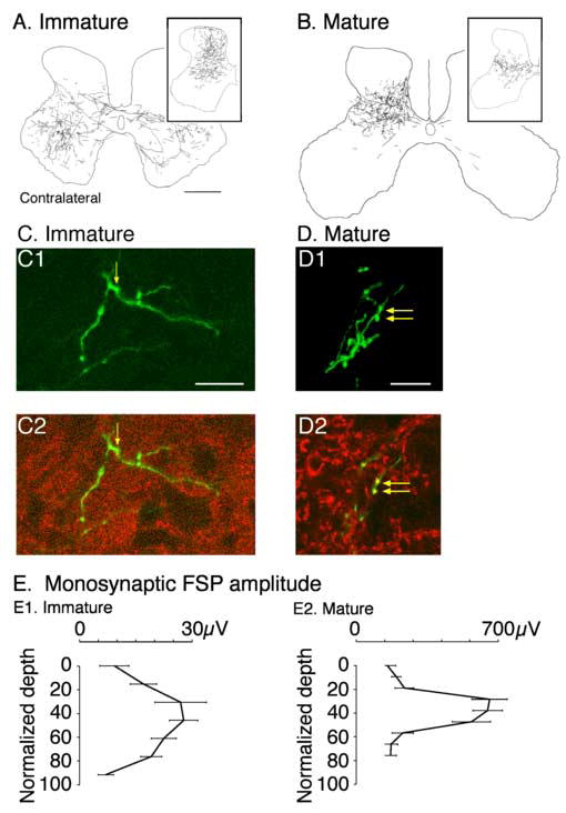
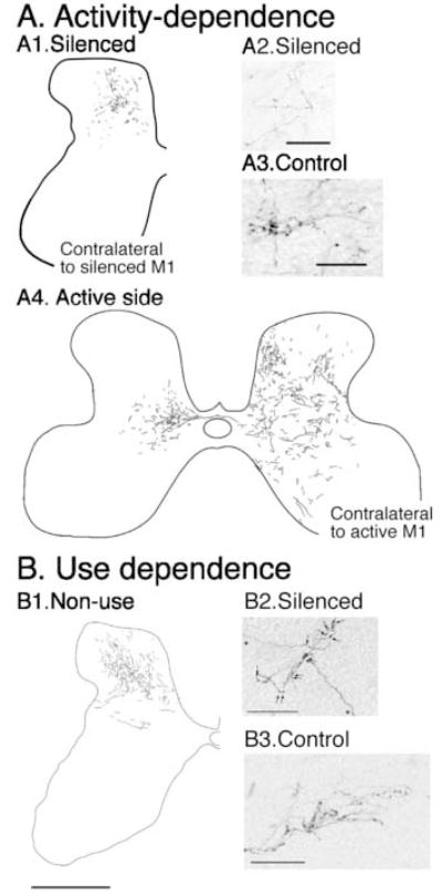
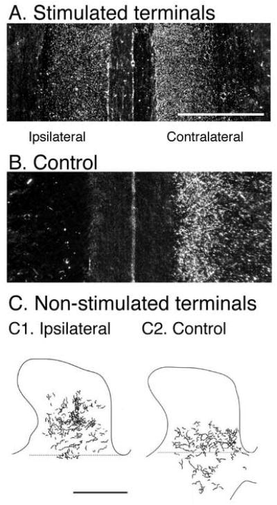
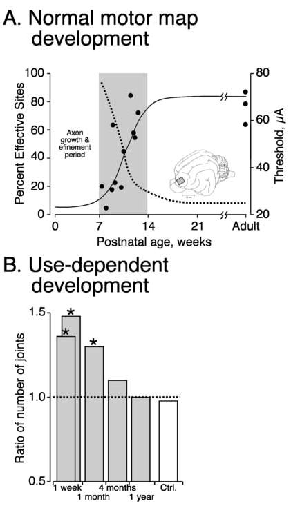
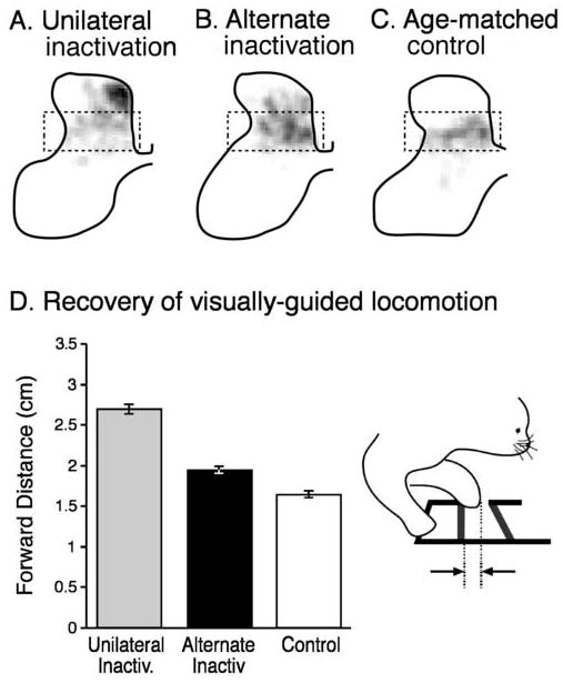
The corticospinal (CS) system, critical for controlling skilled movements, develops during the late prenatal and early postnatal periods in all species examined. In the cat, there is a sequence of development of the mature pattern of terminations of CS tract axons in the spinal gray matter, followed by motor map development of the primary motor cortex. Skilled limb movements begin to be expressed as the map develops. Development of the proper connections between CS axons and spinal neurons in cats depends on CS neural activity and motor behavioral experience during a critical postnatal period. Reversible CS inactivation or preventing limb use produces an aberrant distribution of CS axon terminations and impairs visually guided movements. This altered pattern of CS connections after inactivation in cats resembles the aberrant pattern of motor responses evoked by transcranial magnetic stimulation in hemiplegic cerebral palsy patients. Left untreated in the cat, these impairments do not resolve. We have found that activity-dependent processes can be harnessed in cats to reestablish normal CS connections and function. This finding suggests that aspects of normal CS connectivity and function might some day be restored in hemiplegic cerebral palsy.


Figures

Figure 1
Figure 1
Development of the CS tract axon spinal terminations in the cervical cord of the cat. A. Terminations in 5 week animal, showing a bilateral pattern. Tracer was injected into the rostral M1 subregion, which results in a ventral bias to the termination field. The inset shows an example of terminations from the caudal M1, which has a dorsal bias. B. Distribution of terminations in the adult. The main figure shows terminations from caudal M1 and the inset, from rostral M1 (Li and Martin, 2000). C. Confocal micrographs of immature (C) and mature (D) CS terminal. Part 1 of each panel is a projection image of the labeled terminal. The labeled terminal is green. Part 2 shows 1 μm optical slices within the projection image. Red corresponds to synaptophysin immuno-label and green, anterograde axonal label. Double labeled presynaptic boutons are indicated by the yellow arrows; yellow corresponds to overlay of green and red labeling (Meng et al. 2004). E. Dorsoventral distribution of the CS monosynaptic focal synaptic potential (FSP). Note that the potential is smaller and dorsoventrally more extensive in the immature animal (Meng and Martin, 2003). Calibrations. A, B. 1 mm; C. 25 μm; D. 50 μm.
Figure 2
Figure 2
Activity (A)- and use-dependent (B) development of CS terminals in the cervical cord. A1. Terminations from the silenced M1. A2, 3. Morphology of silenced and control CS terminations. A4. Terminations from the active M1. (Friel and Martin, 2005) B1. Terminations on the non-used side of the cervical cord. B2, 3. Morphology of non-used and control CS terminations. (Martin et al. 2004) Calibrations. A1. 1 mm (bar in B1); A2, A3. 50 μm; B1. 1 mm; B2, B3. 50 μm.
Figure 3
Figure 3
Effects of CS system electrical stimulation on developing CS axon terminations. A, B. Horizontal sections through the deep dorsal horn. CS axon terminals are labeled using horseradish peroxidase conjugated to wheat germ agglutinin (white dots). Stimulation (A) results in a bilateral distribution of terminations while only contralateral terminations are in the control (B). C. Effect of unilateral stimulation on the non-stimulated CS system (C1) showing a dorsal shift in the terminations compared with the control (C2). BDA was used to label the non-stimulated axons. (Salimi and Martin, 2004) Calibrations. A, B, C. 1 mm.
Figure 4
Figure 4
Development of the motor map in the M1. A. Reduction in threshold for evoking movement by microstimulation (dotted line) and the increase in effective sites (solid line) with age.(Chakrabarty and Martin, 2000) B. effect of prehension training between weeks 7 and 11 on the incidence of sites where stimulation produced muscle contraction around multiple contralateral forelimb joints (Martin et al. 2005).
Figure 5
Figure 5
Alternate inactivation restores connectivity and function. A, B, C. Distribution of contralateral CS axon terminal label in 15 week old animals shown as a gray scale, where white is no label and black is maximal label per square micrometer. The same gray scale was used for each part. The normal distribution is highlighted by the dashed box, which corresponds to the control (C). Unilateral inactivation results in dorsal label (A) while after the alternate inactivation (B), label is more ventral, like the control. D. Effect of alternate inactivation on forward step distance. Inset illustrates the step measurement. (Friel et al. 2006).

Similar articles

Cited by

References

    1. Alisky JM, Swink TD, Tolbert DL. The postnatal spatial and temporal development of corticospinal projections in cats. Exp Brain Res. 1992;88:265–276. - PubMed
    1. Arlotta P, Molyneaux BJ, Chen J, Inoue J, Kominami R, Macklis JD. Neuronal subtype-specific genes that control corticospinal motor neuron development in vivo. Neuron. 2005;45:207–221. - PubMed
    1. Armand J, Kuypers HGJM. Cells of origin of crossed and uncrossed corticospinal fibers in the cat. A quantitative horseradish peroxidase study. Exp Brain Res. 1980;40:23–34. - PubMed
    1. Armand J, Olivier E, Edgley SA, Lemon RN. Postnatal development of corticospinal projections from motor cortex to the cervical enlargement in the macaque monkey. J Neurosci. 1997;17:251–266. - PMC - PubMed
    1. Baldissera F, Hultborn H, Illert M. Integration in spinal neuronal systems. In: Brooks VB, editor. Handbook of Physiology, Section I: The Nervous System, Vol II, Motor Control. American Physiological Society; Bethesda: 1981. pp. 509–596.

Publication types