Review
. 2007 Nov;101(11-12):1913-21.
doi: 10.1016/j.jinorgbio.2007.05.001. Epub 2007 May 17.

DNA repair glycosylases with a [4Fe-4S] cluster: a redox cofactor for DNA-mediated charge transport?

Affiliations
Review

DNA repair glycosylases with a [4Fe-4S] cluster: a redox cofactor for DNA-mediated charge transport?

Amie K Boal et al. J Inorg Biochem. 2007 Nov.

Abstract

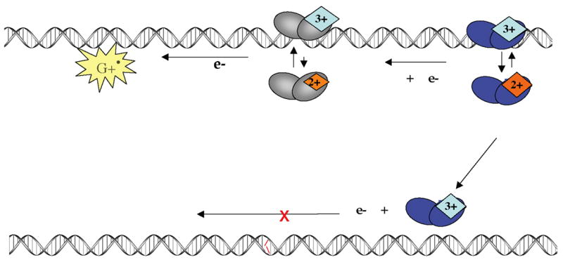
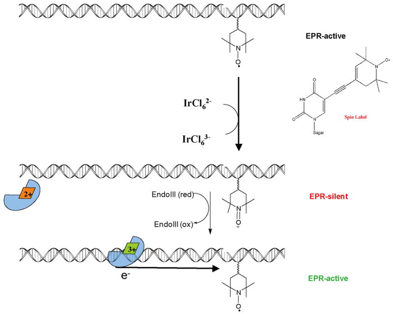
The [4Fe-4S] cluster is ubiquitous to a class of base excision repair enzymes in organisms ranging from bacteria to man and was first considered as a structural element, owing to its redox stability under physiological conditions. When studied bound to DNA, two of these repair proteins (MutY and Endonuclease III from Escherichia coli) display DNA-dependent reversible electron transfer with characteristics typical of high potential iron proteins. These results have inspired a reexamination of the role of the [4Fe-4S] cluster in this class of enzymes. Might the [4Fe-4S] cluster be used as a redox cofactor to search for damaged sites using DNA-mediated charge transport, a process well known to be highly sensitive to lesions and mismatched bases? Described here are experiments demonstrating the utility of DNA-mediated charge transport in characterizing these DNA-binding metalloproteins, as well as efforts to elucidate this new function for DNA as an electronic signaling medium among the proteins.


Figures

Figure 1
Figure 1
A schematic illustration showing the general strategy for protein electrochemistry experiments at DNA-modified electrodes. (a) Modified DNA self-assembles into a monolayer on an Au or HOPG electrode surface. (b) The DNA-modified surface is then passivated with an alkane or alkanethiol to prevent any interactions between the protein and the bare electrode surface. (c) A protein solution is introduced and monitored electrochemically with cyclic or square-wave voltammetry.
Figure 2
Figure 2
Endo III potentials are measured electrochemically with and without DNA. The top panel shows a square wave voltammogram (right) for Endo III at a DNA-modified HOPG electrode. A peak with a midpoint potential of +20 mV versus NHE is evident. At top left is a cartoon representation this electrode setup. The bottom panel (right) shows square wave voltammetry at a bare HOPG electrode where a peak at +250 mV is apparent. Note that a peak is not observed at +20 mV in the absence of DNA. At bottom left is a cartoon representation of Endo III analyzed at a bare HOPG electrode.
Figure 3
Figure 3
A model for DNA CT in DNA repair. Upon binding to DNA, a protein with an FeS cluster in the 2+ oxidation state can become oxidized to the 3+ state (top left). If the surrounding DNA is undamaged, the released electron can reduce another oxidized protein bound at a distal site (top right) causing the second protein to lose affinity for DNA. As described, DNA CT between two BER enzymes allows for a rapid search of the intervening region of the genome. If a damaged site is present between the two proteins, the DNA-mediated CT event does not occur among these two proteins; both enzymes remain oxidized and bound close to the aberrant site. This process allows proteins to be redistributed from undamaged sites to locations containing lesions.
Figure 4
Figure 4
The flash-quench technique is used to generate Ru(III) and then guanine radicals by DNA-mediated CT. The dppz complex of Ru(II) intercalates into the DNA helix. Upon irradiation, the Ru(II) complex is excited to Ru(II)* which can then be quenched by Q in solution to generate Ru(III). This Ru(III) species has sufficient driving force to oxidize guanine in DNA to form the guanine radical cation and the original Ru(II) species. Guanine radical, a signal of oxidative stress, then can serve as an oxidant for the [4Fe-4S] cluster of the repair protein. Note that back electron transfer pathways are shown with dashed arrrows.
Figure 5
Figure 5
A model for guanine radical initiated searching via DNA CT. Guanine radical, one of the first signs of oxidative stress, can initiate scanning by oxidizing the [4Fe-4S]2+ cluster in MutY. The first oxidized protein can then be reduced by a second protein via the DNA base-pair stack. Again, if a lesion is present, the proteins stay oxidized and bound in the vicinity of the damaged site. Oxidative stress thus provides the driving force for the DNA CT search.
Figure 6
Figure 6
Strategy for electron trapping from the DNA-bound repair protein. (Top) A stable, EPR-active, nitroxyl radical is incorporated as a modified base in the DNA duplex. (Middle) A mild Ir(IV) oxidant is used to generate the EPR-silent N-oxo-ammonium ion. (Bottom) Protein binding and cluster oxidation releases the electron, which reduces the modified base back to the EPR-active nitroxide radical.

Similar articles

Cited by

References

    1. Friedberg EC, Walker GC, Siede W, Wood RD, Schultz RA, Ellenberger T, editors. DNA Repair and Mutagenesis. ASM Press; Washington DC: 2006.
    1. David SS, Williams SD. Chem Rev. 1998;98:1221–1262. - PubMed
    1. Blainey PC, van Oijen AM, Banerjee A, Verdine GL, Xie XS. Proc Natl Acad Sci USA. 2006;103:5752–5757. - PMC - PubMed
    1. Francis AW, David SS. Biochemistry. 2003;42:801–810. - PubMed
    1. Halford SE, Szczelkun MD. Eur Biophys J. 2002;31:257–267. - PubMed

Publication types

LinkOut - more resources