. 2007 Aug;206(2):280-7.
doi: 10.1016/j.expneurol.2007.05.017. Epub 2007 Jun 2.

Reperfusion accelerates acute neuronal death induced by simulated ischemia

Affiliations

Reperfusion accelerates acute neuronal death induced by simulated ischemia

Dongdong Li et al. Exp Neurol. 2007 Aug.

Abstract

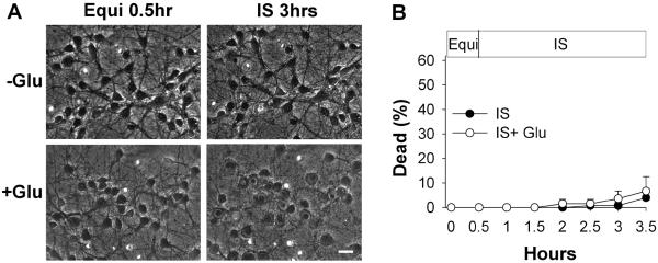
Observations in real time can provide insights into the timing of injury and the mechanisms of damage in neural ischemia-reperfusion. Continuous digital imaging of morphology and cell viability was applied in a novel model of simulated ischemia-reperfusion in cultured cortical neurons, consisting of exposure to severe hypoxia combined with glucose deprivation, mild acidosis, hypercapnia, and elevated potassium, followed by return of oxygenated, glucose-containing physiological saline. Substantial acute injury resulted following 1 h of simulated ischemia, with 36+/-8% neurons dying within 2 h of reperfusion. Inclusion of moderate glutamate elevation (30 microM) in the simulation of ischemia increased the acute neuronal death to 51+/-6% at 2 h of reperfusion. While some swelling and neuritic breakdown occurred during ischemia, particularly with inclusion of glutamate, neuronal death, as marked by loss of somatic membrane integrity, was entirely restricted to the reperfusion phase. Morphological and cytoskeletal changes suggested a predominance of necrotic death in the acute phase of reperfusion, with more complete delayed death accompanied by some apoptotic features occurring over subsequent days. Prolonged simulated ischemia, without reperfusion, did not induce significant acute neuronal death even when extended to 3 h. We conclude that while morphological changes suggesting initiation of neuronal injury appear during severe simulated ischemia, the irreversible injury signaled by membrane breakdown is accelerated by the events of reperfusion itself.


Figures

Figure 1
Figure 1. Ischemia-reperfusion injury in cultured neurons
A) Representative digital images (combined phase-contrast and propidium fluorescence imaging) of cortical neurons (10 days in vitro) subjected to ischemia-reperfusion. Left-hand column, cell images after 0.5 hour baseline equilibration by perfusion with physiological balanced salt solution (BSS). Middle column, images after an additional 1 hour perfusion with BSS (control), with glutamate, or with simulated ischemia, without or with glutamate (30 μM). Right-hand column, images after simulated reperfusion with oxygenated BSS for an additional 2 hours. In control experiments (`Cont', top row), neurons remained normal in morphology during the entire 3.5 hour protocol. Application of 30μM glutamate alone for 60 minutes (`Glu', second row) produced considerable cell swelling, but no significant increase in cell death within 3 hours. With application of simulated ischemia solution, without addition of glutamate (`IR', third row), only subtle morphologic changes occurred during ischemia (arrow), but a progressively increasing fraction of neurons died with reperfusion as marked by newly acquired nuclear propidium fluorescence (arrowhead). During application of simulated ischemia including glutamate (`IR/G', bottom row), dramatic cell swelling and neuritic breakdown occurred during ischemia when glutamate was included (arrow), but again cell death, marked by propidium uptake, took place only upon reperfusion (arrowhead). Scale bar in lower right panel represents 20 microns for all images. B) Cell death, defined by propidium uptake, in ischemia-reperfusion, comparing results from parallel experiments in controls, ischemia alone, and ischemia with glutamate (n = 6, 9, and 5 independent replicates respectively, with 25–53 neurons imaged per field; *significantly different from parallel control value, and significantly different from parallel value in ischemia without glutamate condition, p< 0.05). C) Cell death in experiments comparing survival with 60 minute exposures to glutamate (30 μM) alone to simulated ischemia plus glutamate, in an independent set of replicates of the latter condition performed in parallel (n = 4 and 3 replicates, respectively, with 44–58 neurons imaged per field; *significantly different, p< 0.05).
Figure 2
Figure 2. Morphological changes during ischemia-reperfusion in cortical neurons marked by MAP-2 immunostaining
Representative digital images of MAP-2 immunofluorescence in cortical neurons subjected to simulated ischemia alone (`− Glu', left column) or simulated ischemia plus 30 μM glutamate (`+ Glu', right column) at baseline after 0.5 hour equilibration (`Equi 0.5 hr'), after 0.5 or 1 hour simulated ischemia (`IS'), and following 1 or 2 hours of simulated reperfusion (`RE'). Scale bar, 20 microns for all panels. In ischemia without glutamate, subtle neuronal swelling could be seen during ischemia (arrows), as well as scattered areas of breakdown of dendrites to a beaded appearance, becoming more prominent with reperfusion (arrowheads). In ischemia with glutamate, prominent somatic swelling and dendritic beading was seen during ischemia, followed by widespread dendritic breakdown and loss of cellular MAP-2 immunoreactivity during reperfusion.
Figure 3
Figure 3. Delayed injury to cortical neurons after simulated ischemia
A) Hoechst staining for nuclear morphology showed pyknotic changes of most nuclei at 2 or 24 hours following ischemia with or without glutamate (arrowheads, examples). A fraction of nuclei showed severe swelling instead of condensation, especially after ischemia with glutamate (arrow). Only rare nuclei showed nuclear fragmentation characteristic of apoptosis (open arrow). B) Relative survival, determined by live/dead counts and normalized to parallel controls, showed significant declines in survival from 2 to 24 and 72 hours following ischemia-reperfusion (IR). At each time point, survival in reperfusion following ischemia plus glutamate (IR/G) was significantly less than that following ischemia alone (n = 3 independent replicates with 134–206 cells counted per condition; two-way ANOVA, effects of time and treatment both significant at p<0.05; post-hoc pairwise comparisons: *different than corresponding 2 hour survival, †different than 24 hour survival). C) At 2 hours of reperfusion, TUNEL staining revealed frequent TUNEL-positive pyknotic nuclei both in cells with apparently intact surrounding membranes and without evident surrounding cell membranes; few TUNEL-positive nuclei had a fragmented appearance. By 24 hours after IR/G, few intact cells could be seen but many TUNEL-positive as well as TUNEL-negative bare pyknotic nuclei remained. D) Quantitation of percentages of neurons staining by the TUNEL method. As this staining method does not provide a sensitive determination of preservation of intact somatic membrane, nuclei without evident surrounding cells were included in the counts. Proportions of cells staining for DNA strand breaks were increased at 2 and 24 hours following simulated ischemia alone and with glutamate (n = 3 independent replicates with 167–223 cells counted per condition; two-way ANOVA, effects of time and treatment both significant at p<0.05; post-hoc pairwise comparisons: *different than corresponding control; †different than ischemia alone condition).
Figure 4
Figure 4. Survival of cortical neurons after prolonged simulated ischemia
A) Representative digital images (combined phase-contrast and fluorescence) of cortical neurons (10 days in vitro) at baseline after equilibration (`Equi') and after continuous simulated ischemia without or with glutamate for 3 hours. Scale bar, 20 microns for all panels. B) Cell death during continuous simulated ischemia, without (filled circles) or with 30 μM glutamate (open circles); n = 4 replicates, with 21–36 neurons imaged per field (error bars are hidden by symbols).

References

    1. Abele AE, Scholz KP, Scholz WK, Miller RJ. Excitotoxicity induced by enhanced excitatory neurotransmission in cultured hippocampal pyramidal neurons. Neuron. 1990;4:413–419. - PubMed
    1. Abella BS, Zhao D, Alvarado J, Hamann K, Vanden Hoek TL, Becker LB. Intra-arrest cooling improves outcomes in a murine cardiac arrest model. Circulation. 2004;109:2786–2791. - PubMed
    1. Abramov AY, Scorziello A, Duchen MR. Three distinct mechanisms generate oxygen free radicals in neurons and contribute to cell death during anoxia and reoxygenation. J. Neurosc. 2007;27:1129–1138. - PMC - PubMed
    1. Ambrosio G, Flaherty JT. Effects of the superoxide radical scavenger superoxide dismutase, and of the hydroxyl radical scavenger mannitol, on reperfusion injury in isolated rabbit hearts. Cardiovasc. Drugs Ther. 1992;6:623–632. - PubMed
    1. Aronowski J, Strong R, Grotta J. Reperfusion injury: Demonstration of brain damage produced by reperfusion after transient focal ischemia in rats. J. Cereb. Blood Flow Metab. 1997;17:1048–1056. - PubMed

Publication types

MeSH terms