Comparative Study
. 2007 Jul 10;104(28):11706-11.
doi: 10.1073/pnas.0700544104. Epub 2007 Jun 28.

Negative regulation of MDA5- but not RIG-I-mediated innate antiviral signaling by the dihydroxyacetone kinase

Affiliations
Comparative Study

Negative regulation of MDA5- but not RIG-I-mediated innate antiviral signaling by the dihydroxyacetone kinase

Feici Diao et al. Proc Natl Acad Sci U S A. .

Abstract

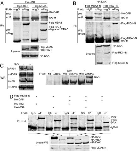
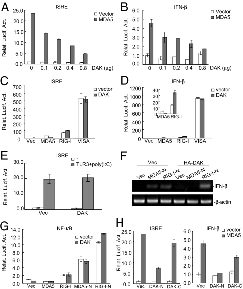
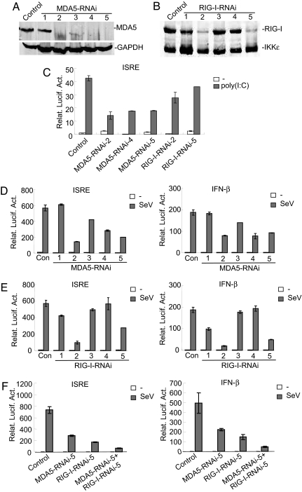
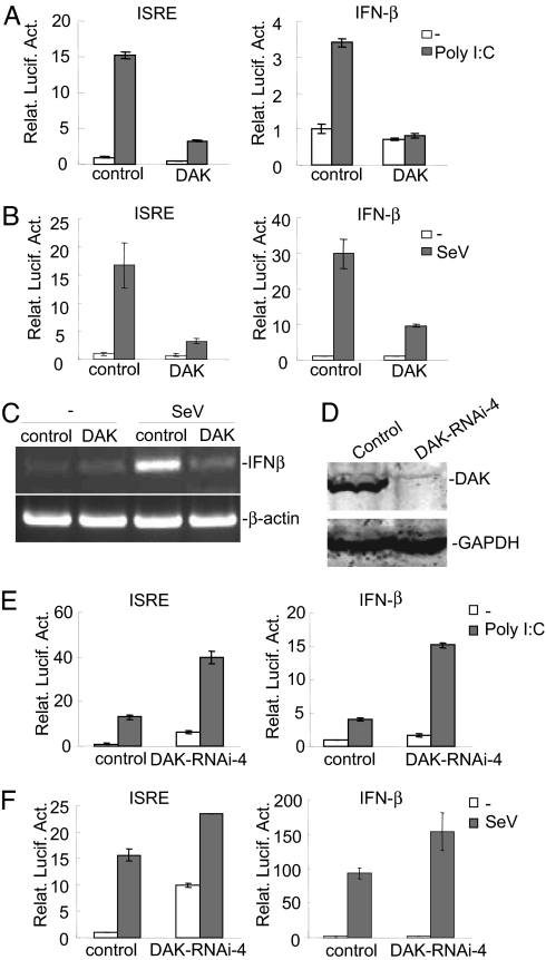
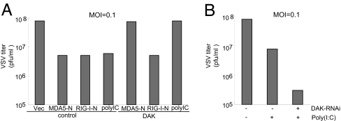
Viral infection leads to activation of the transcription factors interferon regulatory factor-3 and NF-kappaB, which collaborate to induce type I IFNs. The RNA helicase proteins RIG-I and MDA5 were recently identified as two cytoplasmic viral RNA sensors that recognize different species of viral RNAs produced during viral replication. In this study, we identified DAK, a functionally unknown dihydroacetone kinase, as a specific MDA5-interacting protein. DAK was associated with MDA5, but not RIG-I, under physiological conditions. Overexpression of DAK inhibited MDA5- but not RIG-I- or TLR3-mediated IFN-beta induction. Overexpression of DAK also inhibited cytoplasmic dsRNA and SeV-induced activation of the IFN-beta promoter, whereas knockdown of endogenous DAK by RNAi activated the IFN-beta promoter, and increased cytoplasmic dsRNA- or SeV-triggered activation of the IFN-beta promoter. In addition, overexpression of DAK inhibited MDA5- but not RIG-I-mediated antiviral activity, whereas DAK RNAi increased cytoplasmic dsRNA-triggered antiviral activity. These findings suggest that DAK is a physiological suppressor of MDA5 and specifically inhibits MDA5- but not RIG-I-mediated innate antiviral signaling.


Conflict of interest statement

The authors declare no conflict of interest.

Figures

Fig. 1.
Fig. 1.
Identification of DAK as an MDA5-associated protein. (A) DAK interacts with MDA5 but not RIG-I in mammalian overexpression system. The 293 cells (2 × 10⁶) were cotransfected with the indicated plasmids (5 μg each), and cell lysates were immunoprecipitated with anti-Flag antibody (αFlag) or control mouse IgG (mIgG). The immunoprecipitates were analyzed by Western blots with anti-HA (Top) or anti-Flag (Middle) antibody. Expression of the transfected proteins was analyzed by Western blots with anti-HA and anti-Flag antibodies (Bottom). (B) DAK interacts with N-terminal domain of MDA5. The experiments were similarly performed as in A. The asterisk (Middle) indicates the band of Flag-MDA5-N, which is close to the IgG light-chain band. (C) Endogenous association of DAK with MDA5. The 293 cells (1 × 10⁸) were infected with SeV or left uninfected for 4 h. Cell lysate was immunoprecipitated with mouse anti-MDA5 or rabbit anti-RIG-I antiserum or control IgG. The immunoprecipitates were analyzed by Western blots with anti-DAK antibody (Right). The expression levels of the endogenous proteins were analyzed by Western blots with anti-MDA5, anti-DAK, and anti-RIG-I antibodies (Left). Each lane was loaded with whole-cell lysate from 5 × 10⁶ cells. (D) Interactions between MDA5-N and DAK, IKKε and VISA. The 293 cells (2 × 10⁶) were cotransfected with the indicated plasmids (5 μg of each). Coimmunoprecipitation and Western blot analysis were performed as in A. WB, Western blot; IP, immunoprecipitation; IB, immunoblot.
Fig. 2.
Fig. 2.
DAK inhibits MDA5- but not RIG-I-mediated activation of ISRE and the IFN-β promoter. (A and B) DAK inhibits MDA5-mediated activation of ISRE (A) and the IFN-β promoter (B) in a dose-dependent manner. The 293 cells (1 × 10⁵) were transfected with ISRE (A) or the IFN-β promoter (B) luciferase reporter plasmid (0.05 μg), an expression plasmid for Flag-MDA5 (0.4 μg, gray bars) or an empty control plasmid (0.4 μg, white bars) and increased amounts of an expression plasmid for HA-DAK (0–0.8 μg as indicated). Luciferase assays were performed 16 h after transfection. (C–E) DAK has no significant effects on activation of ISRE and the IFN-β promoter mediated by RIG-I, TLR3, and VISA. The 293 cells (1 × 10⁵) were transfected with the indicated reporter plasmid (0.05 μg) and expression plasmids (0.4 μg of each). Reporter assays were performed 16 h after transfection. In E, transfected cells were treated with poly I:C (40 μg/ml) for 6 h before reporter assays were performed. (F) DAK inhibits MDA5-N- but not RIG-I-N-induced expression of IFN-β. The 293 cells (5 × 10⁵) were transfected with Flag-MDA5-N or Flag-RIG-I-N plasmid (1 μg) and HA-DAK or empty control plasmid (2 μg). RT-PCR was performed with human IFN-β or β-actin primers 18 h after transfection. (G) DAK does not inhibit MDA5-N and RIG-I-N-mediated activation of NF-κB. The 293 cells (1 × 10⁵) were transfected with NF-κB luciferase reporter (0.1 μg) and the indicated plasmids (0.4 μg of each). Reporter assays were performed 16 h after transfection. (H) DAK-N was sufficient to inhibit MDA5-mediated activation of ISRE and the IFN-β promoter. The experiments were performed as in C and D. Relat. Lucif. Act., relative luciferase activity; Vec, vector.
Fig. 3.
Fig. 3.
Both MDA5 and RIG-I are involved in cytoplasmic poly(I:C)- and SeV-triggered signaling. (A) Effects of MDA5 RNAi plasmids on the expression of endogenous MDA5. The 293 cells (1 × 10⁶) were transfected with a control or MDA5 RNAi plasmids (10 μg of each). Forty-eight hours after transfection, cell lysates were analyzed by Western blots with anti-MDA5 (Upper) or anti-GAPDH (Lower) antibody. (B) Effects of RIG-I RNAi plasmids on the expression of RIG-I. The 293 cells (5 × 10⁵) were transfected with HA-tagged RIG-I and IKKε plasmid (1 μg) and a control or RIG-I RNAi plasmids (2 μg of each). Forty-eight hours after transfection, cell lysates were analyzed by Western blot with anti-HA antibody. (C) Effects of MDA5 and RIG-I RNAi plasmids on cytoplasmic poly(I:C)-triggered ISRE activation. The 293 cells (1 × 10⁵) were transfected with ISRE luciferase reporter plasmid (0.05 μg), control, or MDA5 or RIG-I RNAi plasmids (0.5 μg of each) as indicated. Thirty-six hours after transfection, cells were further transfected with poly(I:C) (4 μg, gray bars) or buffer (white bars) by Lipofectamine for 12 h before luciferase assays were performed. (D) Effects of MDA5 RNAi plasmids on SeV-triggered activation of ISRE and the IFN-β promoter. The 293 cells (1 × 10⁵) were transfected with ISRE or the IFN-β promoter luciferase reporter plasmid (0.05 μg) as indicated and MDA5 RNAi plasmids (0.5 μg of each). Twenty-four hours after transfection, cells were infected with SeV (gray bars) or left untreated (white bars) for 12 h before luciferase assays were performed. (E) Effects of RIG-I RNAi plasmids on SeV-triggered activation of ISRE and the IFN-β promoter. The experiments were performed as in D. (F) Effects of a combination of MDA5 and RIG-I RNAi plasmids on SeV-triggered activation of ISRE and the IFN-β promoter. The experiments were performed as in D. The 293 cells were transfected with 0.5 μg of MDA5 or RIG-I RNAi plasmid alone, or a combination of 0.25 μg of each. Relat. Lucif. Act., relative luciferase activity.
Fig. 4.
Fig. 4.
DAK negatively regulates IFN-β signaling triggered by cytoplasmic poly(I:C) and SeV in 293 cells. (A) DAK inhibits cytoplasmic poly(I:C)-induced activation of ISRE and the IFNβ promoter. The 293 cells (1 × 10⁵) were transfected with the indicated reporter (0.05 μg) and expression plasmids (0.5 μg of each). Sixteen hours after transfection, cells were further transfected with poly(I:C) (4 μg) (gray bars) or buffer (white bars) for 12 h before luciferase assays were performed. (B) DAK inhibits SeV-triggered activation of ISRE and the IFN-β promoter. The 293 cells (1 × 10⁵) were transfected with the indicated reporter (0.05 μg) and expression plasmids (0.5 μg of each). Sixteen hours after transfection, cells were infected with SeV or left uninfected for 12 h before luciferase assays were performed. (C) DAK inhibits SeV-induced expression of IFN-β. The 293 cells (1 × 10⁵) were transfected with empty control plasmid or an expression plasmid for DAK (0.5 μg of each). Twelve hours after transfection, cells were infected with SeV or left uninfected for 12 h before RT-PCR was performed with human IFNβ and β-actin primers. (D) Effect of DAK RNAi #4 plasmid on the expression of DAK. The 293 cells (1 × 10⁶) were transfected with a control or DAK RNAi plasmid (5 μg) for 60 h before Western blot analysis was performed with the indicated antibodies. (E) DAK RNAi potentiates cytoplasmic poly(I:C)-induced activation of ISRE and the IFN-β promoter in 293 cells. The 293 cells (1 × 10⁵) were transfected with the indicated reporter (0.05 μg) and DAK RNAi (0.5 μg) plasmid. Thirty-six hours after transfection, cells were further transfected with poly(I:C) (4 μg) (gray bars) or buffer (white bars) for 12 h before luciferase assays were performed. (F) DAK RNAi potentiates SeV-triggered activation of ISRE and the IFN-β promoter in 293 cells. The 293 cells were transfected as in A. The transfected cells were infected with SeV or left uninfected for 12 h before luciferase assays were performed. Relat. Lucif. Act., relative luciferase activity.
Fig. 5.
Fig. 5.
Effects of DAK and DAK RNAi on MDA5-mediated antiviral response. (A) DAK inhibits MDA5-mediated antiviral response. The 293 cells (2 × 10⁵) were transfected with the indicated expression plasmids (1 μg of each). Twenty-four hours after transfection, cells were infected with VSV [multiplicity of infection (MOI) = 0.1], and supernatants were harvested at 12 h after infection. Supernatants were analyzed for VSV production by using standard plaque assays. Plaques were counted, and titers were calculated as plaque-forming units per milliliter. (B) DAK RNAi potentiates cytoplasmic poly(I:C)-triggered antiviral response. The 293 cells (2 × 10⁵) were transfected with a control or DAK RNAi plasmid (1 μg of each). Thirty-six hours after transfection, cells were further transfected with poly(I:C) (20 μg) or buffer. Twelve hours later, cells were infected with VSV (MOI = 0.1), and supernatants were harvested at 12 h after infection for plaque assays.

Similar articles

Cited by

References

    1. Durbin JE, Fernandez-Sesma A, Lee CK, Rao TD, Frey AB, Moran TM, Vukmanovic S, Garcia-Sastre A, Levy DE. J Immunol. 2000;164:4220–4228. - PubMed
    1. Levy DE, Garcia-Sastre A. Cytokine Growth Factor Rev. 2001;12:143–156. - PubMed
    1. Levy DE, Marie IJ. Nat Immunol. 2004;5:699–701. - PubMed
    1. Taniguchi T, Ogasawara K, Takaoka A, Tanaka N. Annu Rev Immunol. 2001;19:623–655. - PubMed
    1. Honda K, Takaoka A, Taniguchi T. Immunity. 2006;25:349–360. - PubMed

Publication types