

Re-expression of a developmentally restricted potassium channel in autoimmune demyelination: Kv1.4 is implicated in oligodendroglial proliferation
- PMID: 17600124
- PMCID: PMC1934532
- DOI: 10.2353/ajpath.2007.061241
Re-expression of a developmentally restricted potassium channel in autoimmune demyelination: Kv1.4 is implicated in oligodendroglial proliferation
Abstract
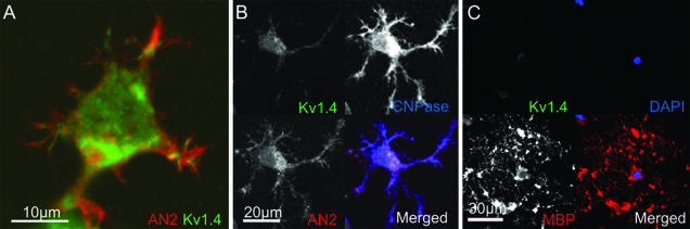
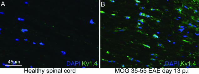
Mechanisms of lesion repair in multiple sclerosis are incompletely understood. To some degree, remyelination can occur, associated with an increase of proliferating oligodendroglial cells. Recently, the expression of potassium channels has been implicated in the control of oligodendrocyte precursor cell proliferation in vitro. We investigated the expression of Kv1.4 potassium channels in myelin oligodendrocyte glycoprotein-induced experimental autoimmune encephalomyelitis, a model of multiple sclerosis. Confocal microscopy revealed expression of Kv1.4 in AN2-positive oligodendrocyte precursor cells and premyelinating oligodendrocytes in vitro but neither in mature oligodendrocytes nor in the spinal cords of healthy adult mice. After induction of myelin oligodendrocyte glycoprotein-induced experimental autoimmune encephalomyelitis, Kv1.4 immunoreactivity was detected in or around lesions already during disease onset with a peak early and a subsequent decrease in the late phase of the disease. Kv1.4 expression was confined to 2',3'-cyclic nucleotide 3'-phosphodiesterase-positive oligodendroglial cells, which were actively proliferating and ensheathed naked axons. After a demyelinating episode, the number of Kv1.4 and 2',3'-cyclic nucleotide 3'-phosphodiesterase double-positive cells was greatly reduced in ciliary neurotrophic factor knockout mice, a model with impaired lesion repair. In summary, the re-expression of an oligodendroglial potassium channel may have a functional implication on oligodendroglial cell cycle progression, thus influencing tissue repair in experimental autoimmune encephalomyelitis and multiple sclerosis.
Figures


Similar articles
-
Limited remyelination in Theiler's murine encephalomyelitis due to insufficient oligodendroglial differentiation of nerve/glial antigen 2 (NG2)-positive putative oligodendroglial progenitor cells.Neuropathol Appl Neurobiol. 2008 Dec;34(6):603-20. doi: 10.1111/j.1365-2990.2008.00956.x. Epub 2008 May 5. Neuropathol Appl Neurobiol. 2008. PMID: 18466224
-
Functional role of endogenous Kv1.4 in experimental demyelination.J Neuroimmunol. 2020 Jun 15;343:577227. doi: 10.1016/j.jneuroim.2020.577227. Epub 2020 Mar 24. J Neuroimmunol. 2020. PMID: 32247877
-
Fumaric acid esters exert neuroprotective effects in neuroinflammation via activation of the Nrf2 antioxidant pathway.Brain. 2011 Mar;134(Pt 3):678-92. doi: 10.1093/brain/awq386. Brain. 2011. PMID: 21354971
-
Nudging oligodendrocyte intrinsic signaling to remyelinate and repair: Estrogen receptor ligand effects.J Steroid Biochem Mol Biol. 2016 Jun;160:43-52. doi: 10.1016/j.jsbmb.2016.01.006. Epub 2016 Jan 14. J Steroid Biochem Mol Biol. 2016. PMID: 26776441 Free PMC article. Review.
-
Function of neurotrophic factors beyond the nervous system: inflammation and autoimmune demyelination.Crit Rev Immunol. 2009;29(1):43-68. doi: 10.1615/critrevimmunol.v29.i1.20. Crit Rev Immunol. 2009. PMID: 19348610 Review.
Cited by
-
Evaluating epigenetic landmarks in the brain of multiple sclerosis patients: a contribution to the current debate on disease pathogenesis.Prog Neurobiol. 2008 Dec 11;86(4):368-78. doi: 10.1016/j.pneurobio.2008.09.012. Epub 2008 Sep 26. Prog Neurobiol. 2008. PMID: 18930111 Free PMC article. Review.
-
Role of glial 14-3-3 gamma protein in autoimmune demyelination.J Neuroinflammation. 2015 Oct 6;12:187. doi: 10.1186/s12974-015-0381-x. J Neuroinflammation. 2015. PMID: 26438180 Free PMC article.
-
Kv1.1-dependent control of hippocampal neuron number as revealed by mosaic analysis with double markers.J Physiol. 2012 Jun 1;590(11):2645-58. doi: 10.1113/jphysiol.2012.228486. Epub 2012 Mar 12. J Physiol. 2012. PMID: 22411008 Free PMC article.
-
Kir4.1 channels in NG2-glia play a role in development, potassium signaling, and ischemia-related myelin loss.Commun Biol. 2018 Jun 28;1:80. doi: 10.1038/s42003-018-0083-x. eCollection 2018. Commun Biol. 2018. PMID: 30271961 Free PMC article.
-
Fingolimod effects in neuroinflammation: Regulation of astroglial glutamate transporters?PLoS One. 2017 Mar 8;12(3):e0171552. doi: 10.1371/journal.pone.0171552. eCollection 2017. PLoS One. 2017. PMID: 28273090 Free PMC article.
References
-
- Ludwin SK. The pathogenesis of multiple sclerosis: relating human pathology to experimental studies. J Neuropathol Exp Neurol. 2006;65:305–318. - PubMed
-
- Patrikios P, Stadelmann C, Kutzelnigg A, Rauschka H, Schmidbauer M, Laursen H, Sorensen PS, Bruck W, Lucchinetti C, Lassmann H. Remyelination is extensive in a subset of multiple sclerosis patients. Brain. 2006;129:3165–3172. - PubMed
-
- Brück W, Kuhlmann T, Stadelmann C. Remyelination in multiple sclerosis. J Neurol Sci. 2003;206:181–185. - PubMed
-
- Prineas JW, Connell F. Remyelination in multiple sclerosis. Ann Neurol. 1979;5:22–31. - PubMed
-
- Prineas JW, Barnard RO, Kwon EE, Sharer LR, Cho ES. Multiple sclerosis: remyelination of nascent lesions. Ann Neurol. 1993;33:137–151. - PubMed
Publication types
MeSH terms
Substances
LinkOut - more resources
Full Text Sources
Medical
Molecular Biology Databases
