. 2007 Aug;171(2):702-11.
doi: 10.2353/ajpath.2007.061251. Epub 2007 Jun 28.

Molecular changes in the vasculature of injured tissues

Affiliations

Molecular changes in the vasculature of injured tissues

Tero A H Järvinen et al. Am J Pathol. 2007 Aug.

Abstract

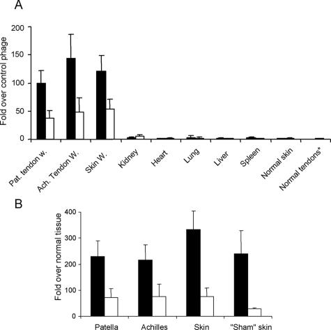
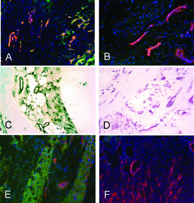
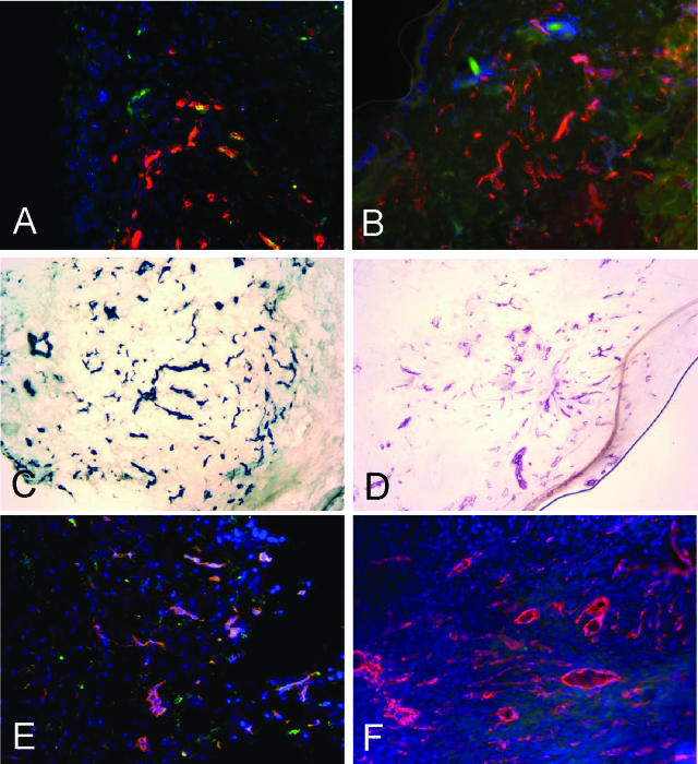
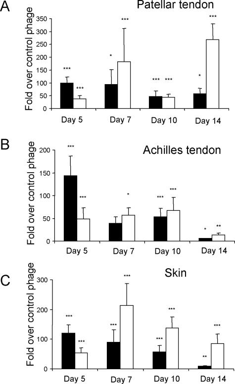
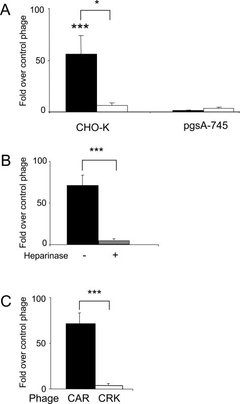
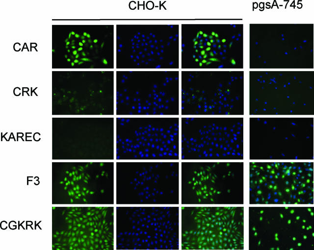
We have explored molecular specialization of the vasculature of regenerating wound tissue in the skin and tendons to identify a different repertoire of markers from that obtained by studying tumor vasculature. We screened a phage-displayed peptide library for peptides that home to wounds in mice and identified two peptides that selectively target phage to skin and tendon wounds: CARSKNKDC (CAR) and CRKDKC (CRK). CAR is homologous to heparin-binding sites in various proteins and binds to cell surface heparan sulfate and heparin. CRK is similar to a segment in thrombospondin type 1 repeat. Intravenously injected CAR and CRK phage, as well as fluorescein-labeled CAR and CRK peptides, selectively accumulated at wound sites, where they partially co-localized with blood vessels. The CAR peptide showed a preference for early stages of wound healing, whereas the CRK favored wounds at later stages of healing. The CAR peptide was internalized into the target cells and delivered the fluorescent label into the cell nuclei. These results identify new molecular markers in wound tissues and show that the expression of these markers in wound vasculature changes as healing progresses. The peptides recognizing these markers may be useful in delivering treatments into regenerating tissues.


Figures

Figure 1
Figure 1
Homing specificity of two wound-homing phage clones. CAR phage (solid bars) or CRK phage (open bars) were injected into rats via tail vein, tissues were dissected 12 minutes later, and phage was recovered and quantified from wounds in patellar and Achilles tendons and skin. Sham-operated (exposed) tendon, normal skin, and other normal tissues were used as controls as indicated. Results are expressed as fold titers relative to that of similarly injected nonrecombinant T7 phage (A) or sham-operated contralateral tendon or normal skin after adjusting the output for the wet weight of the tissue samples (B). Sham skin refers to skin incision wounds made on the lateral side of the right knee, when the patellar tendon was exposed in the sham-operation. Error bars represent mean ± SEM of 11 experiments for wounds and 4 experiments for control tissues. A: Two-way analysis of variance; B: paired Student’s t-test. Both the CAR and CRK phage homed to each of the wound tissues significantly more strongly (P < 0.001 or smaller) than the control (nonrecombinant) phage. The CAR phage showed significantly stronger homing than CRK phage to all wounds (P < 0.01 or smaller). Normal tendons* indicates combined data from normal Achilles and patellar tendons.
Figure 2
Figure 2
CAR peptide homes to blood vessels and granulation tissue in tendon and skin wounds. Fluorescein-conjugated peptides CAR (A, C, and E) or CAR2 (B, D, and F) were intravenously injected into rats (A–D) or mice (E and F) with 5-day-old wounds in Achilles tendons (A–D) or skin (E and F). Wound tissue was collected 4 hours later, which allowed the peptide to penetrate into the tissue, and examined for the presence of the peptides. In A–D, rabbit anti-FITC followed by FITC-conjugated (A and B) or biotin-conjugated anti-rabbit IgG and streptavidin peroxidase (C and D; brown), were used to detect the signal from the fluorescein-labeled peptide. Blood vessels were stained with CD-31 antibody (magenta in C and D) and the nuclei were stained with DAPI (blue). Original magnifications, ×200.
Figure 3
Figure 3
CRK peptide homes to blood vessels and granulation tissue in wounds. Fluorescein-conjugated peptides CRK (A, C, and E) or KAREC (control peptide: B, D, and F), were intravenously injected as in Figure 2 into rats (A and B) or mice (C–F) with 5-day-old wounds in the skin (A–D) or patellar tendons (E and F). The peptides, blood vessels, and nuclei were detected as in Figure 2. Original magnifications, ×200.
Figure 4
Figure 4
Healing stage dependence of phage homing to wounds. CAR phage (filled bars) or CRK phage (open bars) was intravenously injected into rats 5, 7, 10, and 14 days after wounding. The phage was recovered 12 minutes after the injection and quantified. Results are expressed as fold titers relative to that of similarly injected nonrecombinant T7 phage after adjusting the output to the wet weight of the tissue samples. A: Patellar tendon wounds; B: Achilles tendon wounds; C: skin wounds. The CAR and CRK phage homing shows a different healing time dependence. Error bars represent mean ± SEM for 11, four, seven, and five separate experiments at each time point. *P < 0.05, **P < 0.01, ***P < 0.001 versus nonrecombinant T7 phage, two-way analysis of variance.
Figure 5
Figure 5
Binding of CAR phage to cell surface heparan sulfate and heparin. A: CAR phage (filled bars) specifically binds to CHO-K cells, but not to the glycosaminoglycan-deficient pgsA-745 cells. CRK phage (open bars) does not bind significantly to either cell line. B: Heparinase treatment of the CHO-K cells (gray bar) suppresses the binding of the CAR phage to these cells. C: CAR phage, but not CRK phage, binds to heparin-coated beads. Error bars represent mean ± SEM for three or more separate experiments performed in duplicate. *P < 0.05; ***P < 0.001. A and B: Two-way analysis of variance; C: unpaired Student’s t-test. Brackets indicate the difference between CAR and CRK phages.
Figure 6
Figure 6
The CAR peptide is internalized into CHO-K cells. Fluorescein-conjugated peptides as indicated were incubated at 10 μmol/L concentration with CHO-K and pgsA-745 cells for 24 hours. The cells were washed, fixed, stained with the nuclear stain DAPI (blue; CHO-K panels, middle), and examined for green fluorescence from the labeled peptides. The CAR peptide produces strong green fluorescence in the CHO-K cells that mostly overlaps with nuclear DAPI staining (right). This peptide does not appear in the glycosaminoglycan-deficient pgsA-745 cells. The CRK peptide binds weakly to the CHO-K cells. No overlap with nuclei is evident. KAREC control peptide gives no detectable cellular fluorescence, whereas F3 and CGKRK internalize in both CHO-K and pgsA-745 cells, overlapping with nuclei. Original magnifications, ×400.

References

    1. Folkman J. Angiogenesis. Annu Rev Med. 2006;57:1–18. - PubMed
    1. Falanga V. Wound healing and its impairment in the diabetic foot. Lancet. 2005;366:1736–1743. - PubMed
    1. Martin P. Wound healing—aiming for perfect skin regeneration. Science. 1997;276:75–81. - PubMed
    1. Wickström S, Keski-Oja J, Alitalo K. Matrix reloaded to circulation hits the tumor target. Cancer Cell. 2003;3:513–514. - PubMed
    1. St. Croix B, Rago C, Velculescu V, Traverso G, Romans KE, Montgomery E, Lal A, Riggins GJ, Lengauer C, Vogelstein B, Kinzler KW. Genes expressed in human tumor endothelium. Science. 2000;289:1197–1202. - PubMed

Publication types

MeSH terms