. 2007 Nov 15;85(15):3267-83.
doi: 10.1002/jnr.21376.

Astrocytic connexin distributions and rapid, extensive dye transfer via gap junctions in the inferior colliculus: implications for [(14)C]glucose metabolite trafficking

Affiliations

Astrocytic connexin distributions and rapid, extensive dye transfer via gap junctions in the inferior colliculus: implications for [(14)C]glucose metabolite trafficking

Kelly K Ball et al. J Neurosci Res. .

Abstract

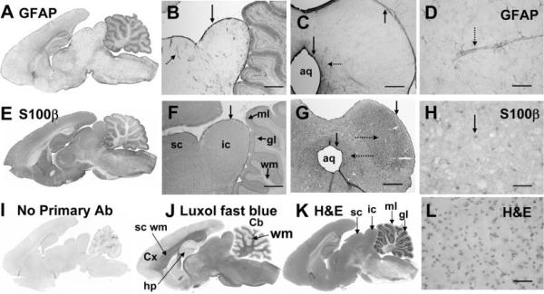
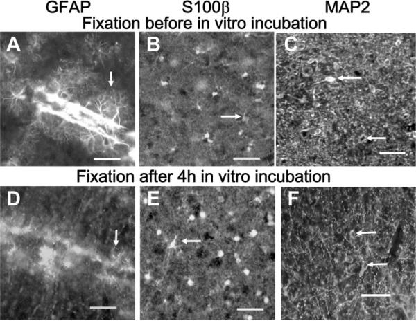
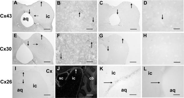
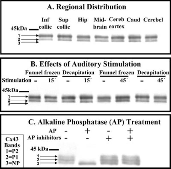
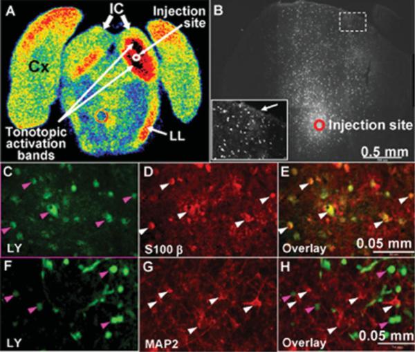
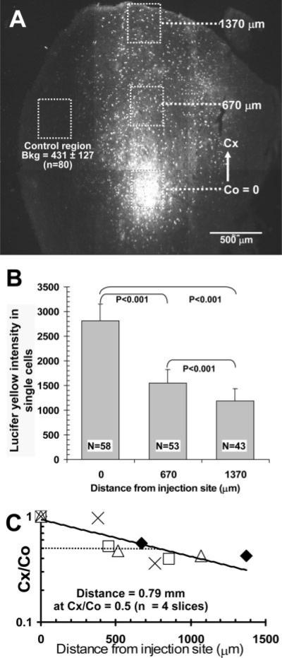
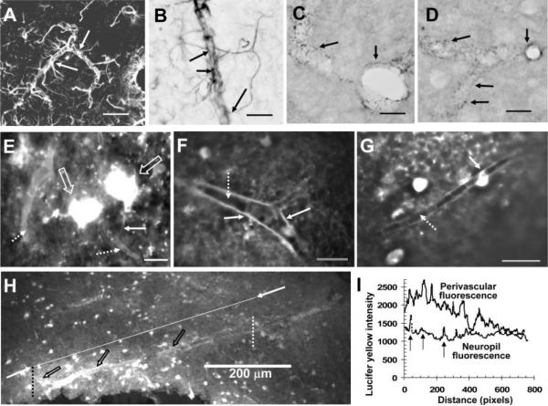
The inferior colliculus has the highest rates of blood flow and metabolism in brain, and functional metabolic activity increases markedly in response to acoustic stimulation. However, brain imaging with [1- and 6-(14)C]glucose greatly underestimates focal metabolic activation that is readily detected with [(14)C]deoxyglucose, suggesting that labeled glucose metabolites are quickly dispersed and released from highly activated zones of the inferior colliculus. To evaluate the role of coupling of astrocytes via gap junctions in dispersal of molecules within the inferior colliculus, the present study assessed the distribution of connexin (Cx) proteins in the inferior colliculus and spreading of Lucifer yellow from single microinjected astrocytes in slices of adult rat brain. Immunoreactive Cx43, Cx30, and Cx26 were heterogeneously distributed; the patterns for Cx43 and Cx 30 differed and were similar to those of immunoreactive GFAP and S100beta, respectively. Most Cx43 was phosphorylated in resting and acoustically stimulated rats. Dye spreading revealed an extensive syncytial network that included thousands of cells and perivasculature endfeet; with 8% Lucifer yellow VS and a 5-min diffusion duration, about 6,100 astrocytes (range 2,068-11,939) were labeled as far as 1-1.5 mm from the injected cell. The relative concentration of Lucifer yellow fell by 50% within 0.3-0.8 mm from the injected cell with a 5-min diffusion interval. Perivascular dye labeling was readily detectable and often exceeded dye levels in nearby neuropil. Thus, astrocytes have the capability to distribute intracellular molecules quickly from activated regions throughout the large, heterogeneous syncytial volume of the inferior colliculus, and rapid trafficking of labeled metabolites would degrade resolution of focal metabolic activation.


Figures

Fig. 1
Fig. 1
Immunoreactive glial fibrillary acidic protein (GFAP) and S100β in adult rat brain. Sagittal sections of adult rat brain showing the heterogeneous distributions of immunoreactive GFAP (A) and S100β (E) compared with 1) low staining by the secondary antibody when the primary antibody was omitted (I), 2) staining of myelin by luxol fast blue (J), and 3) cellular staining by hematoxylin and eosin (H&E; K). Higher-power sagittal (B,F) and coronal (C,D and G,H) sections compare GFAP (B–D) and S100β (F–H) staining with H&E cell stain (L). Note the intense immunostaining of both GFAP and S100β in the meninges along the surface of the inferior colliculus (vertical arrows, B,C,F,G) and in the cells adjacent to the aqueduct (aq; vertical arrows, C,G). The parenchyma surrounding the aqueduct (leftward-facing horizontal dotted arrows, C,G) is also more heavily stained by both antibodies, and immunoreactive S100β is somewhat more dense in the central zone of the inferior colliculus (rightward-facing dotted arrow, G). GFAP staining is most intense around blood vessels (dotted arrows, B,D) and is much lower in neuropil (A–D). S100β staining is more uniform throughout the neuropil but is more intense in nuclei (arrow, H) compared with cytoplasm; it is also evident in perivascular structures (not shown, see Fig. 7). All immunostaining used the PAP procedure with DAB (see Materials and Methods). Cx, cerebral cortex; sc wm, subcortical white matter; hp, hippocampus; Cb, cerebellum; wm, white matter; sc, superior colliculus; ic, inferior colliculus; ml, molecular layer of cerebellum; gl, granular layer of cerebellum; aq, aqueduct. Scale bars = 800 μm in B,C,F,G; 50 μm in D,H,L.
Fig. 2
Fig. 2
Loss of immunoreactive GFAP after in vitro incubation of slices of inferior colliculus. Tissue slices (250 μm thick) were immersion fixed in 4% paraformaldehyde either immediately after slicing (A–C) or 4 hr after slicing and in vitro incubation (D–F), then assayed for immunoreactive GFAP (A,D), S100β (B,E), or MAP2 (C,F; all images were obtained by use of fluorescent secondary antibodies; see Materials and Methods). Note loss of fine immunoreactive GFAP-positive processes when fixation is delayed (compare A and D, arrows), whereas staining of nuclei and processes by anti-S100β (arrows, B,E) and neuronal staining by anti-MAP2 (arrows, C,F) were not substantially altered by the slice-recovery incubation procedure. Scale bars = 50 μm.
Fig. 3
Fig. 3
Immunoreactive Cx43, Cx30, and Cx26 in the inferior colliculus of the adult rat. Coronal sections at the level of midinferior colliculus (except the sagittal section, J) illustrate the distributions of immunoreactive Cx43 (A,B), Cx30 (E,F), and Cx26 (I–K) compared with the respective controls in which the primary antibody was omitted (C,D,G,H,L). Vertical arrows indicate highest staining in the meninges, cells surrounding the aqueduct and around blood vessels (A,B,E,F,I,J), which is low in the control sections (C,D,G,H,L); note that immunoreactive Cx30 is somewhat higher in the central zone of inferior colliculus (dotted arrow, E). The PAP method was used for immunostaining, except in J, where a fluorescent secondary antibody was used to visualize the very faint staining of Cx26 in the meninges and in cells around the aqueduct (arrows, I–K). Because Nagy et al. (2001) reported 1) very high levels of Cx26 protein in Western blots of extracts from superior and inferior colliculus after separation by gel electrophoresis, 2) immunofluorescence labeling of superficial layers of superior colliculus and the cerebellar granular layer, and 3) sparse distribution in the molecular layer that contrasted the low immunostaining in superior and inferior colliculus (but note that cortical and cerebellar staining observed in the present study, Fig. 3J was similar to the Nagy et al. findings), Cx26 was also assayed with various primary antibody dilutions (1:12.5, 1:20, 1:50, 1:100, 1:200) and with different preparative methods (perfusion-fixed tissue, in slices fixed immediately by immersion, air-dried fresh-frozen slices that were not fixed or were fixed in methanol or acetone), but no improvement in staining of the inferior colliculus could be obtained (data not shown). aq, Aqueduct; ic, inferior colliculus; sc, superior colliculus; Cx, cerebral cortex; cb, cerebellum. Scale bars = 800 μm in A,C,E,G,I,J; 50 μm in B,D,F,H,K,L.
Fig. 4
Fig. 4
Highly phosphorylated Cx43 is the predominant isoform in the inferior colliculus during rest and after acoustic stimulation. A: Cx43 isoforms in seven brain regions. B: Phosphorylation state of Cx43 before (dashes indicated “resting” rats) and after 15 or 45 min of acoustic stimulation (40 Hz to 8 kHz broadband stimulus) of conscious rats; tissue was sampled from inferior colliculus by dissection from brain tissue that was frozen in situ (funnel frozen) or obtained after decapitation (see Materials and Methods). C: Brain homogenates were incubated in vitro in the presence or absence of alkaline phosphatase (AP) or AP inhibitors (fluoride plus vanadate) prior to separation on SDS-PAGE gels, Western blotting, and immunostaining (see Materials and Methods). Pretreatment of the Zymed antibody with the Zymed Cx43 peptide completely blocked Cx43 immunostaining (not shown). NP denotes nonphosphorylated Cx43, whereas P1 and P2 denote phosphorylated isoforms; the horizontal bar indicates the position of 45-kDa molecular weight standard. Inf collic, inferior colliculus; Sup collic, superior colliculus; Hip, hippocampus; Cereb, cerebral; Caud, caudate; Cerebel, cerebellum; AP, alkaline phosphatase, Cx43, connexin 43.
Fig. 5
Fig. 5
Rapid, extensive transfer of Lucifer yellow among astrocytes in slices of inferior colliculus. A: Autoradiograph of coronal section of rat brain at the level of the inferior colliculus after in vivo metabolic imaging with the [14C]DG method during unilateral monotonic auditory stimulation (8 kHz at 103 dB, one ear blocked) to stimulate glucose utilization in the auditory pathway in the right hemisphere (see Cruz et al., 2007). Color coding represents glucose utilization rate (high to low: black, red, orange, yellow, green, blue). Note the high rates of glucose utilization represented by the two black tonotopic activation bands in the right inferior colliculus (IC); the red band in the lower medial region of the left inferior colliculus probably arose from crossover of auditory fibers. Activation is also evident in the right lateral lemniscus (LL). Cx denotes cerebral cortex. B: A single astrocyte in a brain slice was microinjected with Lucifer yellow (LY; 4% LYVS + 4% LYCH), approximately in the center of the inferior colliculus as indicated in A, and the dye was allowed to diffuse from the pipette for 5 min. LY-labeled cells coupled to an injected astrocyte via gap junctions are shown in B, C, and F; the inset in B is a higher magnification view of a region distant from the injection site (dashed box) near the meninges. Note the bright labeling at the tissue boundary (arrow in inset, B), suggesting a diffusion barrier. Immunohistochemical staining identified LY-labeled cells (green) in C and F as astrocytes by colocalization with S100β (D,E) but not MAP2 (G,H). Arrowheads identify specific cells in these panels; similar results were obtained with immunostaining of three replicate preparations. Scale bars = 0.05 mm.
Fig. 6
Fig. 6
Relative fluorescence intensity of Lucifer yellow in gap junction-coupled astrocytes as function of distance from injection site. A: Representative montage showing the site where a single astrocyte in a slice of inferior colliculus was microinjected with 4% Lucifer yellow (LY) VS + 4% LYCH and allowed to diffuse for 5 min. Boxes (300 × 300 μm) were drawn around the dye-labeled cells and at various distances from the injection site. Cx denotes the distance from the center of the box surrounding the injection site (Co) to the center of a box located at various distances ranging from about 0.4 to1.4 mm from the injection site in different brain slices; in this representative figure, Cx = 0.67 and 1.37 mm. B: Single LY-labeled astrocytes within each box in A were outlined in MetaVue software to determine their fluorescence intensities. The maximal intensity to saturate the camera is about 4,000 fluorescence units. By visual inspection, the peak intensity in the cells assayed was below the saturation value, but the possibility of differential fluorescence quenching in the labeled cells as a function of distance was not evaluated. Values are means for the number of cells indicated and correspond to cells located in the respective boxes shown in A; vertical lines = 1 SD. The fluorescence intensities among all groups were statistically significantly different (ANOVA, Bonferroni test). Note that the mean intensity of cells located about 1.4 mm from the injection site was about three times higher than background (Bkg.) fluorescence determined in a control region (dotted box, A) that had few, if any, cells labeled by dye transfer; the parenchyma and blood vessels in this region had low levels of autofluorescence after paraformaldehyde fixation. C: Semi-log plot of the Cx/Co ratio for the mean LY fluorescence intensities in cells located at various distances from the injection site in four slices. In each slice (indicated by a different symbol), cells were counted in each of three boxes as shown in A,B, and Cx/Co ratios were calculated for each slice using mean values for cells in each box. The distance at which the fluorescence intensity fell by 50% (Cx/Co = 0.5) was calculated to be 0.79 mm, from the equation of the regression line, i.e., y = 0.937e−0.0008x (R2 = 0.749), based on the data from four slices. If the cells with the high levels of LY did have some quenching, the Co values would be underestimated, causing overestimation of the Cx/Co ratios and of the calculated distance at which Cx/Co = 0.5; this distance estimate is, therefore, a maximal value. For example, if all Co values were underestimated by a factor of two, the regression line in C would be y = 0.809e−0.0014x (R2 = 0.791), and calculated distance for Cx/Co = 0.5 is 0.34 mm.
Fig. 7
Fig. 7
Perivascular spread of Lucifer yellow after injection of a single astrocyte. Astrocytes in close contact with the microvasculature were readily identified via their contents of immunoreactive glial fibrillary acidic protein (GFAP), using either a fluorescent secondary antibody (A, arrows indicate two astrocytes with processes along the vessel and in the parenchyma) or the PAP method with DAB staining (B, arrows indicate endfeet attached to astrocytic processes near a vessel). Punctate staining of gap junction proteins (arrows) around blood vessels shown in cross-section or longitudinal section was readily detected with the PAP method and DAB staining of immunoreactive Cx43 (C) and Cx30 (D; see Materials and Methods). E–H illustrate perivascular localization of Lucifer yellow after microinjection of a single astrocyte. E: One of the highly fluorescent astrocytes (large arrows) has a fine process (solid arrow) that extends toward a blood vessel that is one of two vessels surrounded by Lucifer yellow (dotted arrows). Perivascular labeling of vessels in cross-section show outlining at a bifurcation (F, solid arrow), with heterogeneous fluorescence intensity along the wall of the vessel that appears to be highest in the vicinity of an astrocytic process (G, solid arrow); note the lack of labeling in the lumen of the vessels (F,G, dotted arrows). The relative intensity of perivascular labeling by Lucifer yellow compared with adjacent tissue (H) after injection of a single astrocyte is illustrated by the intensity line scans determined with MetaVue software (I) that were drawn from left to right along the upper boundary of the vessel wall (open arrows; denoted as perivascular fluorescence in I) between the dotted vertical lines in H or through the parenchyma along the line connecting the two arrows shown in H and denoted as neuropil fluorescence. Note that the fluorescence intensity along the vessel wall decreased with distance left to right; it was heterogeneous and, over a substantial distance, was about twice that of parenchyma (I); higher perivascular vs. parenchymal labeling is also evident in E–G. Note the intermittent “spike” increases in fluorescence intensity (vertical arrows) as the tissue scan line passes from left to right through highly labeled astrocytes in the neuropil (compare H and I). In line scans made along seven additional blood vessels located within the range of about 0.7–1.2 mm from the injection site, the perivascular Lucifer yellow fluorescence varied in intensity as illustrated in H,I; the perivascular regions with high fluorescence intensity exceeded that of the adjacent neuropil by a factor of 1.7 ± 0.3 (mean ± SD, n = 7). Scale bars = 50 μm in A; 20 μm in B–G; 200 μm in H.

References

    1. Ackermann RF, Lear JL. Glycolysis-induced discordance between glucose metabolic rates measured with radiolabeled fluorodeoxyglucose and glucose. J Cereb Blood Flow Metab. 1989;9:774–785. - PubMed
    1. Adachi K, Cruz NF, Sokoloff L, Dienel GA. Labeling of metabolic pools by [6-14C]glucose during K+-induced stimulation of glucose utilization in rat brain. J Cereb Blood Flow Metab. 1995;15:97–110. - PubMed
    1. Binmöller FJ, Müller CM. Postnatal development of dye-coupling among astrocytes in rat visual cortex. Glia. 1992;6:127–137. - PubMed
    1. Brightman MW. The brain's interstitial clefts and their glial walls. J Neurocytol. 2002;31:595–603. - PubMed
    1. Bruzzone R, White TW, Paul DL. Connections with connexins: the molecular basis of direct intercellular signaling. Eur J Biochem. 1996;238:1–27. - PubMed

Publication types