

Primary angiosarcoma of the testis: report of a rare entity and review of the literature
- PMID: 17601346
- PMCID: PMC1919353
- DOI: 10.1186/1746-1596-2-23
Primary angiosarcoma of the testis: report of a rare entity and review of the literature
Abstract
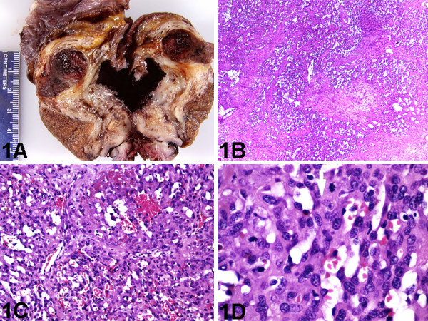
Background: Primary testicular angiosarcomas are extremely rare, and their clinicopathologic features are not well described. Our objective was to further define the clinical features and pathologic spectra of primary testicular angiosarcomas.
Methods: Six previously reported case reports were identified in the English language medical literature using MEDLINE and a subsequent bibliographic search of all pertinent reports and reviews was performed. After excluding 2 cases because they did not involve the testis, we identified 4 previously reported cases of true primary testicular angiosarcoma. We also searched the electronic medical archival records of our institution and identified one additional unreported case of true primary testicular angiosarcomas. Data were extracted on the demographics, predisposing factors, clinical presentation, gross pathology, microscopic pathology, immunophenotype, therapy, and outcomes of each of these 5 cases of true primary testicular angiosarcomas.
Results: Primary testicular angiosarcomas were found at a mean age of 43.4 years. None of the cases was associated with exposure to radiation, arsenic, thorium dioxide, or vinyl chloride. However, 1 case was associated with hydrocele. It typically presented with painless mass (mean size, 6.3 cm). Histologically, all showed classic anastomosing channels lined by plump hyperchromatic cells, though most showed epithelioid cytology and some showed solid architectural pattern. One patient had multiple metastatic recurrences but eventual outcome was not available, and 1 patient died a month after diagnosis from stroke but no autopsy was performed. The remaining 3 patients were alive at the time of publication of their respective cases (mean, 17 months).
Conclusion: Primary testicular angiosarcomas are typically rare tumors of men of all ages that appear to segregate into 2 groups; one associated with teratoma and occurring in young people, and the other occurring in the elderly and not associated with germ cell neoplasm, but may be associated with chronic hydrocele. They present with advanced disease and show a wide histologic spectrum. However, their prognosis may be better than previously thought.
Figures

Similar articles
-
Primary carcinoid tumor arising within mature teratoma of the kidney: report of a rare entity and review of the literature.Diagn Pathol. 2007 May 17;2:15. doi: 10.1186/1746-1596-2-15. Diagn Pathol. 2007. PMID: 17509135 Free PMC article.
-
Oral and salivary gland angiosarcoma: a clinicopathologic study of 29 cases.Mod Pathol. 2003 Mar;16(3):263-71. doi: 10.1097/01.MP.0000056986.08999.FD. Mod Pathol. 2003. PMID: 12640107
-
Primary angiosarcoma of the bladder.Arch Pathol Lab Med. 2006 Oct;130(10):1543-7. doi: 10.5858/2006-130-1543-PAOTB. Arch Pathol Lab Med. 2006. PMID: 17090199
-
Primary uterine angiosarcoma.Gynecol Oncol. 1999 Nov;75(2):272-6. doi: 10.1006/gyno.1999.5566. Gynecol Oncol. 1999. PMID: 10525385 Review.
-
Angiosarcoma of the breast: a clinicopathologic analysis of cases from the last 10 years.Ann Diagn Pathol. 2009 Jun;13(3):147-50. doi: 10.1016/j.anndiagpath.2009.02.001. Epub 2009 Apr 1. Ann Diagn Pathol. 2009. PMID: 19433291 Review.
Cited by
-
Primary angiosarcoma of the breast: a case report.Diagn Pathol. 2013 Apr 22;8:66. doi: 10.1186/1746-1596-8-66. Diagn Pathol. 2013. PMID: 23607567 Free PMC article.
-
Oncological outcomes in patients with stage I testicular seminoma and nonseminoma: pathological risk factors for relapse and feasibility of surveillance after orchiectomy.Diagn Pathol. 2013 Apr 8;8:57. doi: 10.1186/1746-1596-8-57. Diagn Pathol. 2013. PMID: 23566361 Free PMC article.
-
Sclerosing rhabdomyosarcoma presenting in the masseter muscle: a case report.Diagn Pathol. 2013 Feb 4;8:18. doi: 10.1186/1746-1596-8-18. Diagn Pathol. 2013. PMID: 23379991 Free PMC article.
-
Angiosarcoma of the breast: a new therapeutic approach?Int J Surg Case Rep. 2015;13:30-2. doi: 10.1016/j.ijscr.2015.06.004. Epub 2015 Jun 6. Int J Surg Case Rep. 2015. PMID: 26092711 Free PMC article.
-
Primary pure angiosarcoma of the testis: a vanishingly rare malignancy. Case report and literature review.BMC Urol. 2020 Oct 31;20(1):175. doi: 10.1186/s12894-020-00747-7. BMC Urol. 2020. PMID: 33129286 Free PMC article. Review.
References
-
- Sahoo S, Ryan CW, Recant WM, Yang XJ. Angiosarcoma masquerading as embryonal carcinoma in the metastasis from a mature testicular teratoma. Arch Pathol Lab Med. 2003;127:360–363. - PubMed
LinkOut - more resources
Full Text Sources
