. 2007;9(3):209.
doi: 10.1186/bcr1679.

Inflammation and breast cancer. Microenvironmental factors regulating macrophage function in breast tumours: hypoxia and angiopoietin-2

Affiliations

Inflammation and breast cancer. Microenvironmental factors regulating macrophage function in breast tumours: hypoxia and angiopoietin-2

Claire E Lewis et al. Breast Cancer Res. 2007.

Abstract

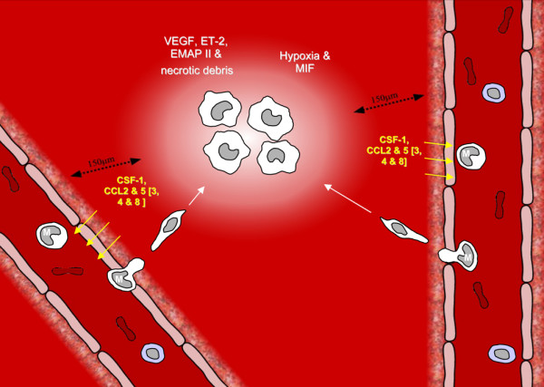
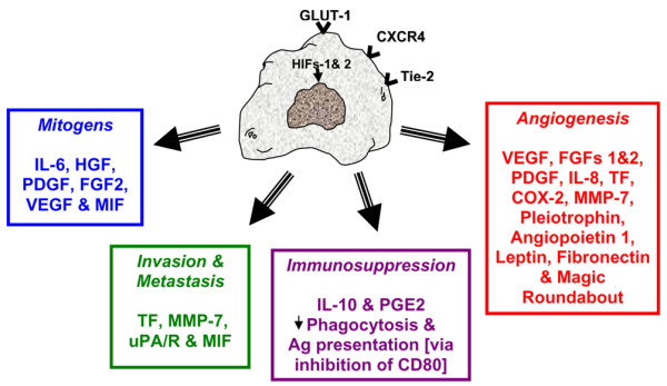
Considerable evidence has now accumulated for tumour-associated macrophages stimulating key aspects of tumour progression, including the proliferation, survival and metastasis of tumour cells, tumour angiogenesis and suppression of the anti-tumour functions of other immune effectors at the tumour site. Tumour micro-environmental factors such as hypoxia have profound, direct effects on these cells, stimulating many of their pro-tumour functions. Hypoxia also does so indirectly by stimulating the release of the cytokine angiopoietin-2 from tumour cells and tumour blood vessels. This in turn then recruits Tie-2-expressing monocytes into tumours from the bloodstream and inhibits their production of anti-apoptotic and anti-angiogenic cytokines.


Figures

Figure 1
Figure 1
Mechanisms responsible for the accumulation of tumour-associated macrophages in hypoxic areas of solid tumours. A microenronment within a human tumour is shown containing two blood vessels and a hypoxic area (white) that has formed because it is more than 100 to 150 μm from either vessel – the critical distance for oxygenation in such tissues. Monocytes (M) pass through these vessels and are recruited into tumours by the release of the monocyte chemoattractants CCL2, 3, 4, 5 and 8 and colony-stimulating factor-1 (CSF-1) by the tumour. Once monocytes have moved across the tumour vasculature, many are attracted into hypoxic areas by the hypoxia-induced release of other monocyte attractants such as vascular endothelial growth factor (VEGF), endothelin-2 (ET-2) and endothelial-monocyte-activating polypeptide (EMAP) II. These innate cells may also be attracted into sites experiencing chronic hypoxia (and thus cell death) along a trail of necrotic debris emanating from these areas. Hypoxia then acts directly on macrophages to immobilise them and also via the upregulation of macrophage migration inhibitory factor (MIF) by tumour cells, which has a similar effect on macrophage migration (reviewed in [13]).
Figure 2
Figure 2
Hypoxia induces marked changes in the phenotype of macrophages. Macrophages upregulate hypoxia-inducible transcription factor (HIF)-1 and HIF-2 in hypoxia, which translocate to the nucleus to induce the expression of a wide array of target genes. Several important cell-surface receptors are upregulated in hypoxia, including the glucose receptor GLUT-1 (for increased glucose uptake as the cell switches to anaerobic glycolysis to make ATP in the absence of oxygen), the chemokine stromal cell-derived factor-1 (SDF-1) receptor CXCR4, and the angiopoietin receptor Tie-2. Hypoxia also stimulates the expression of a wide array of other pro-tumour cytokines, enzymes and receptors, grouped here according to their known function in tumours. Downregulation of a factor or tumour-associated macrophage function is indicated by an arrow [15, 17, 18]. Ag, antigen; COX, cyclo-oxygenase; FGF, fibroblast growth factor; HGF, hepatocyte growth factor; MIF, macrophage migration inhibitory factor; MMP, matrix metalloproteinase; PDGF, platelet-derived growth factor; PGE2, prostaglandin E2; TF, tissue factor; uPA/R, urokinase plasminogen activator receptor; VEGF, vascular endothelial growth factor.

Similar articles

Cited by

References

    1. Lin EY, Nguyen AV, Russell RG, Pollard JW. Colony-stimulating factor 1 promotes progression of mammary tumors to malignancy. J Exp Med. 2001;193:727–740. doi: 10.1084/jem.193.6.727. - DOI - PMC - PubMed
    1. Bingle L, Brown NJ, Lewis CE. The role of tumour-associated macrophages in tumour progression: implications for new anticancer therapies. J Pathol. 2002;196:254–265. doi: 10.1002/path.1027. - DOI - PubMed
    1. Lewis CE, Pollard JW. Distinct role of macrophages in different tumor microenvironments. Cancer Res. 2006;66:605–612. doi: 10.1158/0008-5472.CAN-05-4005. - DOI - PubMed
    1. Lin EY, Li JF, Gnatovskiy L, Deng Y, Zhu L, Grzesik DA, Qian H, Xue XN, Pollard JW. Macrophages regulate the angiogenic switch in a mouse model of breast cancer. Cancer Res. 2006;66:11238–11246. doi: 10.1158/0008-5472.CAN-06-1278. - DOI - PubMed
    1. Leek RD, Lewis CE, Whitehouse R, Greenall M, Clarke J, Harris AL. Association of macrophage infiltration with angiogenesis and prognosis in invasive breast carcinoma. Cancer Res. 1996;56:4625–4629. - PubMed

Publication types

MeSH terms