. 2007 Sep;293(3):L790-9.
doi: 10.1152/ajplung.00099.2007. Epub 2007 Jun 29.

TGF-beta1 stimulates human AT1 receptor expression in lung fibroblasts by cross talk between the Smad, p38 MAPK, JNK, and PI3K signaling pathways

Affiliations

TGF-beta1 stimulates human AT1 receptor expression in lung fibroblasts by cross talk between the Smad, p38 MAPK, JNK, and PI3K signaling pathways

Mickey M Martin et al. Am J Physiol Lung Cell Mol Physiol. 2007 Sep.

Retraction in

  • Retraction.
    [No authors listed] [No authors listed] Am J Physiol Lung Cell Mol Physiol. 2012 Apr 1;302(7):L719. doi: 10.1152/ajplung.zh5-6097-retr.2012. Am J Physiol Lung Cell Mol Physiol. 2012. PMID: 22467678 Free PMC article. No abstract available.

Abstract

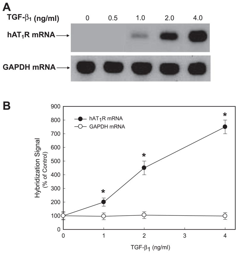
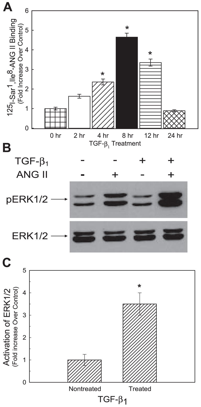
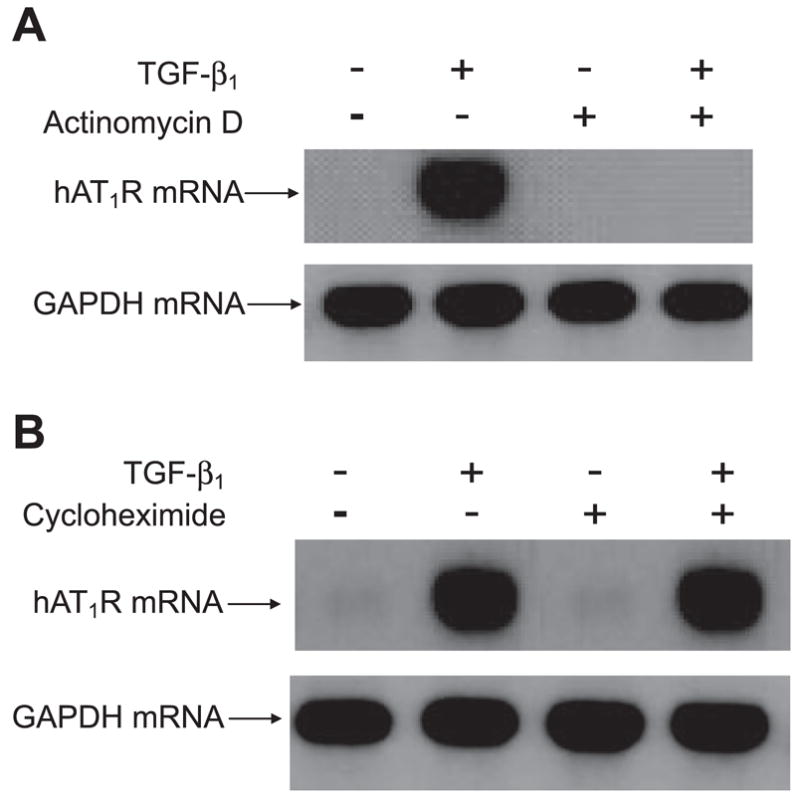
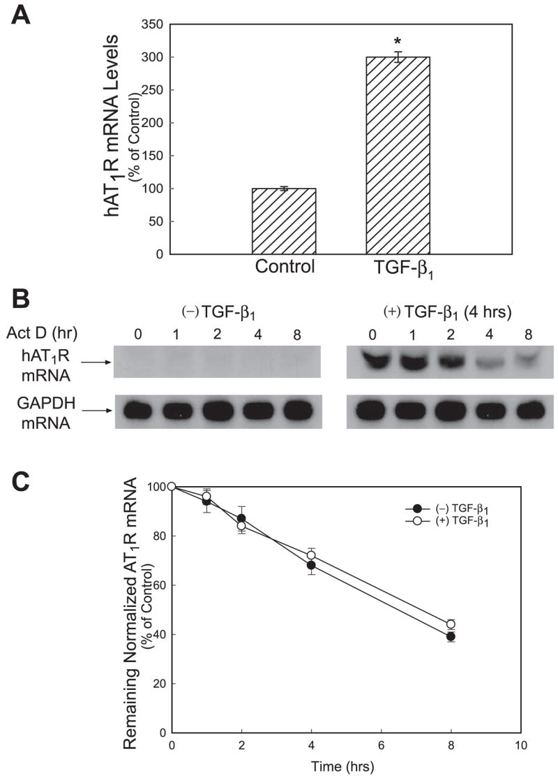
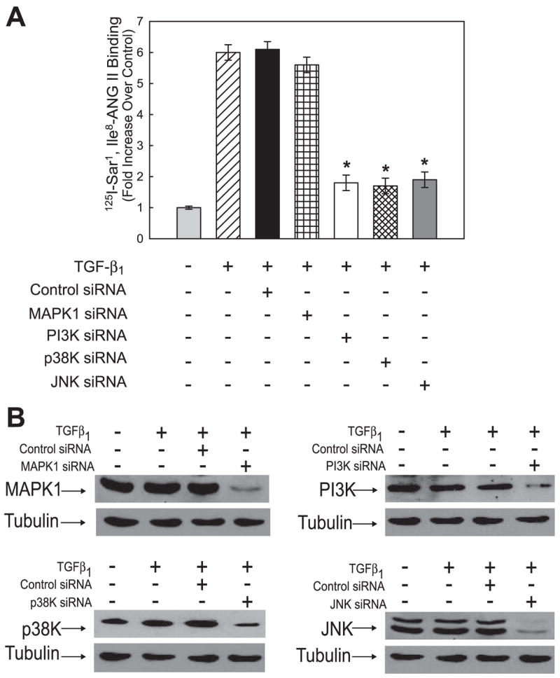
Both angiotensin II (ANG II) and transforming growth factor-beta1 (TGF-beta1) are thought to be involved in mediating pulmonary fibrosis. Interactions between the renin-angiotensin system (RAS) and TGF-beta1 have been well documented, with most studies describing the effect of ANG II on TGF-beta1 expression. However, recent gene expression profiling experiments demonstrated that the angiotensin II type 1 receptor (AT(1)R) gene was a novel TGF-beta1 target in human adult lung fibroblasts. In this report, we show that TGF-beta1 augments human AT(1)R (hAT(1)R) steady-state mRNA and protein levels in a dose- and time-dependent manner in primary human fetal pulmonary fibroblasts (hPFBs). Nuclear run-on experiments demonstrate that TGF-beta1 transcriptionally activates the hAT(1)R gene and does not influence hAT(1)R mRNA stability. Pharmacological inhibitors and specific siRNA knockdown experiments demonstrate that the TGF-beta1 type 1 receptor (TbetaRI/ALK5), Smad2/3, and Smad4 are essential for TGF-beta1-stimulated hAT(1)R expression. Additional pharmacological inhibitor and small interference RNA experiments also demonstrated that p38 MAPK, JNK, and phosphatidylinositol 3-kinase (PI3K) signaling pathways are also involved in the TGF-beta1-stimulated increase in hAT(1)R density. Together, our results suggest an important role for cross talk among Smad, p38 MAPK, JNK, and PI3K pathways in mediating the augmented expression of hAT(1)R following TGF-beta1 treatment in hPFB. This study supports the hypothesis that a self-potentiating loop exists between the RAS and the TGF-beta1 signaling pathways and suggests that ANG II and TGF-beta1 may cooperate in the pathogenesis of pulmonary fibrosis. The synergy between these systems may require that both pathways be simultaneously inhibited to treat fibrotic lung disease.


Figures

Fig. 1
Fig. 1
Transforming growth factor-β1 (TGF-β1) stimulation upregulates human angiotensin II type 1 receptor (hAT1R) steady-state mRNA levels in human fetal pulmonary fibroblasts (hPFBs) in a time-dependent manner. hPFBs were grown to 70–80% confluence, washed twice, and serum-starved for 24 h. A: total RNA was isolated from TGF-β1-treated (4 ng/ml) fibroblasts at the times indicated. RNA (20 μg) was fractionated, blotted, and probed with a radiolabeled cDNA specific for hAT1R mRNA. The Northern blot was stripped and reprobed with a GAPDH control cDNA to ensure that the relative quantity of the total RNA present in each lane was approximately equal. Data are representative of 3 separate experiments. B: TGF-β1 time course of hAT1R and GAPDH Northern hybridization signal intensity was quantitated by densitometric analysis. Each point represents the relative hybridization signal (±SE) normalized to 0-h treatment with vehicle (100%) from 3 separate experiments. *P < 0.01, TGF-β1-treated vs. nontreated hPFBs.
Fig. 2
Fig. 2
TGF-β1 stimulation upregulates hAT1R steady-state mRNA levels in hPFBs in a dose-dependent manner. hPFBs were grown to 70–80% confluence, washed twice, and serum-starved for 24 h. A: hPFBs were treated with TGF-β1 at the concentrations indicated for 4 h, and total RNA was isolated. RNA samples (20 μg) were fractionated, blotted, and probed with a radiolabeled hAT1R cDNA. The Northern blot was stripped and hybridized with a labeled GAPDH cDNA probe. Data are representative of 3 separate experiments. B: TGF-β1 dose response of hAT1R and GAPDH Northern hybridization signal intensity was quantitated by densitometric analysis. Each point represents the relative hybridization signal (±SE) normalized to untreated hPFBs from 3 separate experiments. *P < 0.01, TGF-β1-treated vs. nontreated hPFBs.
Fig. 3
Fig. 3
TGF-β1 stimulation increases AT1R protein levels and ANG II-induced ERK1/2 activation. hPFBs were grown to 70–80% confluence, washed twice, and serum-starved for 24 h. A: hPFBs were subsequently incubated with TGF-β1 (4 ng/ml) for the times indicated, and AT1R radioreceptor binding assays were performed as described in EXPERIMENTAL PROCEDURES. Data are expressed as relative increase over nontreated (i.e., 0 h) hPFBs. Error bars represent SE of 3 independent experiments. *P < 0.01, TGF-β1-treated vs. nontreated hPFBs. B: hPFBs were serum-starved for 24 h, incubated with TGF-β1 (4 ng/ml, 8 h), and further activated with 0.1 μM ANG II for 5 min, and phospho-ERK1/2 activation was determined by Western blot analysis. The blot was stripped and reprobed with an ERK1/2-specific antibody. Data are representative of 3 separate experiments. C: each autoradiograph was quantitated by densitometric analysis, and ERK1/2 phosphorylation (p) was normalized with ERK1/2 protein levels and plotted as relative increase of ANG II-induced pERK1/2 over non-ANG II-induced pERK1/2 values. Error bars represent SE of 3 independent experiments. *P < 0.01, TGF-β1-treated vs. nontreated hPFBs.
Fig. 4
Fig. 4
TGF-β1-enhanced hAT1R mRNA expression requires transcription but does not require protein synthesis. hPFBs were serum-starved for 24 h and then pretreated for 1 h with PBS or 5 μg/ml actinomycin D (Act D) to block transcription of mRNA (A) or with 10 μg/ml cycloheximide to block protein synthesis (B). Cells were subsequently treated with TGF-β1 (4 ng/ml, 4 h), total RNA was isolated, and Northern blot analysis was performed as described. Data are representative of 3 separate experiments.
Fig. 5
Fig. 5
TGF-β1 stimulation of hPFBs increases hAT1R mRNA levels through a transcriptional mechanism and does not affect hAT1R mRNA stability. A: hPFBs were serum-starved for 24 h and incubated with TGF-β1 (4 ng/ml, 4 h), nuclei were isolated and subjected to nuclear run-on analyses, and hAT1R mRNA levels were quantitated utilizing RT-PCR as described in EXPERIMENTAL PROCEDURES. Error bars represent SE of 3 independent experiments. *P < 0.01, TGF-β1-treated vs. nontreated hPFBs. B: hPFBs were serum-starved for 24 h and then incubated with TGF-β1 (4 ng/ml, 4 hr). Cells were then either collected (time 0, control) or treated with 5 μg/ml Act D, to further block transcription of mRNA, and then harvested at 1, 4 and 8 h thereafter. AT1R and GAPDH mRNAs were detected by Northern blot analysis. Data are representative of 3 separate experiments. C: hAT1R and GAPDH mRNA levels were quantitated by densitometric analysis. hAT1R mRNA values were normalized to GAPDH expression at each time point. To obtain hAT1R values for the nontreated hPFBs, autoradiography was performed for 7 days (data not shown). The half-life of hAT1R mRNA in TGF-β1-treated and nontreated cells was calculated as the time required for a given transcript to decrease to 50% of its initial abundance. Error bars represent SE of 3 independent experiments.
Fig. 6
Fig. 6
Inhibition of TGF-β1 type 1 receptor (TβRI) activity abolishes TGF-β1-stimulated increases in AT1R steady-state mRNA levels. A: hPFBs were serum-starved for 24 h, incubated with 1 μM SB 431542, a selective inhibitor of TGF-β1 type 1 receptor kinase (ALK5), for 1 h, and subsequently stimulated with 4 ng/ml TGF-β1 for 4 h. Total RNA was isolated, and Northern blot analysis was performed. Data are representative of 3 separate experiments. B: hPFBs were treated as described in A; however, cells were lysed in RIPA buffer and subjected to immunoblotting with phosphospecific or Smad2 and Smad3 antibodies. Data are representative of 3 separate experiments. C: hPFBs were grown to 30–60% confluence and subsequently transduced (100 multiplicity of infection) with adenovirus expressing constitutively active ALK5 (caALK5) or empty vector. Forty-eight hours after infection, total RNA was isolated and Northern analysis was performed. Data are representative of 3 separate experiments. D: hPFBs were treated as described in C; however, cells were lysed in RIPA buffer and subjected to immunoblotting with phosphospecific or Smad2 and Smad3 antibodies. Data are representative of 3 separate experiments.
Fig. 7
Fig. 7
TGF-β1-induced hAT1R expression is mediated by a Smad-dependent mechanism. A: hPFBs were grown to 30–60% confluence and transiently transfected with control, ALK5-, Smad2-, Smad3-, or Smad4-specific small interference RNAs (siRNAs; 25 nM final concentration). Forty-eight hours after transfection, hPFBs were serum-starved for an additional 24 h and subsequently stimulated with TGF-β1 (4 ng/ml, 8 h), and AT1R radioreceptor binding assays were performed. Error bars represent SE of 3 independent experiments. *P < 0.01, TGF-β1/ALK5, TGF-β1/Smad2, TGF-β1/Smad3, or TGF-β1/Smad4 siRNA-treated vs. TGF-β1-treated hPFBs. B: hPFBs were transfected and treated as described in A; however, cells were lysed and subjected to immunoblotting with the antibodies indicated. Tubulin immunoblots were performed as a protein loading control. Data are representative of 3 separate experiments.
Fig. 8
Fig. 8
TGF-β1-induced hAT1R expression can be attenuated by PI3K, p38 MAPK (p38K), and JNK pharmacological inhibitors. A: hPFBs were grown to 70–80% confluence, washed twice, and serum-starved for 24 h. Cells were preincubated for 30 min with the following inhibitors at the indicated concentrations: PD-98059 (10 μM, MAPK inhibitor), LY-294002 [5 μM, phosphatidylinositol 3-kinase (PI3K) inhibitor], SB 203580 (1 μM, p38K inhibitor), SP6000125 (10 μM, JNK inhibitor), R031-8425 (10 μM, PKC inhibitor), and U0126 (10 μM, ERK inhibitor). After pretreatment with the various inhibitors, cells were stimulated with 4 ng/ml TGF-β1 for 4 h. Total RNA was isolated, and Northern analysis was performed. B: hAT1R steady-state mRNA levels were quantitated by densitometric analysis. All values were normalized to GAPDH mRNA levels. Error bars represent SE of 3 independent experiments. *P < 0.01, TGF-β1 + inhibitor vs. TGF-β1.
Fig. 9
Fig. 9
TGF-β1-induced hAT1R expression can be attenuated by PI3K, p38K, and JNK siRNAs. A: hPFBs were grown to 30–60% confluence and transiently transfected with control, MAPK1-, PI3K-, p38K-, or JNK-specific siRNAs (25 nM final concentration). Forty-eight hours after transfection, hPFBs were serum-starved for an additional 24 h and subsequently stimulated with TGF-β1 (4 ng/ml, 8 h), and AT1R radioreceptor binding assays were performed. Error bars represent SE of 3 independent experiments. *P < 0.01, TGF-β1/PI3K, TGF-β1/p38K, or TGF-β1/JNK siRNA-treated vs. TGF-β1-treated hPFBs. B: hPFBs were transfected and treated as described in A; however, cells were lysed and subjected to immunoblotting with the antibodies indicated. Tubulin immunoblots were performed as a protein loading control. Data are representative of 3 separate experiments.
Fig. 10
Fig. 10
Schematic model of the proposed synergistic interaction among TGF-β1 activation of Smads, PI3K, p38K, and JNK signaling pathways, and augmented hAT1R gene expression. Traditionally, TGF-β1 signaling is initiated by ligand binding to the transmembrane receptors TβRI (i.e., ALK5) and TβRII. The activated TβRI subsequently phosphorylates Smad2 and Smad3 within their conserved COOH-terminal SSXS motif (11, 36). These activated Smad proteins, together with Smad4, translocate to the nucleus and regulate the transcription of target genes. Our study demonstrates that there is intracellular cross talk between the described Smad pathway and the PI3K, p38K, and JNK signaling pathways. We propose that activation of PI3K, p38K, and JNK by TβRI/TβRII leads to phosphorylation (P) of Smad2/3 at additional serine/threonine sites located in the linker region of these proteins. The hyperphosphorylated Smad2/3, together with Smad4, are translocated to the nucleus, specific coactivators are recruited to the transcriptional complex, and hAT1R gene expression is stimulated. Alternatively, TGF-β1 activation of PI3K, p38K, and JNK may result in the direct or indirect phosphorylation of distinct transcription factors, which translocate to the nucleus and merge their signals with the activated R-Smad/Smad4 complex, and hAT1R gene expression is subsequently activated.

Comment in

  • Findings of research misconduct.
    [No authors listed] [No authors listed] NIH Guide Grants Contracts (Bethesda). 2013 Jan 18:NOT-OD-13-023. NIH Guide Grants Contracts (Bethesda). 2013. PMID: 23367540 Free PMC article. No abstract available.

Similar articles

Cited by

References

    1. Bakin AV, Tomlinson AK, Bhowmick NA, Moses HL, Arteaga CL. Phosphatidylinositol 3-kinase function is required for transforming growth factor β-mediated epithelial to mesenchymal transition and cell migration. J Biol Chem. 2000;275:36803–36810. - PubMed
    1. Bartram U, Speer CP. The role of transforming growth factor beta in lung development and disease. Chest. 2004;125:754–765. - PubMed
    1. Brezniceanu ML, Wei CC, Zhang SL, Hsieh TJ, Guo DF, Hebert MJ, Ingelfinger JR, Filep JG, Chan JSD. Transforming growth factor-β1 stimulates angiotensinogen gene expression in kidney proximal tubular cells. Kidney Int. 2006;69:1977–1985. - PubMed
    1. Broekelmann TJ, Limper AH, Colby TV, McDonald JA. Transforming growth factor β1 is present at sites of extracellular matrix gene expression in human pulmonary fibrosis. Proc Natl Acad Sci USA. 1991;88:6642–6646. - PMC - PubMed
    1. Bullock GR, Steyaert I, Bulbe G, Carey RM, Kips J, DePaepe B, Pauwels R, Praet M, Siragy HM, de Gasparo M. Distribution of type-1 and type-2 angiotensin receptors in the normal human lung and in lungs from patients with chronic obstructive pulmonary disease. Histochem Cell Biol. 2001;115:117–124. - PubMed

Publication types

MeSH terms

Substances