. 2007 Jun;19(6):1866-84.
doi: 10.1105/tpc.107.052522. Epub 2007 Jun 29.

Reciprocal phosphorylation and glycosylation recognition motifs control NCAPP1 interaction with pumpkin phloem proteins and their cell-to-cell movement

Affiliations

Reciprocal phosphorylation and glycosylation recognition motifs control NCAPP1 interaction with pumpkin phloem proteins and their cell-to-cell movement

Ken-Ichiro Taoka et al. Plant Cell. 2007 Jun.

Abstract

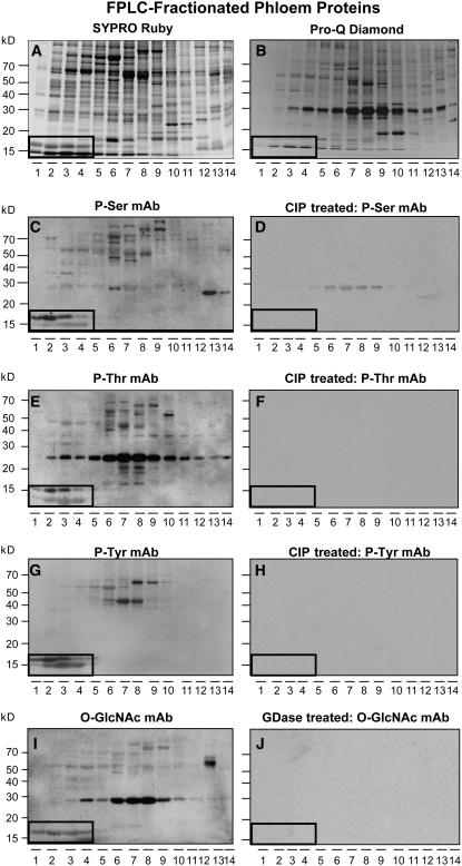
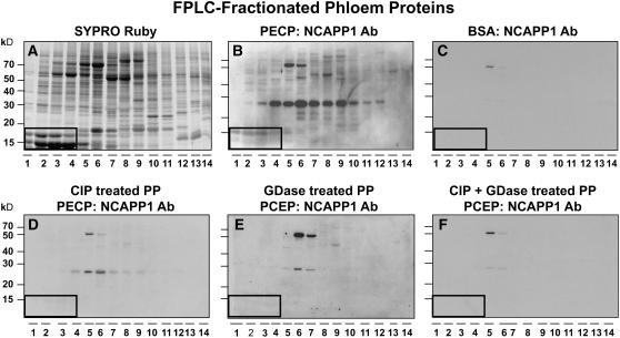
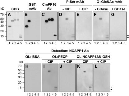
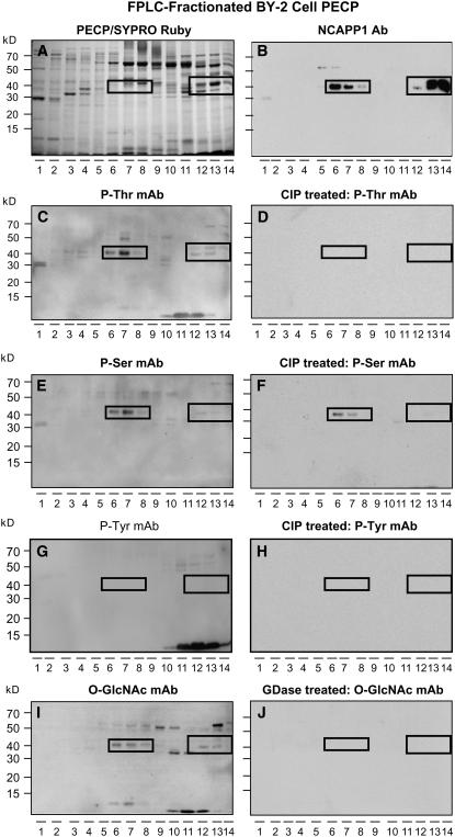
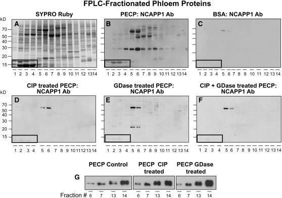
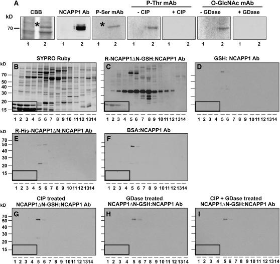
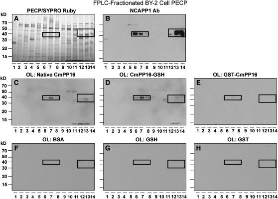
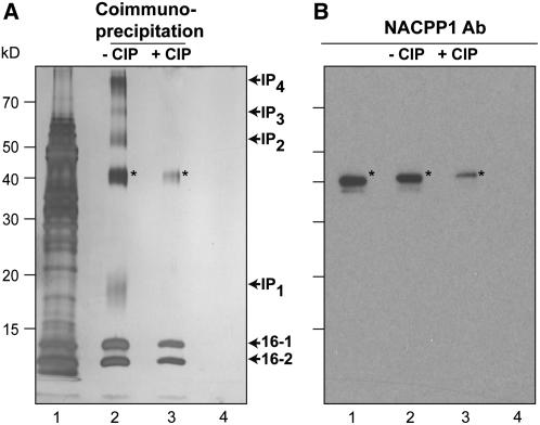
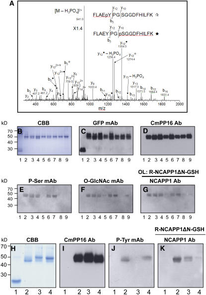
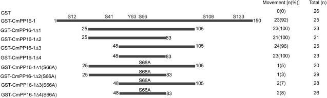
In plants, cell-to-cell trafficking of non-cell-autonomous proteins (NCAPs) involves protein-protein interactions, and a role for posttranslational modification has been implicated. In this study, proteins contained in pumpkin (Cucurbita maxima cv Big Max) phloem sap were used as a source of NCAPs to further explore the molecular basis for selective NCAP trafficking. Protein overlay assays and coimmunoprecipitation experiments established that phosphorylation and glycosylation, on both Nicotiana tabacum NON-CELL-AUTONOMOUS PATHWAY PROTEIN1 (Nt-NCAPP1) and the phloem NCAPs, are essential for their interaction. Detailed molecular analysis of a representative phloem NCAP, Cm-PP16-1, identified the specific residues on which glycosylation and phosphorylation must occur for effective binding to NCAPP1. Microinjection studies confirmed that posttranslational modification on these residues is essential for cell-to-cell movement of Cm-PP16-1. Lastly, a glutathione S-transferase (GST)-Cm-PP16-1 fusion protein system was employed to test whether the peptide region spanning these residues was required for cell-to-cell movement. These studies established that a 36-amino acid peptide was sufficient to impart cell-to-cell movement capacity to GST, a normally cell-autonomous protein. These findings are consistent with the hypothesis that a phosphorylation-glycosylation recognition motif functions to control the binding of a specific subset of phloem NCAPs to NCAPP1 and their subsequent transport through plasmodesmata.


Figures

Figure 1.
Figure 1.
Subpopulations of Pumpkin Phloem Proteins Are Phosphorylated and Glycosylated. (A) Pumpkin phloem sap FPLC-fractionated proteins. Proteins were separated on a 13% SDS-PAGE gel and then stained with SYPRO Ruby reagent. (B) Phosphorylation status of FPLC-fractionated proteins from (A) assessed using Pro-Q Diamond reagent. (C) Detection of phosphoproteins in FPLC-fractionated phloem sap by an antiphosphoserine monoclonal antibody. (D) Aliquots of the phloem fractions used in (C) were pretreated with CIP prior to protein gel blot analysis with the antiphosphoserine monoclonal antibody. Absence of signals detected in (C) establishes the specificity of the antibody reaction. (E) and (F) Detection of phosphoproteins in untreated and CIP-treated FPLC-fractionated phloem proteins by an antiphosphothreonine monoclonal antibody, respectively. (G) and (H) Detection of phosphoproteins in untreated and CIP-treated FPLC-fractionated phloem proteins by an antiphosphotyrosine monoclonal antibody, respectively. (I) and (J) Detection of glycosylated proteins in untreated and GDase-treated FPLC-fractionated phloem proteins by an anti-O-GlcNAc monoclonal antibody, respectively. Boxed areas indicate the location of native Cm-PP16-1 (top band) and Cm-PP16-2 (bottom band).
Figure 2.
Figure 2.
Posttranslational Modification of Pumpkin Phloem Proteins Is Essential for Nt-NCAPP1 Binding. (A) Pumpkin phloem sap FPLC-fractionated proteins stained with SYPRO Ruby. (B) FPLC-fractionated phloem proteins were blotted onto membrane, overlaid with a PECP preparation, and Nt-NCAPP1 interaction partners detected by anti-NCAPP1 polyclonal antibody (Lee et al., 2003). (C) BSA control. FPLC-fractionated phloem proteins were blotted onto membrane, overlaid with BSA, and then probed with anti-NCAPP1 polyclonal antibodies. (D) to (F) Nt-NCAPP1 overlay assays performed on FPLC-fractionated phloem proteins (PP) from (A) that were pretreated with CIP, GDase, or both CIP and GDase, respectively; interaction proteins were detected by anti-Nt-NCAPP1 polyclonal antibodies. Note that phosphorylation and glycosylation are necessary for Nt-NCAPP1 binding. Boxed areas indicate the location of native Cm-PP16-1 (top band) and Cm-PP16-2 (bottom band).
Figure 3.
Figure 3.
Posttranslational Modification on Cm-PP16-1 Controls Its Binding Capacity with Nt-NCAPP1. (A) Recombinant Cm-PP16-1 was expressed in and purified from E. coli as a GST-Cm-PP16-1 fusion protein (Lee et al., 2003) or by transient expression in N. benthamiana as a fusion protein with a GFP-Strep-His (GSH) tag (Cm-PP16-1-GSH). Lane 1, GST (27 kD) expressed in E. coli; lane 2, GST-Cm-PP16-1 (43 kD); lane 3, Cm-PP16-1-GSH (46 kD); lane 4, mCmPP16-1-GSH S-all-A (46 kD); lane 5, native phloem-purified Cm-PP16-1/-2. Protein aliquots (1 μg) were separated on 13% SDS-PAGE and purification verified by Coomassie Brilliant Blue (CBB) staining. (B) and (C) Protein gel blot analyses performed on proteins from (A) using an anti-GST and anti-Cm-PP16-1 polyclonal antibodies, respectively. (D) and (E) Phosphorylation status of E. coli and in planta–expressed Cm-PP16-1 determined using an antiphosphoserine monoclonal antibody. CIP pretreatment confirms the specificity of the antibody reaction. (F) and (G) Glycosylation status of E. coli and in planta–expressed Cm-PP16-1 determined using an anti-O-GlcNAc monoclonal antibody. GDase pretreatment confirms the specificity of the antibody reaction. (H) to (L) Aliquots of proteins from (A) were blotted to membranes and then overlaid with BSA, PECP preparation (100 μg/mL), or NCAPP1ΔN-GSH (5 μg/mL); binding between the various forms of Cm-PP16-1 and NCAPP1 was detected with anti-Nt-NCAPP1 polyclonal antibodies. CIP-treated Cm-PP16-1 controls confirmed the phosphorylation requirement for Cm-PP16-1–Nt-NCAPP1 binding. Arrowheads indicate position of Cm-PP16-1 (top) and Cm-PP16-2 (bottom).
Figure 4.
Figure 4.
Native Nt-NCAPP1 Contained in PECP Preparation Is Both Phosphorylated and Glycosylated. (A) Cation-exchange FPLC-fractionated PECPs separated on a 13% SDS-PAGE gel and stained with SYPRO Ruby. (B) Protein gel blot analysis of FPLC-fractionated proteins from (A) performed with anti-Nt-NCAPP1 polyclonal antibody preparation. Note the presence of strong Nt-NCAPP1 signal in lanes 6 to 8 and 12 to 14. (C) to (H) Phosphorylation status of native Nt-NCAPP1 probed using antiphosphothreonine, antiphosphoserine, or antiphosphotyrosine monoclonal antibodies. CIP pretreatment of the FPLC-fractionated proteins from (A) confirmed the specificity of the immunoreaction. (I) and (J) Glycosylation status of native Nt-NCAPP1 probed using an anti-O-GlcNAc monoclonal antibody. GDase pretreatment of the FPLC-fractionated proteins from (A) confirmed the specificity of the immunoreaction. Boxed areas indicate the location of Nt-NCAPP1 isoforms on the PECP FPLC fractions.
Figure 5.
Figure 5.
Posttranslational Modification on Nt-NCAPP1 Controls Its Binding Capacity with Cm-PP16-1 and Other Phloem Proteins. (A) Pumpkin phloem sap FPLC-fractionated proteins stained with SYPRO Ruby. (B) FPLC-fractionated phloem proteins were blotted onto membrane, overlaid with a PECP preparation, and Nt-NCAPP1 interaction partners detected by anti-NCAPP1 polyclonal antibodies. (C) BSA control. FPLC-fractionated phloem proteins were blotted onto membrane, overlaid with BSA, and then probed with anti-NCAPP1 polyclonal antibodies. (D) to (F) Nt-NCAPP1 overlay assays performed on FPLC-fractionated phloem proteins from (A). PECP preparation was pretreated with CIP, GDase, or both CIP and GDase; interaction proteins were detected by anti-Nt-NCAPP1 polyclonal antibodies. Note that phosphorylation and glycosylation on Nt-NCAPP1 are necessary for binding to Cm-PP16-1 and other phloem proteins. Boxed areas indicate the location of native Cm-PP16-1 (top band) and Cm-PP16-2 (bottom band). (G) Antigenicity of Nt-NCAPP1 contained in the PECP preparation was not affected by CIP or GDase treatment. Cation-exchange FPLC fractions 6, 7, 13, and 14 (see Figure 4B) were CIP or GDase treated and then protein gel blot analyses performed with anti-Nt-NCAPP1 polyclonal antibodies.
Figure 6.
Figure 6.
Involvement of Posttranslational Modification of NCAPP1 for Binding to Phloem Proteins. (A) Phosphorylation and glycosylation status of in planta–expressed and purified recombinant Nt-NCAPP1. Protein gel blot analyses were performed with anti-Nt-NCAPP1 polyclonal antibodies and antiphosphoserine, antiphosphotheronine, and anti-O-GlcNAc monoclonal antibodies. CIP and GDase pretreatment of purified recombinant Nt-NCAPP1 confirmed the specificity of these immunoreactions. Lane 1, partially purified GSH; lane 2, partially purified Nt-NCAPP1ΔN-GSH. (B) FPLC-fractionated pumpkin phloem proteins stained with SYPRO Ruby. (C) FPLC-fractionated phloem proteins were blotted onto membrane, overlaid with recombinant Nt-NCAPP1ΔN-GSH, and Nt-NCAPP1 interaction partners detected by anti-NCAPP1 polyclonal antibodies. (D) GSH control. FPLC-fractionated phloem proteins were blotted onto membrane, overlaid with GSH, and then probed with anti-NCAPP1 polyclonal antibodies. (E) FPLC-fractionated phloem proteins were blotted onto membrane, overlaid with E. coli–expressed recombinant His-tagged Nt-NCAPP1ΔN (R-His-Nt-NCAPP1ΔN), and Nt-NCAPP1 interaction partners detected by anti-NCAPP1 polyclonal antibodies. (F) BSA control. FPLC-fractionated phloem proteins were blotted onto membrane, overlaid with BSA, and then probed with anti-NCAPP1 polyclonal antibodies. (G) to (I) Nt-NCAPP1ΔN-GSH overlay assays performed on FPLC-fractionated phloem proteins. Purified recombinant Nt-NCAPP1ΔN-GSH was pretreated with CIP, GDase, or both CIP and GDase, respectively; interaction proteins were detected by anti-Nt-NCAPP1 polyclonal antibodies. Note that phosphorylation and glycosylation on Nt-NCAPP1 is necessary for binding to Cm-PP16-1 and other phloem proteins. Boxed areas indicate the location of native Cm-PP16-1 (top band) and Cm-PP16-2 (bottom band).
Figure 7.
Figure 7.
Native and in Planta–Expressed, but Not E. coli–Produced, Cm-PP16-1 Interacts Strongly with Nt-NCAPP1. (A) Cation-exchange FPLC-fractionated PECPs separated on a 13% SDS-PAGE gel and stained with SYPRO Ruby. (B) Protein gel blot analysis of FPLC-fractionated proteins from (A) performed with anti-Nt-NCAPP1 polyclonal antibody preparation. (C) to (E) FPLC-fractionated PECP preparations were blotted and then overlaid with native phloem-purified Cm-PP16-1, in planta–expressed recombinant Cm-PP16-1-GSH, and E. coli–expressed recombinant GST-Cm-PP16-1; interaction partners were detected using anti-Cm-PP16-1–specific polyclonal antibodies. Note that the GST-Cm-PP16-1 interaction was limited to Nt-NCAPP1 present in lane 7. (F) to (H) FPLC-fractionated PECP preparations were blotted and then overlaid with BSA, in planta–expressed GSH, or E. coli–expressed GST. Signals were detected by an anti-Cm-PP16-1–specific polyclonal antibodies. Boxed areas show the location of Nt-NCAPP1 isoforms.
Figure 8.
Figure 8.
Nt-NCAPP1 Coimmunoprecipitates with Cm-PP16-1. (A) Phloem-purified native Cm-PP16-1 and Cm-PP16-2 were mixed with a PECP preparation (lane 1, input proteins), and co-IP was performed using either anti-Cm-PP16-1 antibody (lanes 2 and 3) or preimmnune serum (lane 4). A comparison of lanes 2 and 3 illustrates the importance of Cm-PP16-1 phosphorylation on protein complex formation. Proteins detected by silver stain. Note that the co-IP complex is comprised of Cm-PP16-1, Cm-PP16-2, interaction protein 1 (IP1), Nt-NCAPP1, IP2, IP3, and IP4. (B) Presence of Nt-NCAPP1 (asterisks) detected with Nt-NCAPP1 polyclonal antibodies. Lanes are as in (A). These co-IP experiments were performed in duplicate with identical results.
Figure 9.
Figure 9.
Identification and Mutational Analysis of Posttranslational Modification Sites on Cm-PP16-1. (A) LC-MS/MS analysis of the phosphorylation sites on phloem-purified Cm-PP16-1. The MS/MS spectrum is a mixture of Ser or Tyr phosphorylation of the same peptide, FLAEpYPGSGGDFHILK and FLAEYPGpSGGDFHILK. These two phosphopeptides have the same parent mass-to-charge ratio of 990.1, and most fragments are the same, but fragments y10, y12, and b5 distinguish the two, as denoted by and asterisks for phosphotyrosine and phosphoserine, respectively. (B) to (G) Analysis of phosphorylation and O-GlcNacylation on Cm-PP16-1 Ser residues. (B) CBB staining of recombinant purified proteins. Lane 1, GSH; lane 2, Cm-PP16-1-GSH; lanes 3 to 7, Cm-PP16-1-GSH mutants in which Ser-12, Ser-41, Ser-66, Ser-108, and Ser-133, respectively, were replaced with Ala; lane 8, Cm-PP16-1-GSH mutant in which all the above five Ser residues were replaced with Ala (mCmPP16-1-GSH S-all-A); lane 9, Cm-PP16-1-GSH mutant in which Ser-66 was replaced with Asp to produce a phosphorylation mimic (mCmPP16-1-GSH S66D). (C) Protein gel blot analysis performed with anti-GFP antibody. (D) Protein gel blot analysis performed with anti-CmPP16-1 polyclonal antibodies. (E) Protein gel blot analysis performed with an anti-phosphoserine monoclonal antibody. (F) Protein gel blot analysis performed with an anti-O-GlcNAc monoclonal antibody. (G) Protein overlay assay (OL) performed with recombinant purified Nt-NCAPP1ΔN-GSH; interacting proteins were detected with anti-Nt-NCAPP1 polyclonal antibodies. (H) to (K) Analysis of the involvement of Cm-PP16-1 Tyr and Ser residues on binding to Nt-NCAPP1. (H) CBB staining of the recombinant purified proteins. Lane 1, GSH; lane 2, Cm-PP16-1-GSH; lane 3, Cm-PP16-1-GSH mutant in which Tyr-63 was replaced with Ala (mCmPP16-1-GSH Y63A); lane 4, mCmPP16-1-GSH S-all-A. (I) Protein gel blot analysis performed with anti-Cm-PP16-1 polyclonal antibodies. (J) Protein gel blot analysis performed with an antiphosphotyrosine monoclonal antibody. (K) Protein overlay assay performed with recombinant purified Nt-NCAPP1ΔN-GSH; interacting proteins were detected with anti-Nt-NCAPP1 polyclonal antibodies.
Figure 10.
Figure 10.
Cell-to-Cell Movement of GST Is Conferred by a Peptide Containing the Cm-PP16-1 Recognition Motif. Microinjection experiments were performed with the illustrated series of GST-Cm-PP16-1 fusion proteins. GST-Cm-PP16-1 N- and C-terminal deletion mutant proteins established that a peptide, comprised of 36 amino acids spanning the identified posttranslational modification sites, was sufficient to impart gain-of-function movement capacity to GST. Test proteins were microinjected into N. benthamiana mesophyll cells, and their movement was detected by coinjection of fluorescein isothiocyanate–labeled 10 kD dextran. Total number of injections (n) and percentage of intercellular movement associated with each test probe are shown on the right.

Similar articles

Cited by

References

    1. Aoki, K., Kragler, F., Xoconostle-Cázares, B., and Lucas, W.J. (2002). A subclass of plant heat shock cognate 70 chaperones carries a motif that facilitates trafficking through plasmodesmata. Proc. Natl. Acad. Sci. USA 99 16342–16347. - PMC - PubMed
    1. Aoki, K., Suzui, N., Fujimaki, S., Dohmae, N., Yonekura-Sakakibara, K., Fujiwara, T., Hayashi, H., Yamaya, T., and Sakakibara, H. (2005). Destination-selective long-distance movement of phloem proteins. Plant Cell 17 1801–1814. - PMC - PubMed
    1. Balachandran, S., Xiang, Y., Schobert, C., Thompson, G.A., and Lucas, W.J. (1997). Phloem sap proteins from Cucurbita maxima and Ricinus communis have the capacity to traffic cell to cell through plasmodesmata. Proc. Natl. Acad. Sci. USA 94 14150–14155. - PMC - PubMed
    1. Banerjee, A.K., Chatterjee, M., Yu, Y., Suh, S.G., Miller, W.A., and Hannapel, D.J. (2006). Dynamics of a mobile RNA of potato involved in a long-distance signaling pathway. Plant Cell 18 3443–3457. - PMC - PubMed
    1. Cao, Q., Kim, J.H., and Richter, J.D. (2006). CDK1 and calcineurin regulate Maskin association with eIF4E and translational control of cell cycle progression. Nat. Struct. Mol. Biol. 13 1128–1134. - PubMed

Publication types

MeSH terms