. 2007 Jun;19(6):1980-93.
doi: 10.1105/tpc.107.050526. Epub 2007 Jun 29.

LPA2 is required for efficient assembly of photosystem II in Arabidopsis thaliana

Affiliations

LPA2 is required for efficient assembly of photosystem II in Arabidopsis thaliana

Jinfang Ma et al. Plant Cell. 2007 Jun.

Retraction in

  • RETRACTION.
    [No authors listed] [No authors listed] Plant Cell. 2016 Dec;28(12):3061. doi: 10.1105/tpc.16.00881. Epub 2016 Nov 28. Plant Cell. 2016. PMID: 27895223 Free PMC article. No abstract available.

Abstract

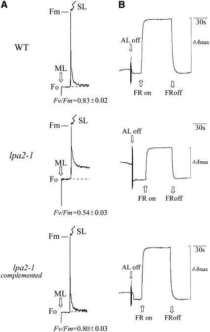
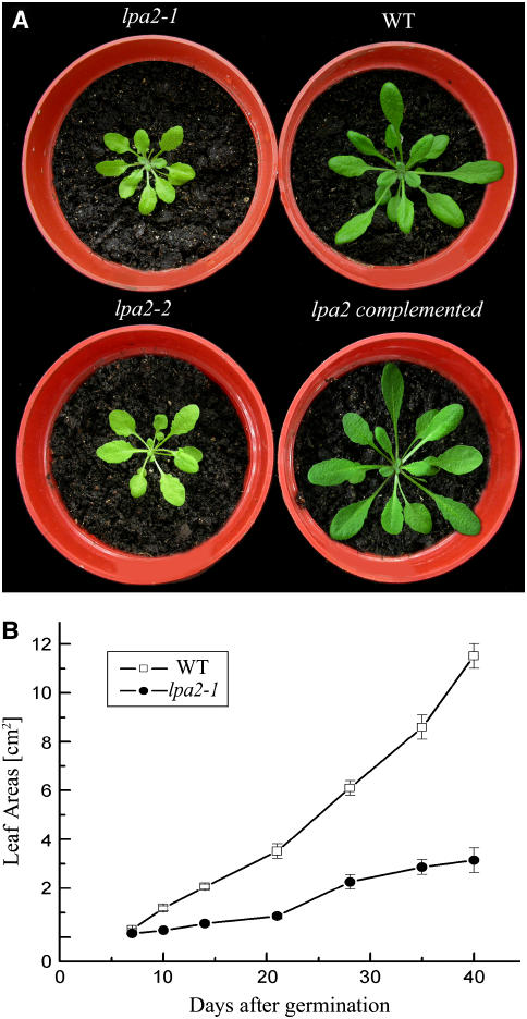
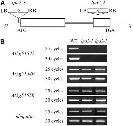
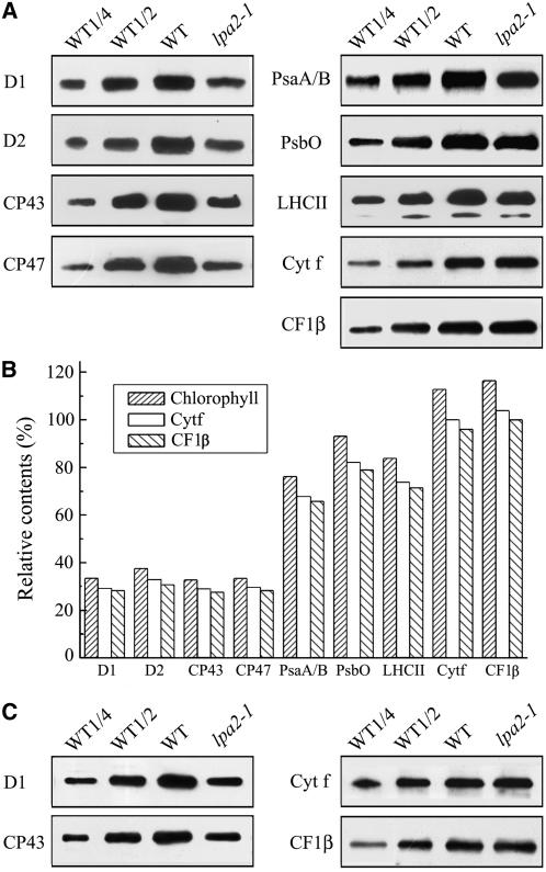
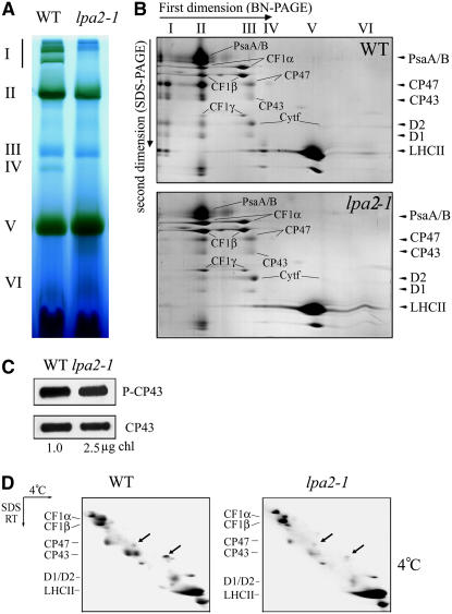
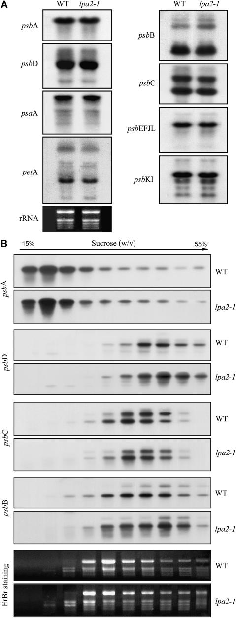
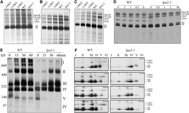
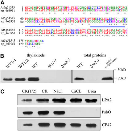
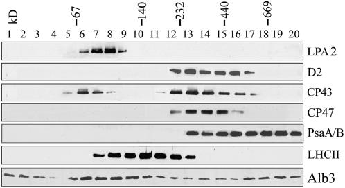
To elucidate the molecular mechanism of photosystem II (PSII) assembly, we characterized the low psii accumulation2 (lpa2) mutant of Arabidopsis thaliana, which is defective in the accumulation of PSII supercomplexes. The levels and processing patterns of the RNAs encoding the PSII subunits are unaltered in the mutant. In vivo protein-labeling experiments showed that the synthesis of CP43 (for chlorophyll a binding protein) was greatly reduced, but CP47, D1, and D2 were synthesized at normal rates in the lpa2-1 mutant. The newly synthesized CP43 was rapidly degraded in lpa2-1, and the turnover rates of D1 and D2 were higher in lpa2-1 than in wild-type plants. The newly synthesized PSII proteins were assembled into PSII complexes, but the assembly of PSII was less efficient in the mutant than in wild-type plants. LPA2 encodes an intrinsic thylakoid membrane protein, which is not an integral subunit of PSII. Yeast two-hybrid assays indicated that LPA2 interacts with the PSII core protein CP43 but not with the PSII reaction center proteins D1 and D2. Moreover, direct interactions of LPA2 with Albino3 (Alb3), which is involved in thylakoid membrane biogenesis and cell division, were also detected. Thus, the results suggest that LPA2, which appears to form a complex with Alb3, is involved in assisting CP43 assembly within PSII.


Figures

Figure 1.
Figure 1.
Spectroscopic Analysis of Wild-Type and lpa2-1 Plants. (A) Chlorophyll fluorescence induction. The minimal level of fluorescence (Fo) of dark-adapted whole plants with all PSII reaction centers open was determined using a pulsed measuring beam of red light. The Fm level with all PSII reaction centers closed was determined using a saturating pulse and dark-adapted leaves. SL, saturating light; ML, measuring light. (B) Redox kinetics of P700. The oxidation of P700 was investigated by measuring absorbance changes of P700 at 820 nm induced by far-red light (FR; 720 nm). ΔAmax, maximum oxidation induced by far-red light. AL, actinic light.
Figure 2.
Figure 2.
Phenotypes of lpa2-1, lpa2-2, Wild-Type, and lpa2-1 Transformant Plants Complemented with the Open Reading Frame of the At5g51545 Gene. (A) Five-week-old plants grown in the growth chamber. (B) Growth kinetics of the lpa2-1 mutant compared with wild-type plants. Values shown are averages ± se of six replicate experiments.
Figure 3.
Figure 3.
Identification of the lpa2 Mutation. (A) Schematic diagram of the LPA2 gene. Exons (white boxes) and introns (lines) are indicated. The positions of the T-DNA insertions corresponding to lpa2-1 and lpa2-2 are shown. ATG indicates the start codon, and TGA indicates the stop codon. LB, left border; RB, right border. The diagram is not drawn to scale. (B) RT-PCR analysis of mutant plants. RT-PCR was performed with specific primers for At5g51545, At5g51540, and At5g51550 and ubiquitin-specific primers.
Figure 4.
Figure 4.
Immunoblot Analysis of Chloroplast Proteins in lpa2-1 and Wild-Type Plants. (A) Immunodetection of chloroplast proteins. Thylakoid membrane proteins (1 μg of chlorophyll) from 5-week-old wild-type and lpa2-1 leaves were separated by SDS-urea-PAGE, and the blots were probed with specific anti-D1, anti-D2, anti-CP43, anti-CP47, anti-PsbO, anti-LHCII, anti-PsaA/B, anti-cytochrome f, and anti-CF1β antibodies. (B) Semiquantitative analysis of chloroplast proteins. X-ray films were scanned and analyzed using an AlphaImager 2200 documentation and analysis system. The protein contents (per unit of chlorophyll) of the thylakoid membrane were determined and compared. The signal intensities of the immunoblot of the mutant, relative to those in the wild type, were also normalized to the cytochrome f and CF1β blot signals. (C) Immunodetection of chloroplast proteins (1 μg of chlorophyll) from young seedlings of wild-type and lpa2-1 plants with specific anti-D1, anti-CP43, anti-cytochrome f, and anti-CF1β antibodies.
Figure 5.
Figure 5.
Accumulation of Chloroplast Proteins, Phosphorylation of CP43, and Binding of Chlorophyll to CP43 in Wild-Type and lpa2-1 Plants. (A) BN gel analysis of thylakoid membrane protein complexes. Thylakoid membranes (10 μg of chlorophyll) from 5-week-old wild-type and lpa2-1 leaves were solubilized with 1% DM and separated by BN gel electrophoresis. The positions of protein complexes representing PSII supercomplexes (band I), monomeric PSI and dimeric PSII (band II), monomeric PSII (band III), CP43 minus PSII (band IV), trimeric LHCII/PSII reaction center (band V), and monomeric LHCII (band VI) were identified by immunodetection and matrix-assisted laser-desorption ionization time-of-flight (MALDI-TOF) mass spectrometry (Guo et al., 2005; Peng et al., 2006). (B) Two-dimensional separation of protein complexes in the thylakoid membrane. BN-PAGE–separated thylakoid proteins in a single lane from a BN gel were separated in a second dimension by 15% SDS-urea-PAGE and stained with Coomassie blue. Names of the proteins resolved by the second-dimensional SDS-PAGE, previously identified, are indicated by arrowheads (Peng et al., 2006). (C) Immunodetection of the phosphorylation of CP43. Thylakoid membranes were isolated from 5-week-old Arabidopsis lpa2-1 (2.5 μg of chlorophyll) and wild-type (1 μg of chlorophyll) plants grown under normal growth conditions, separated by SDS-PAGE, and immunodetected with anti-phosphothreonine (top panel; P-CP43) and anti-CP43 (bottom panel; CP43) antibodies. (D) Analysis of chlorophyll binding to CP43. Thylakoid membranes isolated from 5-week-old wild-type and lpa2-1 plants were solubilized at 4°C. After solubilization, the thylakoid membranes were separated in the first dimension on a 7.5 to 15% polyacrylamide gradient gel at 4°C followed by a second-dimensional 12 to 18% polyacrylamide gel in the presence of 6 M urea at room temperature. CP47 and CP43, on the diagonal, are indicated by arrows. The proteins identified by MALDI-TOF mass spectrometry are indicated at left.
Figure 6.
Figure 6.
mRNA Expression and Polysome Accumulation in Chloroplasts. (A) RNA gel blot hybridization with total RNA from the leaves of wild-type and lpa2-1 plants. Preparations (10 μg) of total leaf RNA from 5-week-old wild-type and lpa2-1 plants were size-fractionated by agarose gel electrophoresis, transferred to a nylon membrane, and probed with 32P-labeled cDNA probes for the genes psbA, psbB, psbC, psbD, psbEFJL, psbKI, petA, and psaA. rRNA was visualized by staining with ethidium bromide as an equal loading control. (B) Association of psbA, psbB, psbC, and psbD mRNAs with polysomes. Ten fractions of equal volume were collected from the top to the bottom of the 15 to 55% sucrose gradients, and equal proportions of the RNA purified from each fraction were analyzed by gel blot hybridization. rRNAs were detected by ethidium bromide (EtBr) staining.
Figure 7.
Figure 7.
In Vivo Synthesis and Assembly of Chloroplast Proteins. (A) Pulse labeling of thylakoid membrane proteins in 5-week-old leaves. After pulse labeling in Arabidopsis 5-week-old leaves in the presence of cycloheximide for 20 min, the thylakoid membranes were isolated, and the proteins were separated by SDS-urea-PAGE and visualized autoradiographically. (B) and (C) Pulse labeling of thylakoid membrane proteins in young seedlings. After pulse labeling in young Arabidopsis seedlings in the presence of cycloheximide for 20 min (B) or 10 min (C), thylakoid membranes were isolated, and the proteins were separated by SDS-urea-PAGE and visualized autoradiographically. (D) Pulse and chase labeling of thylakoid membrane proteins. Twenty minutes of pulse labeling in 12-d-old young Arabidopsis seedlings in the presence of cycloheximide was followed by 0.5, 1, 2, or 4 h of chase with cold Met. After translation, thylakoid membranes were isolated, separated by SDS-urea-PAGE, and visualized autoradiographically. (E) and (F) Two-dimensional BN-SDS-PAGE analysis of the incorporation of [35S]Met into thylakoid membrane protein complexes. A 20-min pulse in Arabidopsis young seedlings in the presence of cycloheximide was followed by a chase of cold Met for 15, 30, or 60 min. After translation, the thylakoid membranes were isolated and solubilized with DM, and the protein complexes were separated by BN-PAGE and visualized by autoradiography (E). Bands corresponding to various PSII assembly complexes of PSII supercomplexes (band I), monomeric PSI superimposed on the PSII dimer (band II), monomeric PSII (band III), CP43-free PSII monomer (band IV), reaction center (band V), and free proteins (band VI) are indicated at right. The BN-PAGE gel–separated protein complexes were further subjected to SDS-PAGE in the second dimension and visualized by autoradiography (F). Designations of proteins resolved by the second dimension SDS-PAGE, previously identified, are indicated at right (Peng et al., 2006).
Figure 8.
Figure 8.
Amino Acid Sequence Alignment and Immunolocalization of LPA2. (A) The amino acid sequence of the At5g51545 protein was compared with a homologous sequence from O. sativa. Identical amino acids are marked with asterisks, and conserved exchanges are marked with colons. Two possible transmembrane domains are shown by blue bars. The sequences were aligned with ClustalW (Thompson et al., 1994). (B) Immunoblot analysis of LPA2. Samples from wild-type, lpa2-1, lpa2-2, and complemented plants, consisting of total leaf proteins and thylakoids (equivalent to 3 μg of chlorophyll), were separated by SDS-PAGE and immunodetected with antibodies raised against LPA2. (C) Salt washing of the thylakoid membranes. The thylakoid membranes were sonicated in the presence of 250 mM NaCl, 1 M CaCl2, and 6 M urea for 30 min at 0°C, then incubated for another 30 min at 0°C. PsbO, the 33-kD luminal protein of PSII, and the PSII core protein CP47 were used as markers. Membranes that had not been subjected to any salt treatment (CK) were used as controls.
Figure 9.
Figure 9.
Sucrose Gradient Fractionation Analysis of Thylakoid Proteins. Thylakoid membranes (0.5 mg chlorophyll/mL) were solubilized with 1% DM and separated by centrifugation on a linear 0.1 to 1 M sucrose gradient at 180,000g for 22 h. Twenty fractions were collected from the top to the bottom of the gradients, and the proteins from each fraction were separated by SDS-PAGE and immunodetected with anti-LPA2, anti-D2, anti-CP47, anti-CP43, anti-LHCII, anti-PsaA/B, and anti-Alb3 antibodies. The positions of 669-kD (thyroglobulin), 440-kD (ferritin), 232-kD (catalase), 140-kD (lactate dehydrogenase), and 66-kD (BSA) molecular mass markers are indicated.
Figure 10.
Figure 10.
LPA2 Interactions. (A) Growth of cells expressing X-Cub-LexA-VP16 (where X represents CP43, D1, D2, Alb3, or LPA1) with NubG-LPA2 on SD-His-Leu-Trp plates. Cells were grown to logarithmic phase, and 5-mL portions of 1:10 serial dilutions were spotted on SD-His-Leu-Trp plates and incubated at 30°C for 2 d. As a positive control, NMY32 containing CP43-Cub-LexA-VP16 was transformed with the NubI-Alg5 expression plasmid, and as a negative control, NMY32 containing CP43-Cub-LexA-VP16 was transformed with the plasmid expressing NubG-Alg5. (B) β-Gal activity of transformants expressing X-Cub-LexA-VP16 (where X represents CP43, D1, D2, Alb3, or LPA1) as bait constructs and NubG-LPA2 as prey construct. Cells were grown on SD-His-Leu-Trp plates, transferred to Whatman filters, permeabilized, and incubated in the presence of 5-bromo-4-chloro-3-indolyl-β-d-galactopyranoside.

Similar articles

Cited by

References

    1. Adir, N., Schochat, S., and Ohad, I. (1990). Light-dependent D1 protein synthesis and translocation is regulated by reaction centre II. Reaction centre II serves as an acceptor for the D1 precursor. J. Biol. Chem. 265 12563–12568. - PubMed
    1. Barkan, A. (1988). Proteins encoded by a complex chloroplast transcription unit are each translated from both monocistronic and polycistronic mRNAs. EMBO J. 7 2637–2644. - PMC - PubMed
    1. Barkan, A., and Goldschmidt-Clermont, M. (2000). Participation of nuclear genes in chloroplast gene expression. Biochimie 82 559–572. - PubMed
    1. Bellafiore, S., Ferris, P., Naver, H., Göhre, V., and Rochaix, J.-D. (2002). Loss of Albino3 leads to the specific depletion of the light-harvesting system. Plant Cell 14 2303–2314. - PMC - PubMed
    1. Boekema, E.J., Hankamer, B., Bald, D., Kruip, J., Nield, J., Boonstra, A.F., Barber, J., and Rogner, M. (1995). Supramolecular structure of the photosystem II complex from green plants and cyanobacteria. Proc. Natl. Acad. Sci. USA 92 175–179. - PMC - PubMed

Publication types

MeSH terms

Substances