Review
. 2007 Aug;13(8):1141-54.
doi: 10.1261/rna.620507. Epub 2007 Jun 29.

RNA-based regulation of genes of tryptophan synthesis and degradation, in bacteria

Affiliations
Review

RNA-based regulation of genes of tryptophan synthesis and degradation, in bacteria

Charles Yanofsky. RNA. 2007 Aug.

Abstract

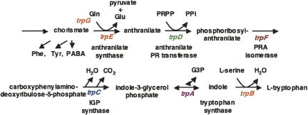
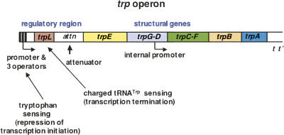
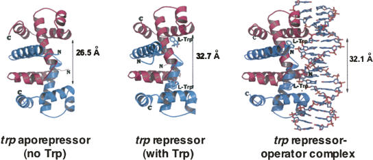
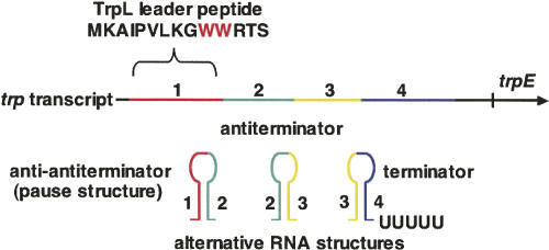
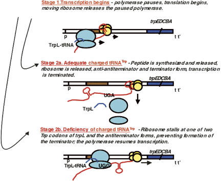
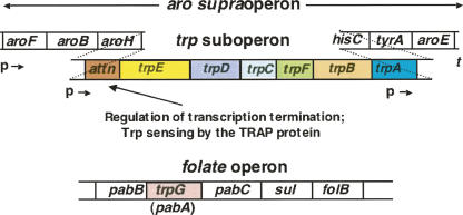
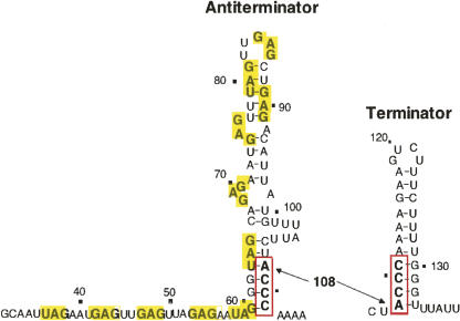
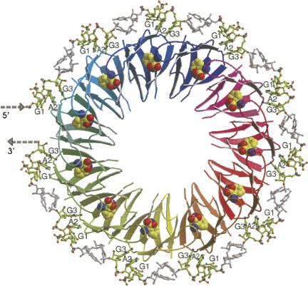
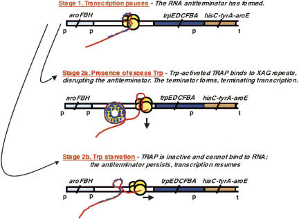
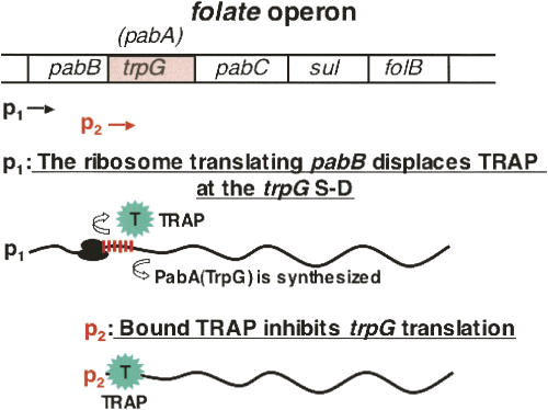
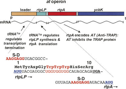
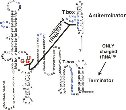
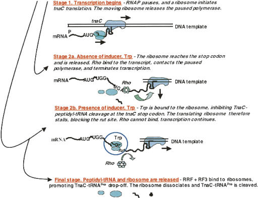
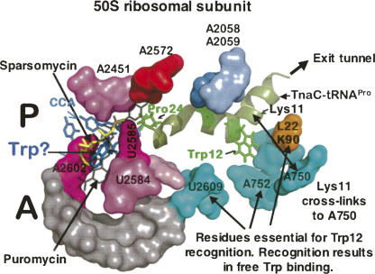
We are now aware that RNA-based regulatory mechanisms are commonly used to control gene expression in many organisms. These mechanisms offer the opportunity to exploit relatively short, unique RNA sequences, in altering transcription, translation, and/or mRNA stability, in response to the presence of a small or large signal molecule. The ability of an RNA segment to fold and form alternative hairpin secondary structures -- each dedicated to a different regulatory function -- permits selection of specific sequences that can affect transcription and/or translation. In the present paper I will focus on our current understanding of the RNA-based regulatory mechanisms used by Escherichia coli and Bacillus subtilis in controlling expression of the tryptophan biosynthetic operon. The regulatory mechanisms they use for this purpose differ, suggesting that these organisms, or their ancestors, adopted different strategies during their evolution. I will also describe the RNA-based mechanism used by E. coli in regulating expression of its operon responsible for tryptophan degradation, the tryptophanase operon.


Figures

FIGURE 1.
FIGURE 1.
The genes, enzymes, and reactions of the tryptophan biosynthetic pathway. The seven genes, or genetic segments, seven enzymes, or enzyme domains, and seven reactions, involved in tryptophan formation are shown (Yanofsky and Crawford 1987). Only one of the reactions is reversible. The products of four other pathways contribute carbon and/or nitrogen during tryptophan formation. Two of the tryptophan pathway enzymes often function as polypeptide complexes: anthranilate synthase, consisting of the TrpG and TrpE polypeptides, and tryptophan synthase, consisting of the TrpB and TrpA polypeptides.
FIGURE 2.
FIGURE 2.
Organization of the trp operon of E. coli. The genes of E. coli required for tryptophan biosynthesis from chorismate are organized in a single operon, or transcriptional unit (Yanofsky and Crawford 1987; Yanofsky 2004). Two pairs of genes are fused: trpG and trpD, and trpC and trpF. The structures of these two bifunctional polypeptides are known, and separate polypeptide segments are concerned with catalysis of each reaction. The relative order of the seven genetic segments, trpEGDFCBA, corresponds roughly to the relative order of the respective enzymatic reactions. The trp operon's regulatory region, located at the beginning of the operon, is designed to sense two signals, L-tryptophan, and charged vs. uncharged tRNATrp (Landick and Yanofsky 1987; Yanofsky 2004). Tryptophan, when in excess, activates the trp aporepressor, while charged and uncharged tRNATrp determine whether transcription will or will not be terminated in the operon's leader region. A poorly expressed internal promoter provides transcripts producing low levels of the last few enzymes of the pathway. This promoter is useful when the principal promoter is turned off. (p = promoter; t = terminator). (Modified from Figs. 1 and 2 in Yanofsky 2004 and reprinted with permission from Elsevier © 2004.)
FIGURE 3.
FIGURE 3.
The basic structures of the trp aporepressor, trp repressor, and trp repressor-operator complex. The aporepressor is a dimer of identical polypeptide chains; it has two tryptophan binding sites (Joachimiak and Zhang 1989; Shakked et al. 1994; Gryk et al. 1996). When tryptophan is bound, the repressor's two helix-turn-helix domains assume conformations that prepare them for binding at specific operator sequences. Binding of the trp repressor at a trp operator site—located within the trp promoter—interferes with RNA polymerase binding, hence binding prevents transcription initiation (Otwinowski et al. 1988). Interactions between trp repressor proteins bound at multiple adjacent operators in the trp promoter-operator region increase the stability of repressor-operator complexes, improving the effectiveness of repression (Fig. 2; Lawson et al. 2004). This figure was kindly drawn, and provided, by Dr. Andrzej Joachimiak.
FIGURE 4.
FIGURE 4.
The trp operon leader transcript and its functions. The initial 141 nt of the trp operon transcript can fold and form three alternative RNA hairpin structures: an anti-antiterminator (1:2), an antiterminator (2:3), and a terminator (3:4) (Landick and Yanofsky 1987; Yanofsky 2004). The anti-antiterminator structure (1:2) also serves as a transcription pause structure. The terminator structure is a typical intrinsic terminator which, when formed, directs the transcribing RNA polymerase to terminate transcription. However, whenever the preceding antiterminator structure has formed and persists, it prevents formation of the terminator. Hence transcription is not terminated. The initial leader RNA sequence has an additional role: It encodes a 14 nt leader peptide, TrpL. Translation of the two Trp codons of trpL is used to sense the availability of charged tRNATrp. Whenever the charged tRNATrp level is adequate for rapid translation of the two Trp codons of trpL, translation of trpL is completed, the translating ribosome dissociates, and the anti-antiterminator (1:2) and terminator (3:4) structures form. This results in transcription termination in the leader region of the operon. When the tRNATrp in the cell is largely uncharged, the translating ribosome stalls at one of the trpL Trp codons. The antiterminator RNA structure (2:3) then forms, preventing terminator formation and transcription termination. (See Fig. 5 for more details). Pausing following formation of the anti-antiterminator structure is essential for the coupling of translation of trpL mRNA with transcription of the leader region. The anti-antiterminator, if allowed to form and persist, would prevent formation of the antiterminator, thus the transcription terminator would form. (Modified from Fig. 2 in Yanofsky 2004 and reprinted with permission from Elsevier © 2004.)
FIGURE 5.
FIGURE 5.
The sequential, alternative events regulating transcription termination in the leader region of the trp operon of E. coli. Stage 1: The RNA polymerase molecule that initiates transcription of the trp operon pauses after synthesizing the initial segment of the transcript—the segment that forms the anti-antiterminator pause structure (Landick and Yanofsky 1987; Yanofsky 2004). While the polymerase is paused, a ribosome binds at the trpL mRNA start codon and initiates synthesis of the leader peptide. This translating ribosome then disrupts the anti-antiterminator pause structure, releasing the paused polymerase and allowing it to resume transcription. Stage 2a: When there is sufficient charged tRNATrp in the cell to allow rapid completion of synthesis of the leader peptide, the translating ribosome is released. The anti-antiterminator and terminator structures then form, promoting transcription termination. Stage 2b: When there is a deficiency of charged tRNATrp, the ribosome translating trpL mRNA stalls at one of its two Trp codons. This permits the RNA antiterminator structure to form, which prevents formation of the terminator. Transcription then continues into the operon's structural genes. (Modified from Fig. 2 in Yanofsky 2004 and reprinted with permission from Elsevier © 2004.)
FIGURE 6.
FIGURE 6.
Organization of the trp suboperon within the aro supraoperon, and the location of trpG in the folate operon, in B. subtilis. Six of the seven trp genes of B. subtilis are organized as a trp suboperon within the aro supraoperon (Gollnick et al. 2005). The three upstream and three downstream genes of this supraoperon encode proteins that participate in the common aromatic pathway or in phenylalanine, tyrosine, or histidine biosynthesis. One of the trp genes, trpG, resides within the folate operon. There is logical, since trpG, or more correctly trpG/pabA, specifies a polypeptide (TrpG/PabA) that serves as a component of two enzyme complexes. This polypeptide is the amino-group donor during the biosynthesis of both o-aminobenzoic acid (anthranilic acid), in tryptophan formation, and p-aminobenzoic acid, in folate formation. Two promoters within the aro supraoperon are used to transcribe the trp suboperon. Transcripts initiated at either promoter can form the alternative RNA structures described in Fig. 7. There is a third promoter—within trpA—that provides a transcript of the terminal three genes of the operon. This transcript is used to provide the polypeptides encoded by these three genes whenever transcription from the two upstream promoters is terminated in the trp suboperon leader region. Transcription, and regulation, of the folate operon is described in Fig. 10 (p = promoter; t = terminator). (Modified from Figs. 1 and 2 in Yanofsky 2004 and reprinted with permission from Elsevier © 2004.)
FIGURE 7.
FIGURE 7.
The alternative antiterminator and terminator structures that can form in the transcript of the leader region of the trp suboperon of B. subtilis. The antiterminator and termination hairpin structures that can form in the transcript segment immediately preceding trpE share an ACCC (bold letters, red box) nucleotide segment (Gollnick et al. 2005). Whenever the antiterminator structure forms, and remains stable, it prevents formation of the terminator structure. Therefore transcription termination does not occur in the leader region. A series of (X)AG trinucleotide repeats (bold letters, yellow rectangles) are located in the sequence immediately preceding, and within, the antiterminator sequence. These constitute the binding site for the tryptophan-activated RNA-binding regulatory protein, TRAP. When tryptophan-activated, TRAP binding to the RNA prevents formation of the antiterminator structure—or disrupts it, if it had formed previously. Disruption of the antiterminator structure frees its ACCC-containing 3′ strand, allowing the terminator to form and terminate transcription. Thus TRAP, when tryptophan-activated, binds to leader RNA and promotes transcription termination in the trp suboperon's leader region. Activated TRAP has a second beneficial effect on trp operon expression. When bound to non-terminated trp mRNA, it promotes formation of an RNA secondary structure in trp leader RNA that reduces initiation of translation of trpE. This also reduces trp operon expression (Merino et al. 1995; Du and Babitzke 1998).
FIGURE 8.
FIGURE 8.
Structure of the TRAP–RNA complex. TRAP of B. subtilis is a doughnut-shaped molecule consisting of 11 identical polypeptide subunits (Antson et al. 1999; Gollnick et al. 2005). A tryptophan-binding site is formed between each pair of subunits. In this figure, the atoms of the bound tryptophan molecules are represented as spheres. Tryptophan binding alters TRAP's surface, preparing it for recognition of, and binding to, RNA segments containing XAG repeats. The spacing between the XAG repeats, 2–3 nt, also is crucial (Gollnick et al. 2005). When TRAP binds to trp suboperon leader RNA it wraps the RNA around its periphery, disrupting the antiterminator structure. This allows the alternate, terminator structure to form and terminate transcription. In B. subtilis TRAP also binds to XAG trinucleotide repeats in the transcript segments preceding the start codons for trpG, trpP, and ycbK and inhibits their translation. (Modified from Fig. 3 in Gollnick et al. 2005 with permission from Annual Reviews © 2005; www.annualreviews.org.)
FIGURE 9.
FIGURE 9.
Stages in tryptophan regulation of transcription termination in the leader region of the trp suboperon of B. subtilis. Stage 1: Transcription initiated at either of the two promoters that precede the trp gene cluster pauses in the trp suboperon leader region upon synthesis of the antiterminator structure. Stage 2a: In the presence of excess tryptophan, the TRAP protein is activated and it binds to the mRNA XAG repeats, preventing formation of, or disrupting, the antiterminator structure. This allows the terminator structure to form and terminate transcription. Stage 2b: In the absence of sufficient tryptophan to activate TRAP, TRAP does not bind to RNA, and the polymerase stalled at Stage 1 resumes transcription. The antiterminator then forms, preventing formation of the terminator. Transcription continues into the structural gene region of the trp suboperon (Gollnick et al. 2005). (Modified from Fig. 3 in Yanofsky 2004 and reprinted with permission from Elsevier © 2004.)
FIGURE 10.
FIGURE 10.
TRAP inhibition of initiation of translation of the trpG/pabA coding region. A series of XAG repeats—a TRAP-binding site—is located immediately preceding the trpG/pabA start codon (Gollnick et al. 2005). Two promoters, p1 and p2, are used to transcribe trpG/pabA, one located before pabB and the second before trpG/pabA (Yakhnin et al. 2007). Transcription from p1 provides a transcript on which ribosomes translating pabB can displace bound TRAP or prevent TRAP from binding, thereby allowing trpG/pabA to be translated. On transcripts initiated at promoter p2, bound tryptophan-activated TRAP masks the trpG/pabA Shine–Dalgarno sequence. This prevents ribosome binding and translation of this coding region. When TRAP is tryptophan-free, and inactive, translation of the trpG/pabA coding region on p2 transcripts can proceed (Yakhnin et al. 2007). The dual promoters allow the TrpG/PabA polypeptide to be synthesized whenever it is needed for either o-aminobenzoic acid or p-aminobenzoic acid synthesis.
FIGURE 11.
FIGURE 11.
Organization and features of the at operon of B. subtilis. The at operon consists of a leader regulatory region, a 10-residue leader peptide coding region, rtpLP, and two structural genes, rtpA, encoding the AT protein, and ycbK, encoding what it is believed to be an efflux transport protein (Sarsero et al. 2000; Chen and Yanofsky 2004; Gollnick et al. 2005; Yakhnin et al. 2006a,b). Transcription of the at operon is regulated by the T box mechanism, in response to the accumulation of uncharged tRNATrp. Initiation of translation of rtpA, the structural gene for the AT protein, is also regulated by charged and uncharged tRNATrp. The 10-codon leader peptide coding region, rtpLP, immediately preceding the at coding region, contains three Trp codons. This transcript segment is used to regulate AT synthesis translationally in response to the accumulation of uncharged tRNATrp. Thus AT synthesis is regulated both transcriptionally and translationally, using different mechanisms, upon sensing the levels of charged and uncharged tRNATrp.
FIGURE 12.
FIGURE 12.
Sequence and presumed alternative structures formed in the T box leader RNA segment of the transcript of the at operon of B. subtilis. The transcript of the at operon leader region has segments analogous to those of many T box sequences that recognize an uncharged tRNA as a regulatory signal (Grundy and Henkin 2004; Sarsero et al. 2000: Gutierrez-Preciado et al. 2005). When uncharged tRNATrp binds to the at operon T box leader RNA segment—by codon–anticodon pairing and by binding of its acceptor end to the T box sequence—the antiterminator structure is stabilized, preventing formation of the terminator. When the cellular tRNATrp is largely charged, it cannot bind to and stabilize the T box antiterminator, therefore the terminator forms, and transcription is terminated. Some critical nucleotides in the leader RNA are colored. (Modified from Fig. 3 in Sarsero et al. 2000 and reprinted with permission from the National Academy of Sciences, USA © 2000.)
FIGURE 13.
FIGURE 13.
The alternative regulatory events involved in the synthesis of the AT protein. In cells with sufficient levels of charged tRNATrp, transcription of the at operon is terminated within its leader region (Gollnick et al. 2005). When there is a mild charged tRNATrp deficiency, uncharged tRNA will occasionally pair with the leader transcript T box sequence and permit transcription to continue into the operon. However, under these conditions, the ribosome translating rtpLP will reach its stop codon and inhibit initiation of translation of rtpA. Thus little or no AT protein will be produced. When there is a severe charged tRNATrp deficiency, the ribosome translating rtpLP will stall at one of its three Trp codons. This stalling will expose the rtpA Shine-Dalgarno region for efficient translation initiation, and the AT protein will be synthesized. (Modified from Fig. 4 in Yanofsky 2004 and reprinted with permission from Elsevier © 2004.)
FIGURE 14.
FIGURE 14.
Organization of the trp operon of other Gram-positive bacteria that have the same or different trp gene arrangements and regulation as in B. subtilis. Several Gram-positive bacterial species have a trp suboperon which is organized similarly to the trp suboperon of B. subtilis (Gutierrez-Preciado et al. 2007). These species have a TRAP protein and there is a TRAP binding site immediately preceding trpE in the trp suboperon. Only one of these species, B. lichiniformis, is also known to form an AT protein (Gutierrez-Preciado et al. 2007). In many other Gram-positive species the trp operon is organized differently, with a trpG within the trp operon. Most of these trp operons do not contain genes of the other aromatic amino acid pathways. In many of these species, all seven trp genes are present in the same operon, and the T box mechanism appears to be the predominant form of regulation of trp operon transcription. (Modified from Fig. 2 in Gutiérrez-Preciado et al. 2007 and reprinted with permission from Elsevier © 2007.)
FIGURE 15.
FIGURE 15.
Some common transcription attenuation regulatory mechanisms used in regulating trp operon expression in different bacterial species. Typical leader transcript segments are illustrated that can form alternative RNA hairpin structures, one of which is an intrinsic transcription terminator. The examples shown involve ribosome-mediated, protein-mediated, and tRNA-mediated regulation of formation of either of two alternative RNA hairpin structures. Each of these mechanisms is common in bacteria (Henkin and Yanofsky 2002; Merino and Yanofsky 2005). (Modified from Fig. 1 in Merino and Yanofsky 2005 and reprinted with permission from Elsevier © 2005.)
FIGURE 16.
FIGURE 16.
Features of the leader regulatory region of the tna operon of E. coli. Transcription of the structural gene region of the tna operon is regulated by two mechanisms: catabolite repression, at the tna operon's promoter, and Rho factor-dependent transcription termination, in the operon's leader region (Cruz-Vera et al. 2006). The CAP-dependent promoter is similar to other promoters subject to catabolite repression. Tryptophan induction of tna operon expression is based on relief from Rho-dependent transcription termination in the operon's leader region. The leader transcript segment contains a coding region for a 24-residue peptide, TnaC, that has a crucial Trp residue at position 12. Synthesis of this peptide in the presence of inducer, tryptophan, prevents cleavage of TnaC–peptidyl–tRNA at the tnaC stop codon. The resulting stalled ribosome blocks Rho factor from binding at the rut site, preventing transcription termination. Transcription then continues into the two structural genes of the operon.
FIGURE 17.
FIGURE 17.
Stages in tryptophan induction of tna operon expression in E. coli. When E. coli is grown in a medium lacking a catabolite-repressing carbon source, the CAP protein is activated, and transcription of the tna operon is initiated (Gong et al. 2001; Gong and Yanofsky 2002). Stage 1: An RNA polymerase complex transcribes the initial region of the operon and pauses following synthesis of an RNA segment that forms a pause structure. While the polymerase is paused, a ribosome binds to the nascent tna transcript and initiates synthesis of TnaC. This translating ribosome then releases the paused polymerase, hence both transcription and translation then proceed. Depending on the cellular concentration of free tryptophan, either Stage 2a or Stage 2b occurs. Stage 2a: When free tryptophan is absent, or present at a low concentration, the translating ribosome completes synthesis of TnaC and dissociates from the transcript. Rho factor then binds to the leader RNA. Meanwhile the transcribing RNA polymerase that had transcribed much of the leader region has paused at one of several pause sites located near the end of the leader region. When Rho factor can bind, and act, it contacts this paused polymerase and terminates transcription. Stage 2b: When free tryptophan is plentiful, it binds to ribosomes in the process of TnaC–tRNAPro synthesis and inhibits TnaC–tRNAPro cleavage. These ribosomes remain stalled at the tnaC stop codon, where they mask the mRNA rut site. Rho factor therefore cannot bind, and RNA polymerase molecules paused near the end of leader segment of the tna operon transcript resume transcription into the operon's two structural genes. TnaC–tRNAPro and the stalled ribosome are then released by RRF and RF3 action, the peptidyl–tRNA is hydrolyzed, and the ribosomal subunits are reused for additional protein synthesis (Gong et al. 2007).
FIGURE 18.
FIGURE 18.
Some peptide-ribosomal interactions believed to be responsible for tryptophan induction of tna operon expression. A region of the 50S ribosomal subunit is shown containing bound sparsomycin, puromycin, and presumably tryptophan, in the A site of the translating ribosome (Porse et al. 1999; Hansen et al. 2003; Cruz-Vera et al. 2005, 2007). Also shown is the probable location of TnaC–tRNAPro in the ribosome exit tunnel; identified are likely locations of residues Trp12 and Pro24, residues essential for induction. Residues of 23RNA and of ribosomal protein L22 in the vicinity of Trp12 of TnaC–tRNAPro, some of which have been shown to be required for tryptophan binding and induction, are also labeled. The approximate location of Trp12 of TnaC–tRNAPro has been determined by showing that Lys11 of TnaC–tRNAPro can be cross-linked to nucleotide A750 (Cruz-Vera et al. 2005). This figure is based on the structure of the E. coli ribosome, determined by Schuwirth et al. (2005). (Modified from Fig. 1 in Cruz-Vera et al. 2007 and reprinted with permission from the American Society for Microbiology © 2007.)

Similar articles

Cited by

References

    1. Antson, A.A., Dodson, E.J., Greaves, R.B., Chen, X.-P., Gollnick, P. Structure of the trp RNA-binding attenuation protein, TRAP, bound to RNA. Nature. 1999;401:235–242. - PubMed
    1. Anyanful, A., Dolan-Livengood, J.M., Lewis, T., Sheth, S., DeZalia, M.N., Sherman, M.A., Kalman, L.V., Benian, G.M., Kalman, D. Paralysis and killing of Caenorhabditis elegans by enteropathogenic Escherichia coli requires the bacterial tryptophanase. Mol. Microbiol. 2005;57:988–1007. - PubMed
    1. Babitzke, P. Regulation of transcription attenuation and translation initiation by allosteric control of an RNA-binding protein: The Bacillus subtilis TRAP protein. Curr. Opin. Microbiol. 2004;7:132–139. - PubMed
    1. Babitzke, P., Stults, J.T., Shire, S.J., Yanofsky, C. TRAP, the trp RNA-binding attenuation protein of Bacillus subtilis, is a multisubunit complex that appears to recognize G/UAG repeats in trpEDCFBA and trpG transcripts. J. Biol. Chem. 1994;269:16597–16604. - PubMed
    1. Chen, G.N., Yanofsky, C. Features of a leader peptide coding region that regulate translation initiation for the anti-TRAP protein of Bacillus subtilis . Mol. Cell. 2004;13:703–711. - PubMed

Publication types

LinkOut - more resources