. 2008 Jan;57(1):73-84.
doi: 10.1007/s00262-007-0351-y. Epub 2007 Jun 30.

Intratumoral injection of inactivated Sendai virus particles elicits strong antitumor activity by enhancing local CXCL10 expression and systemic NK cell activation

Affiliations

Intratumoral injection of inactivated Sendai virus particles elicits strong antitumor activity by enhancing local CXCL10 expression and systemic NK cell activation

Atsuko Fujihara et al. Cancer Immunol Immunother. 2008 Jan.

Abstract

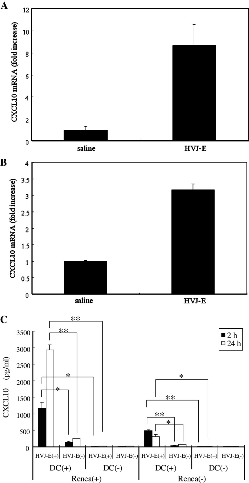
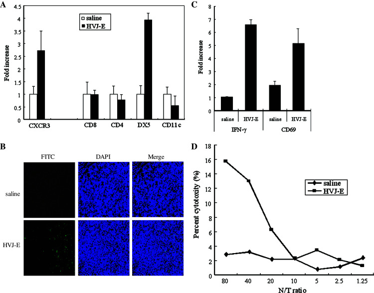
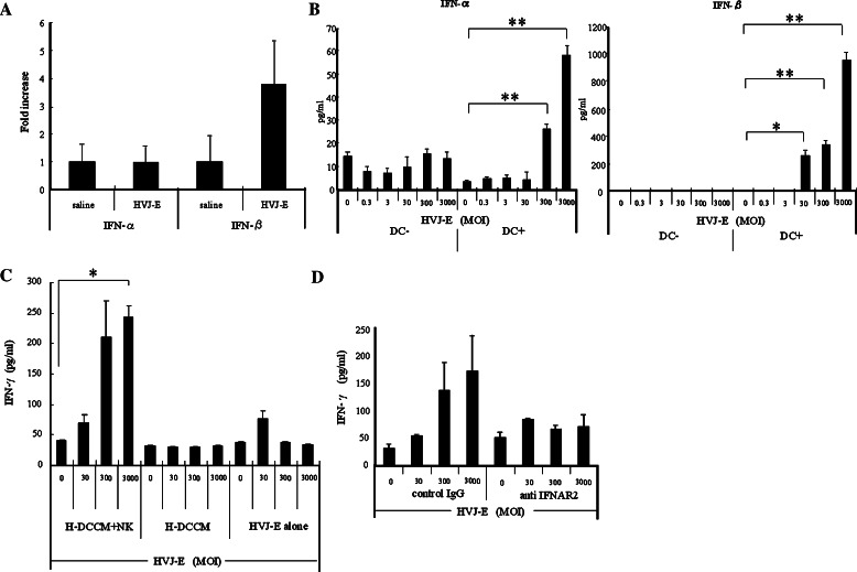
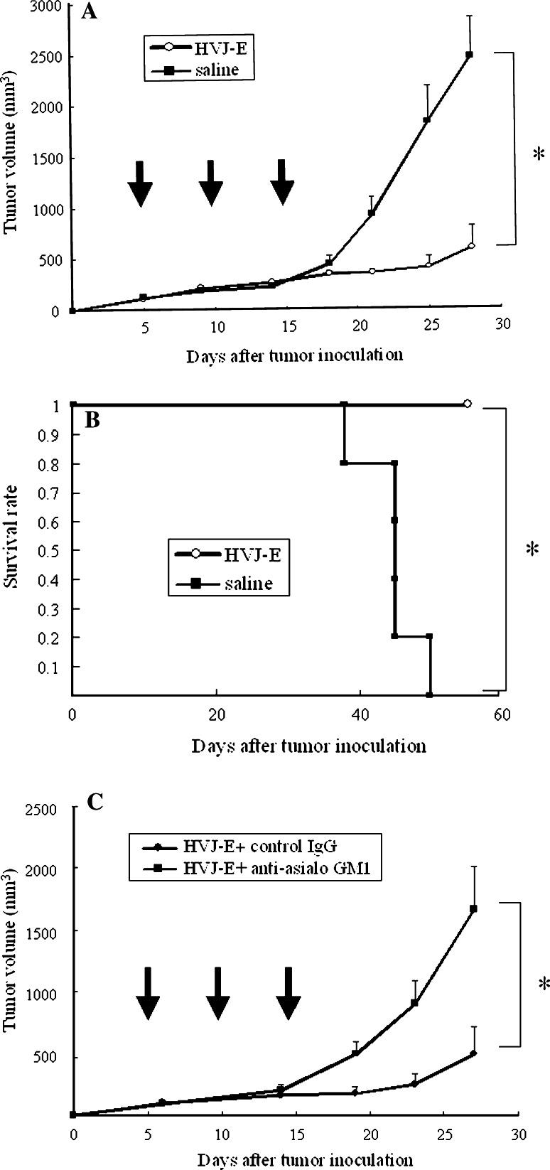
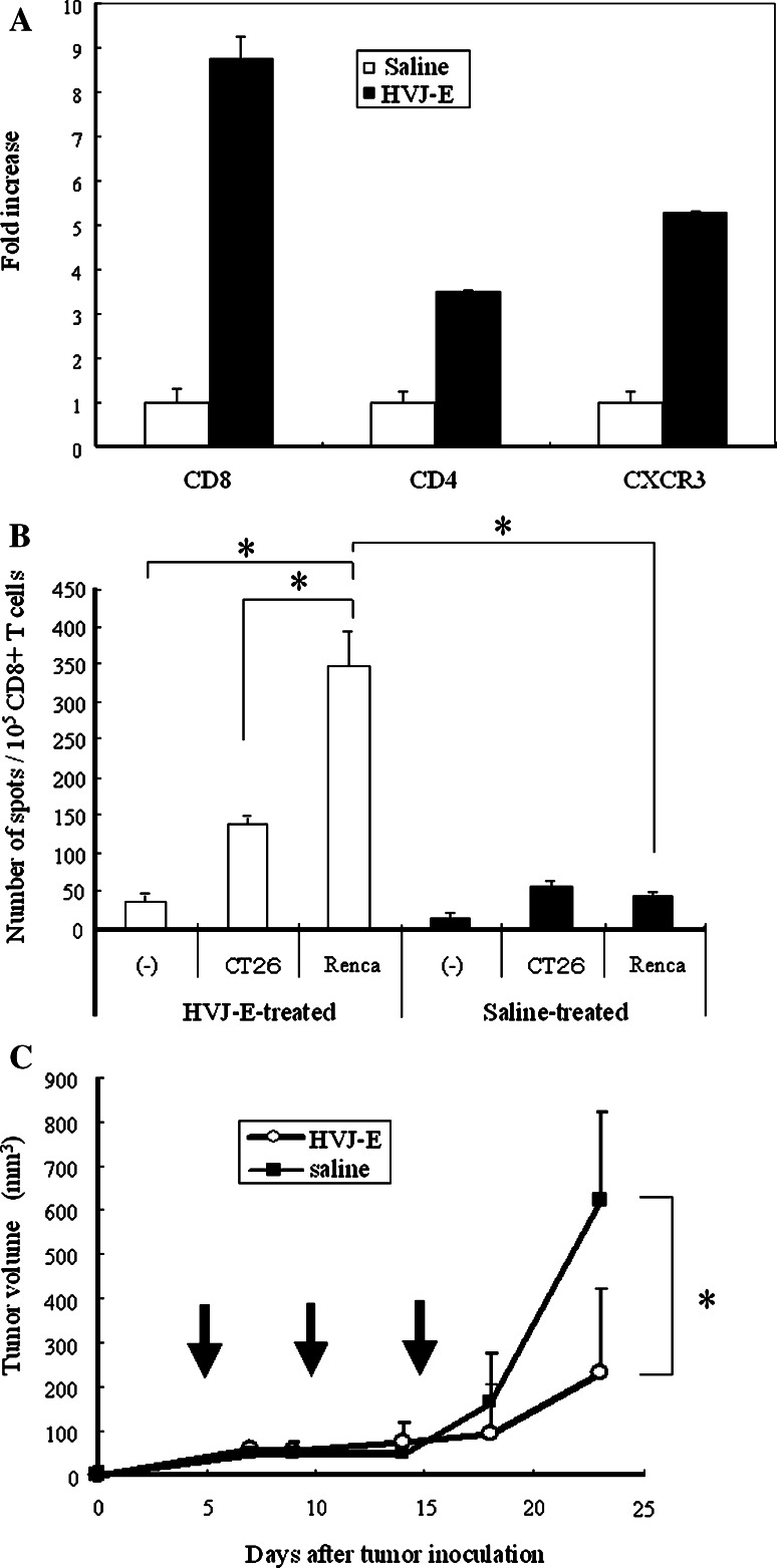
We have already demonstrated that inactivated, replication-defective Sendai virus particles (HVJ-E) have a powerful antitumor effect by both the generation of tumor-specific cytotoxic T cells and inhibition of regulatory T cell activity. Here, we report that HVJ-E also has an antitumor effect through non-T cell immunity. Microarray analysis revealed that direct injection of HVJ-E induced the expression of CXCL10 in established Renca tumors. CXCL10 was secreted by dendritic cells in the tumors after HVJ-E injection. Quantitative real-time RT-PCR and immunohistochemistry revealed that CXCR3+ cells (predominantly NK cells) infiltrated the HVJ-E-injected tumors. Moreover, HVJ-E injection caused systemic activation of NK cells and enhanced their cytotoxity against tumor cells. In an in vivo experiment, approximately 50% of tumors were eradicated by HVJ-E injection, and this activity of HVJ-E against Renca tumors was largely abolished by NK cell depletion using anti-asialo GM1 antibody. Since HVJ-E injection induced systemic antitumor immunity by enhancing or correcting the chemokine-chemokine receptor axis, it might be a potential new therapy for cancer.


Figures

Fig. 1
Fig. 1
Induction of CXCL10 by HVJ-E in vivo and in vitro. CXCL10 mRNA expression in whole tumors (a) or DCs isolated from tumors (b) after injection of HVJ-E or saline was measured by quantitative real-time RT-PCR (n = 5 per group). CXCL10 mRNA expression was increased by injection of HVJ-E. Error bars indicate the SE (<5%). This experiment was repeated three times with similar results. c CXCL10 levels in the medium of cultures with (+) or without (-) Renca cells, HVJ-E, or DCs. When HVJ-E and syngeneic mouse DCs were added to cultured Renca cells, a significant increase of CXCL10 production was observed. A significant increase of CXCL10 was also detected in the medium of DCs cultured with HVJ-E even in the absence of Renca cells, although the level was lower than that obtained with Renca cells (*P < 0.05, **P < 0.01). Data points are the mean ± SE of triplicate wells. SE < 5%. This experiment was repeated four times with similar results
Fig. 2
Fig. 2
Infiltration and activation of NK cells after intratumoral injection of HVJ-E. a Intratumoral infiltration of immune cells was investigated by quantitative real-time RT-PCR (n = 5). DX5 mRNA expression showed a marked increase. SE < 5%. This experiment was repeated three times with similar results. b NK cell infiltration. Immunofluorescence staining using a FITC-conjugated anti-mouse CD49b (DX5)/Pan NK cell monoclonal antibody shows prominent infiltration of DX5-positive cells (green) into an HVJ-E-treated tumor (×200). This experiment was repeated three times with similar results. c Activation of intratumoral NK cells: NK cells were purified from tumors injected with HVJ-E or saline, and IFN-γ and CD69 mRNA expression was assayed by quantitative real-time RT-PCR. Both IFN-γ and CD69 expressions were upregulated in HVJ-E-treated tumors. SE < 5%. This experiment was repeated three times with similar results. d NK cytotoxity in vivo. Cytotoxicity assays were performed with NK cells harvested from the spleens of mice after treatment with HVJ-E (filled square) or saline (filled diamond). NK cells from HVJ-E-treated mice showed an increase of cytotoxity against Renca cells. This experiment was repeated three times with similar results
Fig. 3
Fig. 3
Activation of NK cells by type I IFNs induced from HVJ-E-stimulated DCs. a Intratumor expression of type I IFN mRNAs after in vivo injection of HVJ-E or saline injection measured by quantitative real-time RT-PCR (n = 5/group). This experiment was repeated four times with similar results. b Type I IFNs in the conditioned medium of Renca cells cultured with HVJ-E in the presence or absence of DCs. HVJ-E was added at an MOI of 0.3–3,000. Both IFN-α and IFN-β levels were increased in an HVJ-E-dose-dependent manner (*P < 0.05, **P < 0.01) only in the cultures with DCs. Data points are the mean ± SE of triplicate wells. This experiment was repeated three times with similar results. c A significant increase of IFN-γ was observed in the culture medium of Renca cells after addition of H-DCCM and NK cells (*P < 0.01), while no significant increase of IFN-γ secretion was detected with either HVJ-E or H-DCCM alone. Data points are the mean ± SE of triplicate wells. This experiment was repeated three times with similar results. d IFN-γ secretion was reduced by anti-IFNAR2 in the medium of Renca cells cultured with H-DCCM. When NK cells were preincubated with anti-IFNAR2, the increase of IFN-γ in response to HVJ-E was abolished. Data points are the mean ± SE of triplicate wells. This experiment was repeated three times with similar results. SE < 5%. Results were statistically analyzed using the unpaired t test
Fig. 4
Fig. 4
Suppression of the growth of Renca tumors in mice by intratumoral injection of HVJ-E. a Renca cells were inoculated intradermally into the backs of syngeneic BALB/c mice. Then HVJ-E (open circle) or saline (filled square) (n = 5 per group) was injected three times (on days 5, 10, and 15) into the resulting tumors. Tumor growth was significantly inhibited by HVJ-E injection (*P < 0.01) and approximately 50% of the mice became tumor-free. Data shown are representative of experiments that were repeated five times with similar results. b Kaplan–Meier survival curves for HVJ-E-treated and saline-treated mice. When HVJ-E (open circle) or saline (filled square) (n = 5 per group) was injected three times into the intradermal Renca tumors of BALB/c mice, the survival of HVJ-E-treated mice was significantly better than that of saline-treated mice (*P < 0.01). Data shown are representative of experiments that were repeated four times with similar results. c Loss of the antitumor effect of HVJ-E after neutralization of NK activity. Renca cells were inoculated intradermally into syngeneic BALB/c mice, and then HVJ-E was injected three times into the resulting tumors together with anti-asialo GM1 antibody (filled square) or control IgG (filled circle) (n = 5 per group). Tumor growth was inhibited in the mice treated with HVJ-E plus control IgG, whereas it was not inhibited in mice treated with HVJ-E plus anti-asialo GM1 antibody (*P < 0.05). Data shown are representative of experiments that were repeated three times with similar results. Arrows indicate the timing of injection. SE (<5%). Statistical analysis was done with the unpaired t test (a, c) and the Log-rank test (b)
Fig. 5
Fig. 5
Induction of T cell immunity in Renca tumors in later phase. a Intratumoral infiltration of T cells 48 h after HVJ-E treatment was investigated by quantitative real-time RT-PCR (n = 3). CD8 and CD4 mRNA expressions showed a marked increase. These experiments were repeated three times with similar results. b ELISPOT analysis of HVJ-E-treated mice (open bars) and saline-treated mice (solid bars). Spleen cells were harvested from the mice at 7 days after the last injection of HVJ-E or saline into Renca tumors. Then the spleen cells were stimulated with MMC-treated Renca cells for 5 days, after which CD8+ T cells were isolated. Subsequently, 1 × 105 purified CD8+ T cells were cultured for 48 h with or without MMC-treated Renca cells or CT26 cells and the number of IFN-γ-secreting CD8+ T cells was counted (*P < 0.05). Data points are the mean of triplicate wells. c Antitumor effect of HVJ-E in SCID mice (n = 5). Renca cells were inoculated intradermally into SCID mice, after which HVJ-E (open circle) or saline (filled square) (n = 5/group) was injected three times into the tumors that developed. Tumor growth was significantly inhibited by HVJ-E (*P < 0.05); although the inhibition was less marked compared with that in wild-type mice. Arrows indicate the timing of injection. This experiment was repeated twice with similar results. SE < 5%. Results were analyzed statistically using the unpaired t test (b, c)

References

    1. Asada T. Treatment of human cancer with mumps virus. Cancer. 1974;34:1907–1928. doi: 10.1002/1097-0142(197412)34:6<1907::AID-CNCR2820340609>3.0.CO;2-4. - DOI - PubMed
    1. Balkwill F. Cancer and the chemokine network. Nat Rev Cancer. 2004;4:540–550. doi: 10.1038/nrc1388. - DOI - PubMed
    1. Barber GN. VSV-tumor selective replication and protein translation. Oncogene. 2005;24:7710–9. doi: 10.1038/sj.onc.1209042. - DOI - PubMed
    1. Bluming AZ, Ziegler JL. Regression of Burkitt’s lymphoma in association with measles infection. Lancet. 1971;2:105–106. doi: 10.1016/S0140-6736(71)92086-1. - DOI - PubMed
    1. Cassel WA, Garrett RE. Newcastle disease virus as an antineoplastic agent. Cancer. 1965;18:863–868. doi: 10.1002/1097-0142(196507)18:7<863::AID-CNCR2820180714>3.0.CO;2-V. - DOI - PubMed

LinkOut - more resources