. 2007 Sep;177(1):89-100.
doi: 10.1534/genetics.107.075150. Epub 2007 Jul 1.

Homeostatic mechanisms for iron storage revealed by genetic manipulations and live imaging of Drosophila ferritin

Affiliations

Homeostatic mechanisms for iron storage revealed by genetic manipulations and live imaging of Drosophila ferritin

Fanis Missirlis et al. Genetics. 2007 Sep.

Abstract

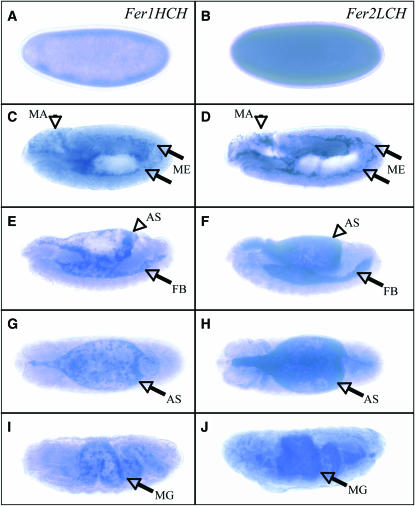
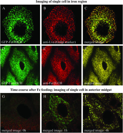
Ferritin is a symmetric, 24-subunit iron-storage complex assembled of H and L chains. It is found in bacteria, plants, and animals and in two classes of mutations in the human L-chain gene, resulting in hereditary hyperferritinemia cataract syndrome or in neuroferritinopathy. Here, we examined systemic and cellular ferritin regulation and trafficking in the model organism Drosophila melanogaster. We showed that ferritin H and L transcripts are coexpressed during embryogenesis and that both subunits are essential for embryonic development. Ferritin overexpression impaired the survival of iron-deprived flies. In vivo expression of GFP-tagged holoferritin confirmed that iron-loaded ferritin molecules traffic through the Golgi organelle and are secreted into hemolymph. A constant ratio of ferritin H and L subunits, secured via tight post-transcriptional regulation, is characteristic of the secreted ferritin in flies. Differential cellular expression, conserved post-transcriptional regulation via the iron regulatory element, and distinct subcellular localization of the ferritin subunits prior to the assembly of holoferritin are all important steps mediating iron homeostasis. Our study revealed both conserved features and insect-specific adaptations of ferritin nanocages and provides novel imaging possibilities for their in vivo characterization.


Figures

F<sc>igure</sc> 1.—
Figure 1.—
Comparison of Drosophila melanogaster Fer1HCH IRE to human L-chain ferritin IRE and to Fer1HCH IREs from different Drosophila species. The IRE-containing alternative exon of Fer1HCH was conserved in all sequenced Drosophila species. Secondary structures from evolutionary divergent species of the Drosophila genus are compared to the D. melanogaster prototype. Nonconserved nucleotides are red. D. melanogaster Fer1HCH IRE was also compared to human L-chain ferritin IRE. Circled nucleotides are mutated in human disease (Cazzola 2002; Rouault 2006). Nucleotides conserved from human to Drosophila are blue.
F<sc>igure</sc> 2.—
Figure 2.—
Characterization of Drosophila strains containing transposable elements in Fer1HCH/Fer2LCH. (A) Fer1HCH and Fer2LCH share the same genetic locus at polytene position 99F on the third chromosome. Open and shaded boxes represent UTRs and the ORFs, respectively. The genomic insertions of P elements are shown as triangles and described in the text. (B) Western blot analysis of whole fly extracts from wild-type flies or the heterozygous Fer1HCH451/+ and Fer2LCH35/+ flies. Antibodies used are indicated at the bottom of the images. Levels of both ferritin subunits are reduced irrespective of which gene is affected by the transposon insertion. (C) Transgene-dependent rescue of the embryonic lethality associated with Fer1HCH451/451 and Fer2LCH35/35 and Fer1HCHG188/451 demonstrates the specific functions provided by the two genes. UAS-Fer1HCH* has no ferroxidase activity and cannot support ferritin function in vivo. (D) Predicted transcripts generated from the chromosome bearing the Fer1HCHG188 P element are depicted, indicating that both IRE- and non-IRE-containing transcripts (Lind et al. 1998; Georgieva et al. 1999) will encode GFP-tagged versions of the Fer1HCH subunit. Note that the predicted cleavage site for generation of the mature polypeptide is contained in the second exon of the Fer1HCH gene, prior to the GFP insertion. (E) Protein extracts from wild-type and Fer1HCHG188/+ adult flies were subjected to reducing SDS–PAGE; Western blots were probed with Fer2LCH and Fer1HCH peptide antibodies. Fer2LCH levels remain unchanged between the two samples, whereas there is less Fer1HCH and a higher-molecular-weight species appearing in Fer1HCHG188/+, migrating at the predicted size for a GFP-Fer1HCH fusion protein. (F) Nonreducing SDS–PAGE separation of lysates from mouse liver and wild-type, Fer1HCH451/+, and Fer1HCHG188/+ Drosophila strains. Flies were allowed to feed for 24 hr on food containing 55FeCl3-NTA. (Left) Western blots; (right) autoradiograph of a portion of the gel run in parallel. All detectable iron in extracts from Fer1HCHG188/+ flies coincides with the ferritin heteropolymers specific to this strain. Higher-molecular-weight species bands most probably reflect different ratios of native and GFP-tagged Fer1HCH subunits within ferritin heteropolymers.
F<sc>igure</sc> 3.—
Figure 3.—
Phenotypes associated with ubiquitous overexpression of ferritin. To achieve ferritin overexpression, we crossed homozygous female flies for the indicated transgene to males from the Actin-Gal4/Cyo stock. The progeny carrying the Actin-Gal4 driver was selected. (A) Western blot analysis from whole fly extracts indicates that overexpression of the single-ferritin chains fails to alter their relative ratio. If a Fer1HCH or a Fer1HCH* subunit is overexpressed in parallel with Fer2LCH, significant overexpression is achieved. (B) To verify that transgenic message is expressed, RT–PCR was performed with primers specific to the UAS-derived transcripts. (C) Nonreducing SDS–PAGE separation of lysates from control, mild, and stronger ferritin overexpression strains, and the Fer1HCH*, Fer2LCH overexpression strain (genotypes are indicated in materials and methods). Flies were allowed to feed for 24 hr on food containing 55FeCl3-NTA. (Left) Western blot probed with anti-Fer2LCH. (Right) An autoradiograph of a portion of the gel run in parallel. Interestingly, similar amounts of total iron are contained in the different size pools of total ferritin. Ferritin containing a mixture of functional endogenous Fer1HCH and nonfunctional transgene-derived Fer1HCH* subunits is still able to bind iron. (D) Survival on 10 mm paraquat is enhanced in female, but not male flies overexpressing ferritin. The solid line represents the control +/+; Actin-Gal4/+flies, the three lightly shaded lines represent flies in which the single Fer1HCH, Fer2LCH, and Fer1HCH* are overexpressed (but not stabilized), while the two darkly shaded lines represent ferritin overexpressors (1H, 2L and 1H*, 2L). (E) Parental crosses were set up in vials containing 180 μm Bathophenanthroline disulfate to cause iron deficiency, regular food or 5 mm ferric ammonium citrate to cause iron overload. Male progeny of flies overexpressing ferritin (i.e., carrying the Actin-Gal4 driver) surviving to adulthood are compared to flies where ferritin overexpression is not induced from the transgene (i.e., carrying the Cyo balancer chromosome). The number of flies counted containing the Cyo balancer chromosome is indicated at the top of each bar. Different levels of ferritin overexpression were achieved by using independent recombinant chromosomes and their combination. Iron deficiency combined with ferritin overexpression can be lethal, whereas ferritin overexpression is well tolerated under conditions of iron overload.
F<sc>igure</sc> 4.—
Figure 4.—
Embryonic expression patterns of the two ferritin genes are similar. Detection of Fer1HCH and Fer2LCH transcripts by in situ hybridization of antisense RNAs to whole-mount preparations of embryos. Probe used for detection is indicated on top right of each column; the two genes are expressed in identical patterns. All images are oriented with anterior to the left. (A and B) High levels of staining in the blastoderm stage suggest that both transcripts are contributed from the mother to the embryo. (C and D) At germ-band elongation, transcripts accumulate in mesoderm (ME) and in the head region, where the macrophage lineage (MA) is specified. (E and F) During germ-band retraction, the fat bodies (FB) and amnioserosa (AS) are stained. (G and H) A dorsal view of the amnioserosa. (I and J) Midgut (MG) expression at late stages of embryogenesis.
F<sc>igure</sc> 5.—
Figure 5.—
Intestinal sites of constitutive and inducible ferritin expression differ. (A and B) Dissected larval midgut from the Fer1HCHG188/+ line. Fluorescence identifies the iron region in the middle midgut (arrows). (C and D) Dissected midgut from larvae of the same genotype raised on food containing 1 mm ferric ammonium citrate. Note that ferritin protein accumulates in the anterior midgut. (E–H) Prussian blue staining of midguts from control or iron-fed larvae: (E) anterior midgut, control diet; (F) iron region, control diet; (G) anterior midgut, iron diet; (H) iron region, iron diet. (I and J) Fer1HCHG188/+ larvae were raised on food containing 1 mm CuSO4 and midguts were imaged using UV excitation and the DAPI filter. (I) Cu-metallothionein fluorescence (orange, asterisks) is prominent in the posterior midgut. (J) Cu-metallothionein fluorescence is also found in a cluster of cells anterior to the iron region, where constitutive metallothionein expression has been reported (Mcnulty et al. 2001). (K and L) Time course of iron-dependent ferritin induction in (K) anterior midgut and (L) middle midgut dissected from third instar larvae. Extracts were subjected to electrophoresis under nonreducing conditions and analyzed by immunoblotting. Ferritin induction in the iron region is much less pronounced.
F<sc>igure</sc> 6.—
Figure 6.—
Ferritin resides in the Golgi apparatus of midgut cells. (A) Section of a single cell in the iron region of a dissected midgut from Fer1HCHG188/+ larvae imaged by confocal microscopy. Note the punctate subcellular localization, consistent with membrane-enclosed ferritin compartments seen by EM in midgut preparations from another insect species (Locke and Leung 1984). (B) The same cell was stained with anti-Lava lamp, a Golgi-associated protein. (C) Yellow identifies the Golgi complex as the primary location of ferritin in cells of the iron region. (D–F) A single cell expressing GFP-Fer1HCH in the iron region was stained with anti-Fer2LCH; colocalization of the ferritin chains was seen in the Golgi. (G–I) Time course of ferritin induction in single cells of the anterior midgut. Merged images of GFP-Fer1HCH in green and Fer2LCH in red are shown. (G) Prior to iron feeding, low levels of the ferritin subunits are expressed. (H) One hour postfeeding, GFP-Fer1HCH is in the endoplasmic reticulum, while Fer2LCH is detected only in Golgi bodies. (I) Four hours postfeeding, the two chains colocalize in the Golgi apparatus.

References

    1. Bauer, M., J. D. Katzenberger, A. C. Hamm, M. Bonaus, I. Zinke et al., 2006. Purine and folate metabolism as a potential target of sex-specific nutrient allocation in Drosophila and its implication for lifespan-reproduction tradeoff. Physiol. Genomics 25: 393–404. - PubMed
    1. Beutler, E., V. Felitti, N. J. Ho and T. Gelbart, 2002. Relationship of body iron stores to levels of serum ferritin, serum iron, unsaturated iron binding capacity and transferrin saturation in patients with iron storage disease. Acta Haematol. 107: 145–149. - PubMed
    1. Brand, A. H., and N. Perrimon, 1993. Targeted gene expression as a means of altering cell fates and generating dominant phenotypes. Development 118: 401–415. - PubMed
    1. Brody, T., C. Stivers, J. Nagle and W. F. Odenwald, 2002. Identification of novel Drosophila neural precursor genes using a differential embryonic head cDNA screen. Mech. Dev. 113: 41–59. - PubMed
    1. Capurro Mde, L., P. Iughetti, P. E. Ribolla and A. G. de Bianchi, 1996. Musca domestica hemolymph ferritin. Arch. Insect Biochem. Physiol. 32: 197–207. - PubMed

Publication types

MeSH terms