. 2007 Aug;176(4):2189-200.
doi: 10.1534/genetics.107.071449. Epub 2007 Jul 1.

Unraveling the genetic complexity of Drosophila stardust during photoreceptor morphogenesis and prevention of light-induced degeneration

Affiliations

Unraveling the genetic complexity of Drosophila stardust during photoreceptor morphogenesis and prevention of light-induced degeneration

Sandra Berger et al. Genetics. 2007 Aug.

Abstract

Drosophila Stardust, a membrane-associated guanylate kinase (MAGUK), recruits the transmembrane protein Crumbs and the cytoplasmic proteins DPATJ and DLin-7 into an apically localized protein scaffold. This evolutionarily conserved complex is required for epithelial cell polarity in Drosophila embryos and mammalian cells in culture. In addition, mutations in Drosophila crumbs and DPATJ impair morphogenesis of photoreceptor cells (PRCs) and result in light-dependent retinal degeneration. Here we show that stardust is a genetically complex locus. While all alleles tested perturb epithelial cell polarity in the embryo, only a subset of them affects morphogenesis of PRCs or induces light-dependent retinal degeneration. Alleles retaining particular postembryonic functions still express some Stardust protein in pupal and/or adult eyes. The phenotypic complexity is reflected by the expression of distinct splice variants at different developmental stages. All proteins expressed in the retina contain the PSD95, Discs Large, ZO-1 (PDZ), Src homology 3 (SH3), and guanylate kinase (GUK) domain, but lack a large region in the N terminus encoded by one exon. These results suggest that Stardust-based protein scaffolds are dynamic, which is not only mediated by multiple interaction partners, but in addition by various forms of the Stardust protein itself.


Figures

F<sc>igure</sc> 1.—
Figure 1.—
Structure of Stardust isoforms and their expression. (A) Four different Sdt isoforms have been identified in embryos: SdtA (also called Sdt-MAGUK1), Sdt-GUK1, and SdtB1 (Bachmann et al. 2001; Hong et al. 2001; Wang et al. 2004). SdtB2 is an isoform expressed in the adult head. Point mutations have been mapped in sdtM120 at position 32639, AAGCAACAT → AAGTAACAT (Q → stop); in sdtK85 at position 44683, CTGCAAA → CTGTAAA (Q → stop); and in sdtE195 at position 44881, ACGCAATC → ACGTAATC (Q → stop) (numbering according to FlyBase). Molecular defects in all alleles sequenced so far lead to the introduction of premature stop codons. (B) Schematic representation of the sdt splice variants discussed here and locations of primers used for RT–PCR (arrowheads). The colors correspond to those in A. Exons with numbers are according to Hong et al. (2001), novel exons are marked in uppercase letters, and untranslated 5′ and 3′ exons are in white. Sequences of primers are depicted in materials and methods. (C–F) Results from RT–PCR experiments using poly(A)+-RNA from adult heads (C–E, lane 2) or embryos (E, lane 1, and F). Primers used for amplification are indicated on top of each lane (for genomic positions see B). In F, developmental stages of embryos are indicated at the bottom of each lane (in hours of development at 25°). (C) Only variants encoding the complete MAGUK domain are expressed in the head. Amplification with the two primer pairs used leads in both cases to a fragment of ∼1.500 bp (arrows), indicating that no RNA variant encoding the GUK isoform is present in the head (which would result in a fragment of ∼300 bp). The larger bands (arrowheads) still contain the intron. (D) All RNA variants expressed in the adult head (arrows) lack exon 3, as revealed by amplification with two different primer pairs. Fragments containing exon 3 would be larger (exon 3 is 1.309 bp in length). (E) RNA encoding the SdtB variant SdtB2 can be detected only in RNA of adult heads, but not in embryos. (F) In the embryo, RNAs containing exon 3 are detectable only between 0 and 4 hr of development (larger band), but not in later stages. RNAs lacking exon 3 are present throughout embryonic development (smaller band). The sequence of all PCR products was determined. Bands marked by asterisks are nonspecific products.
F<sc>igure</sc> 2.—
Figure 2.—
Phenotypic defects in eye clones of different sdt alleles. (A–D′) Cross sections of ommatidia of white flies (A and A′) and flies carrying clones of sdtXP96 (B and B′), sdtN5 (C and C′), and sdtK85 (D and D′), kept for 7–14 days in the dark (A–D) or for 7 days in constant light (A′–D′). (E and F) Stalk membranes of w (E) and sdtN5 (F) photoreceptors, labeled in red. Those in sdtN5 are reduced in length. (G) The length of the stalk membranes in sdtK85 (n = 140) and sdtN5 (n = 224) is reduced by ∼25% compared to that of w flies (n = 210). (H and I) Cross sections of sdtN5 (H) and sdtK70 (I) eyes from flies raised on a medium depleted of vitamin A and kept for 7 days in constant light. The rhabdomeres are smaller due to reduced amounts of rhodopsin, but the mutant PRCs do not degenerate. Bar, 1 μm.
F<sc>igure</sc> 3.—
Figure 3.—
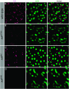
Expression of Sdt protein in pupal eyes of different sdt alleles: optical cross sections of pupal eyes at 40–45% PD, before stalk membrane formation. MARCM clones of different sdt alleles were stained with anti-Sdt-PDZ (red) and anti-DE-cadherin to stain the zonula adherens (blue). Mutant cells are marked by GFP (green). Depending on the level of the optical section, the green staining is sometimes weaker. Cells not expressing GFP are wild type and serve as a control to demonstrate that Sdt localizes apically in PRCs at this stage. In sdtXP96 mutant PRCs (A–A″), Sdt protein is no longer detectable apically, although it is still localized earlier (not shown). sdtN5 mutant PRCs (B–B″) express reduced amounts of Sdt protein, which is, however, still localized apically. No Sdt protein can be detected in sdtK85 mutant PRCs (C and C′).
F<sc>igure</sc> 4.—
Figure 4.—
Expression of Sdt protein in the adult eye of different sdt alleles. Optical cross sections of adult Drosophila eyes were stained with phalloidin to highlight the F-actin-rich rhabdomeres (green) and with anti-Sdt-PDZ (purple). (A) In wild-type (w) eyes, Sdt is restricted to the stalk membrane, adjacent to the rhabdomere. (B) In sdtXP96 eyes, Sdt protein is lost from the stalk membrane. Note the morphological defects in the rhabdomeres (compare the F-actin staining in A′ and B′). (C) In sdtN5 eyes, the shape of the rhabdomeres is normal and Sdt protein can be detected at the stalk membrane, albeit in reduced amounts. (D) sdtK85 eyes show morphological defects in the rhabdomeres and no Sdt protein can be detected.
F<sc>igure</sc> 5.—
Figure 5.—
Expression of Sdt protein and RNA. (A) Western blots of wild-type (w) protein extracts from different developmental stages and organs, probed with an antibody directed against the PDZ domain. (B) Northern blots of poly(A)+-RNA from wild-type (w) heads hybridized with a probe encoding the full-length MAGUK domain (including PDZ, SH3, and GUK domain) or the SH3 domain only. (C) Western blots of extracts from wild-type (w) and mutant eyes (four eyes/lane), probed with anti-Sdt-PDZ antibody. Eyes used for this experiment contained large mutant clones with hardly any wild-type ommatidia (∼1%), corresponding to ∼32 ommatia. (D) Western blot of different amounts of wild-type eyes, probed with anti-Sdt-PDZ antibody. A faint band of Sdt protein can be detected in one-quarter of an eye, which corresponds to ∼200 ommatidia.
F<sc>igure</sc> 6.—
Figure 6.—
Localization of DPATJ and DPar-6 in sdt mutant pupal photoreceptor cells. Optical cross sections of pupal eyes at 40–45% PD., before stalk membrane formation, are shown. MARCM clones of different sdt alleles were stained with anti-DPATJ (A) or anti DPar-6 (B) (red) and anti-DE-cadherin (C) to stain the zonula adherens (blue). Mutant cells are marked by GFP (green). Cells not expressing GFP are wild type and serve as a control.

References

    1. Aartsen, W. M., A. Kantardzhieva, J. Klooster, A. G. van Rossum, S. A. van de Pavert et al., 2006. Mpp4 recruits Psd95 and Veli3 towards the photoreceptor synapse. Hum. Mol. Genet. 15: 1291–1302. - PubMed
    1. Alloway, P. G., L. Howard and P. J. Dolph, 2000. The formation of stable rhodopsin-arrestin complexes induces apoptosis and photoreceptor cell degeneration. Neuron 28: 129–138. - PubMed
    1. Bachmann, A., M. Schneider, F. Grawe, E. Theilenberg and E. Knust, 2001. Drosophila Stardust is a partner of Crumbs in the control of epithelial cell polarity. Nature 414: 638–643. - PubMed
    1. Bachmann, A., M. Timmer, J. Sierralta, G. Pietrini, E. D. Gundelfinger et al., 2004. Cell type-specific recruitment of Drosophila Lin-7 to distinct MAGUK-based protein complexes defines novel roles for Sdt and Dlg-S97. J. Cell Sci. 117: 1899–1909. - PubMed
    1. Bilder, D., M. Schober and N. Perrimon, 2003. Integrated activity of PDZ protein complexes regulates epithelial polarity. Nat. Cell Biol. 5: 53–58. - PubMed

Publication types

MeSH terms

LinkOut - more resources