A comprehensive algorithm for anterior skull base reconstruction after oncological resections
- PMID: 17603642
- PMCID: PMC1852574
- DOI: 10.1055/s-2006-959333
A comprehensive algorithm for anterior skull base reconstruction after oncological resections
Abstract
Objective: To present our method for anterior skull base reconstruction after oncological resections.
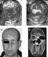
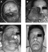
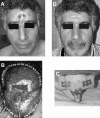
Methods: One hundred nine patients who had undergone 120 anterior skull base resections of tumors (52 malignant [43%], 68 benign [57%]) via the subcranial approach were studied. Limited dural defects were closed primarily or reconstructed using a temporalis fascia. Large anterior skull base defects were reconstructed by a double-layer fascia lata graft. A split calvarial bone graft, posterior frontal sinus wall, or three-dimensional titanium mesh were used when the tumor involved the frontal, nasal, or orbital bones. A temporalis muscle flap was used to cover the orbital socket for cases of eye globe exenteration, and a rectus abdominis free flap was used for subcranial-orbitomaxillary resection. Pericranial flap wrapping of the frontonaso-orbital segment was performed to prevent osteoradionecrosis if perioperative radiotherapy was planned.
Results: The incidence of cerebrospinal fluid (CSF) leak, intracranial infection, and tension pneumocephalus was 5%. Histopathological and immunohistochemical analysis of fascia lata grafts in reoperated patients (n = 7) revealed integration of vascularized fibrous tissue to the graft and local proliferation of a newly formed vascular layer embedding the fascial sheath.
Conclusion: A double-layer fascial graft alone was adequate for preventing CSF leak, meningitis, tension pneumocephalus, and brain herniation. We describe a simple and effective method of anterior skull base reconstruction after resections of both malignant and benign tumors.
Figures


References
-
- Neligan P C, Mulholland S, Irish J, et al. Flap selection in cranial base reconstruction. Plast Reconstr Surg. 1996;98:1159–1166. - PubMed
-
- Simpson D, Robson A. Recurrent subarachnoid bleeding in association with dural substitute. Report of three cases. J Neurosurg. 1984;60:408–409. - PubMed
-
- Clayman G L, DeMonte F, Jaffe D M, et al. Outcome and complications of extended cranial-base resection requiring microvascular free-tissue transfer. Arch Otolaryngol Head Neck Surg. 1995;121:1253–1257. - PubMed
-
- Califano J, Cordeiro P G, Disa J J, et al. Anterior cranial base reconstruction using free tissue transfer: changing trends. Head Neck. 2003;25:89–96. - PubMed
-
- Kiyokawa K, Tai Y, Inoue Y, et al. Efficacy of temporal musculopericranial flap for reconstruction of the anterior base of the skull. Scand J Plast Reconstr Surg Hand Surg. 2000;34:43–53. - PubMed

