. 2007 Sep 15;165(1):111-21.
doi: 10.1016/j.jneumeth.2007.05.024. Epub 2007 May 29.

Volumetric neuroimage analysis extensions for the MIPAV software package

Affiliations

Volumetric neuroimage analysis extensions for the MIPAV software package

Pierre-Louis Bazin et al. J Neurosci Methods. .

Abstract

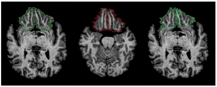
We describe a new collection of publicly available software tools for performing quantitative neuroimage analysis. The tools perform semi-automatic brain extraction, tissue classification, Talairach alignment, and atlas-based measurements within a user-friendly graphical environment. They are implemented as plug-ins for MIPAV, a freely available medical image processing software package from the National Institutes of Health. Because the plug-ins and MIPAV are implemented in Java, both can be utilized on nearly any operating system platform. In addition to the software plug-ins, we have also released a digital version of the Talairach atlas that can be used to perform regional volumetric analyses. Several studies are conducted applying the new tools to simulated and real neuroimaging data sets.


Figures

Fig. 1
Fig. 1
The MIPAV graphical user interface. Painting in a tri-planar view, volume rendering, and contour-based segmentation tools are a few of the capabilities offered by the software package.
Fig. 2
Fig. 2
The skull stripping plug-in: original image, interface, and stripped image mask overlaid on original image (from left to right).
Fig. 3
Fig. 3
The tissue classification plug-in (clockwise from top left): the original image, classified image, inhomogeneity field, dialog box, white matter membership, gray matter membership, CSF membership.
Fig. 4
Fig. 4
The Talairach alignment plug-in: (clockwise from top left) main dialog box, original image, AC-PC aligned image, image after full Talairach transformation, triplanar view for AC-PC alignment landmark selection, AC-PC alignment dialog box.
Fig. 5
Fig. 5
The Talairach atlas for MIPAV: (a) the volumetric images with the structure labels (b) the corresponding VOIs, over a Talairach aligned brain.
Fig. 6
Fig. 6
Volume measurements using the Talairach parcellation and the MIPAV statistics generator.
Fig. 7
Fig. 7
Plot of total brain volumes in cubic centimeters (cc) estimated using the semi-automated approach with our Brainstrip plug-in against the manual approach. The red line plots the result of a linear regression.
Fig. 8
Fig. 8
FANTASM and FAST applied to the Brainweb phantom: (a) phantom image with 3% noise and 20% inhomogeneity, (b) true classification, (c) FANTASM classification (d) FAST classification, (e) true gray matter partial volume, (f) FANTASM gray matter membership, (g) FAST gray matter partial volume, (h) FAST gray matter probability map.
Fig. 9
Fig. 9
Frontal lobe delineations performed manually (left), using Talairach labels (center), and using Talairach labels after transforming back into the original image space (right).
Fig. 10
Fig. 10
Plot of frontal lobe volumes measured using Talairach and manual protocols: (a) left hemisphere, (b) right hemisphere. The red line plots the result of a linear regression.

Similar articles

Cited by

References

    1. Andreasen NC, Rajarethinam R, Stephan Arndt TC, II, VWS, Flashman LA, O’Leary DS, dt JCE, Yuh WT. Automatic atlas-based volume estimation of human brain region s from mr images. Journal of Computer Assisted Tomography. 1996;20(1):98–106. - PubMed
    1. Aylward E, Augustine A, Li Q, Barta P, Pearlson G. Measurement of frontal lobe volume on magnetic resonance imaging scans. Psychiatry Research: Neuroimaging Section. 1997;71:23–30. - PubMed
    1. Barta P, Dhingra L, Royall R, Schwartz E. Improving stereological estimates for the volumes of structures identified in three-dimensional arrays of spatial data. Journal of Neuroscience Methods. 1997;75:111–118. - PubMed
    1. Bassett S, Yousem D, Cristinzio C, Kusevic I, Yassa M, Caffo B, Zeger S. Familial risk for Alzheimer’s disease alters fMRI activation patterns. Brain. 2006;129:1229–1239. - PMC - PubMed
    1. Bazin P-L, McAuliffe M, Gandler W, Pham DL. Free software tools for atlas-based volumetric neuroimage analysis. Proceedings of SPIE Medical Imaging 2005: Image Processing. 2005;5747:1824–1833.

Publication types

LinkOut - more resources