Review
. 2007 Jun;3(6):e78.
doi: 10.1371/journal.ppat.0030078.

Building the perfect parasite: cell division in apicomplexa

Affiliations
Review

Building the perfect parasite: cell division in apicomplexa

Boris Striepen et al. PLoS Pathog. 2007 Jun.

Abstract

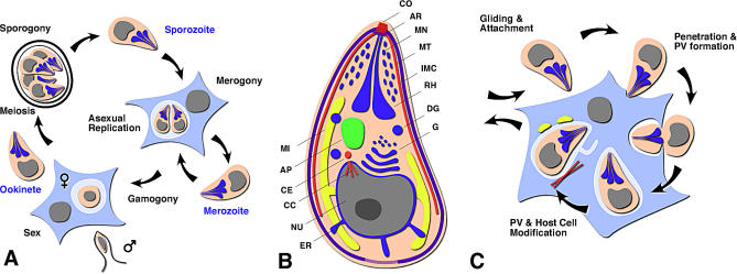
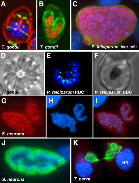
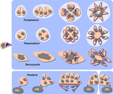
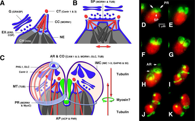
Apicomplexans are pathogens responsible for malaria, toxoplasmosis, and crytposporidiosis in humans, and a wide range of livestock diseases. These unicellular eukaryotes are stealthy invaders, sheltering from the immune response in the cells of their hosts, while at the same time tapping into these cells as source of nutrients. The complexity and beauty of the structures formed during their intracellular development have made apicomplexans the darling of electron microscopists. Dramatic technological progress over the last decade has transformed apicomplexans into respectable genetic model organisms. Extensive genomic resources are now available for many apicomplexan species. At the same time, parasite transfection has enabled researchers to test the function of specific genes through reverse and forward genetic approaches with increasing sophistication. Transfection also introduced the use of fluorescent reporters, opening the field to dynamic real time microscopic observation. Parasite cell biologists have used these tools to take a fresh look at a classic problem: how do apicomplexans build the perfect invasion machine, the zoite, and how is this process fine-tuned to fit the specific niche of each pathogen in this ancient and very diverse group? This work has unearthed a treasure trove of novel structures and mechanisms that are the focus of this review.


Conflict of interest statement

Competing interests. The authors have declared that no competing interests exist.

Figures

Figure 1
Figure 1. Apicomplexa Are Intracellular Parasites
(A) Highly simplified apicomplexan life cycle. Apicomplexans are haplonts, and meiosis (sporogony) immediately follows fertilization. Fertilization might occur within a host cell or extracellularly, giving rise to an oocyst or, less frequently, an invasive stage zygote (ookinete). (B) Schematic representation of a zoite (not all structures are present in all apicomplexans). AP, apicoplast; AR, apical rings; CC, centrocone; CE, centrosome; CO, conoid; DG, dense granule; ER, endoplasmic reticulum; G, Golgi; IMC, inner membrane complex; MI, mitochondrion; MN, microneme; MT, subpellicular microtubule; NU, nucleus; RH, rhoptry. (C) Zoites actively invade the cells of their hosts, establishing a specialized parasitophorous vacuole (PV) (in some species the parasite lyses the vacuole and develops freely in the cytoplasm).
Figure 2
Figure 2. The Diversity of Intracellular Development in Apicomplexans
(A) In T. gondii, two daughters are formed during budding. IMC1, red; MORN1, green (reproduced with permission from [32]). (B) T. gondii. Histone H2, red; IMC3, green (reproduced from [71]). (C) In Plasmodium falciparum liver schizont, budding results in massive numbers of zoites. Image courtesy of Volker Heussler. (D) T. gondii, phase contrast image of parasitophorous vacuole harboring multiple tachyzoites. (E and F) P. falciparum late erythrocyte schizont. Acyl carrier protein (plastid), green. RBC, red blood cell. (G–I) Sarcocystis neurona. Two intracellular stages with polyploid nuclei, one in interphase and one during mitosis. Tubulin, red. (J) S. neurona budding. IMC3, green. (K) A Theileria schizont divides in association with its host cell. Polymorphic immunodominant molecule (parasite surface), green; γ-tubulin (host centrosomes), red. HN, host nucleus. Image courtesy of Dirk Dobbelaere. The DNA dye DAPI is shown in blue throughout. Not to scale.
Figure 3
Figure 3. The Flexibility of Apicomplexan Cell Division
Schematic outline of cell division by Toxoplasma (endodyogeny), Plasmodium (schizogony), and Sarcocystis (endopolygeny). The Theileria schizont is divided in association with host cell division (HN, host nucleus). DNA, grey; IMC, purple; centrosome, red. Note that a centriole as center of the spindle plaque body has not been clearly demonstrated in P. falciparum. Both Sarcocystis and Theileria develop directly in the host cell cytoplasm, while Toxoplasma and Plasmodium are contained within a parasitophorous vacuole (light blue).
Figure 4
Figure 4. The Mechanics of Apicomplexan Mitosis and Budding
(A–C) Schematic representation of the nucleus during interphase (A), mitosis (B), and mid-stage budding (C). Smaller type abbreviations refer to organelle-specific marker proteins in T. gondii (most are available as fluorescent protein in vivo tags, see text for further details and references). AP, apicoplast; AR, apical rings; CC, centrocone; CH, chromosome; CO, conoid; CT, centromere; EX, ER exit site; MT, subpellicular microtubule; NE, nuclear envelope; PR, posterior ring; SP, spindle. (D–K) Time lapse series of nuclear division in T. gondii reproduced from [32]. The nucleus is labeled in red (Histone H2b-RFP) and MORN1 in green (MORN1-YFP).

References

    1. Sibley LD. Intracellular parasite invasion strategies. Science. 2004;304:248–253. - PubMed
    1. Dobrowolski JM, Sibley LD. Toxoplasma invasion of mammalian cells is powered by the actin cytoskeleton of the parasite. Cell. 1996;84:933–939. - PubMed
    1. Meissner M, Schluter D, Soldati D. Role of Toxoplasma gondii myosin A in powering parasite gliding and host cell invasion. Science. 2002;298:837–840. - PubMed
    1. Carruthers V, Boothroyd JC. Pulling together: An integrated model of Toxoplasma cell invasion. Curr Opin Microbiol. 2006;10:83–89. - PubMed
    1. Cowman AF, Crabb BS. Invasion of red blood cells by malaria parasites. Cell. 2006;124:755–766. - PubMed

Publication types