
Characterization of reemerging chikungunya virus

Marion Sourisseau et al. PLoS Pathog. 2007 Jun.

Abstract

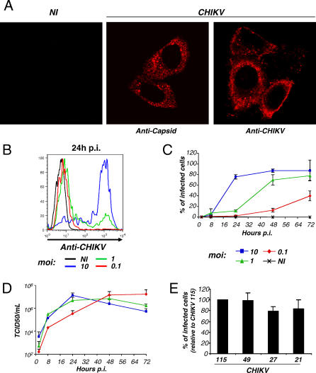
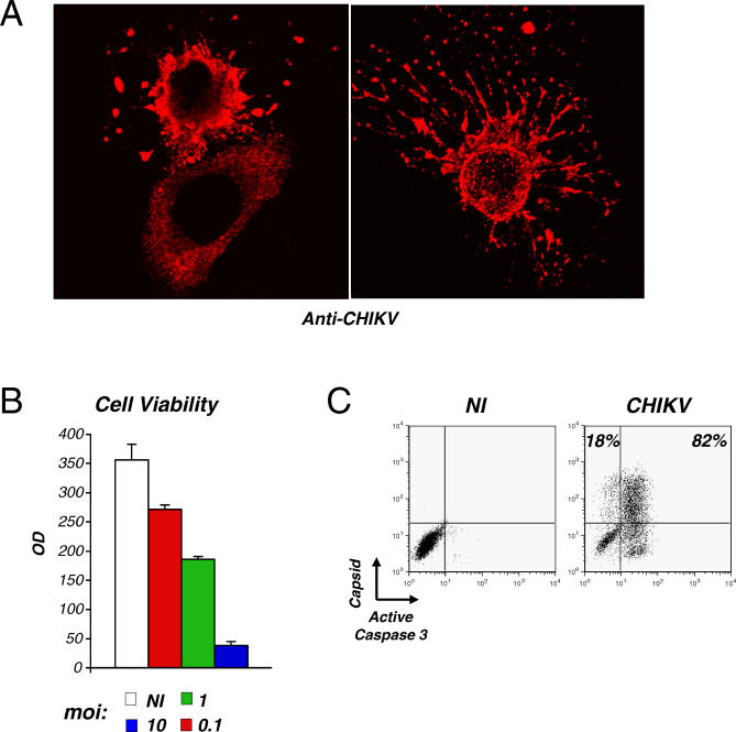
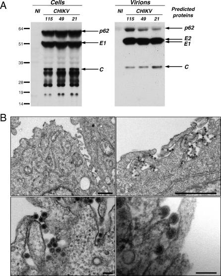
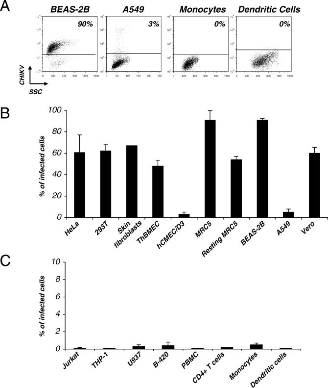
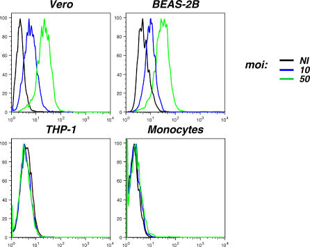
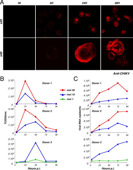
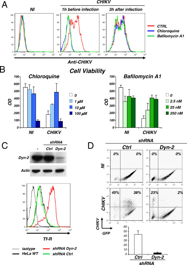
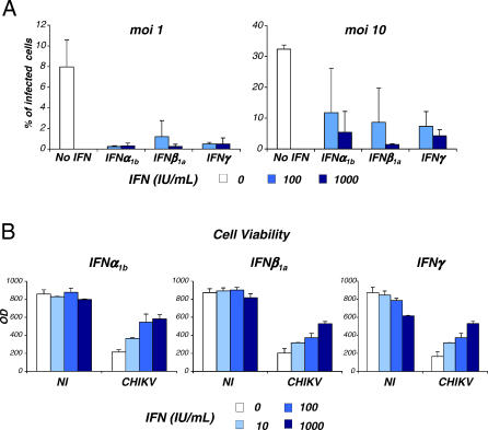
An unprecedented epidemic of chikungunya virus (CHIKV) infection recently started in countries of the Indian Ocean area, causing an acute and painful syndrome with strong fever, asthenia, skin rash, polyarthritis, and lethal cases of encephalitis. The basis for chikungunya disease and the tropism of CHIKV remain unknown. Here, we describe the replication characteristics of recent clinical CHIKV strains. Human epithelial and endothelial cells, primary fibroblasts and, to a lesser extent, monocyte-derived macrophages, were susceptible to infection and allowed viral production. In contrast, CHIKV did not replicate in lymphoid and monocytoid cell lines, primary lymphocytes and monocytes, or monocyte-derived dendritic cells. CHIKV replication was cytopathic and associated with an induction of apoptosis in infected cells. Chloroquine, bafilomycin-A1, and short hairpin RNAs against dynamin-2 inhibited viral production, indicating that viral entry occurs through pH-dependent endocytosis. CHIKV was highly sensitive to the antiviral activity of type I and II interferons. These results provide a general insight into the interaction between CHIKV and its mammalian host.


Conflict of interest statement

Competing interests. The authors have declared that no competing interests exist.

Figures

Figure 1
Figure 1. CHIKV Infection of HeLa Cells
(A) HeLa cells were exposed to CHIKV-115. At 24 h pi, cells were fixed and stained with an anti-alphavirus capsid mAb or with polyclonal anti-CHIKV antibodies, and analyzed by confocal microscopy. (B) Quantification of CHIKV-infected cells by flow cytometry. HeLa cells were exposed to CHIKV-115 at the indicated moi. At 24 h pi, cells were fixed, permeabilized, stained with anti-CHIKV antibodies, and analyzed by flow cytometry. Similar results were obtained with the anti-capsid mAb. (C) Kinetic analysis. HeLa cells were infected as described in (B) and analyzed at the indicated time points. Data are a compilation (with standard deviation [SD]) of three independent experiments. (D) Viral release in supernatants. Levels of infectious virions in supernatants were measured by limiting dilution on Vero cells and are expressed as TCID50/ml. Data are a compilation (with SD) of three independent experiments. (E) Comparison of the infectivity of four CHIKV strains. Mean ±SD of five independent experiments are depicted, with 100% corresponding to values obtained with CHIKV-115, at days 1 and 2 pi, with mois of 10 and 1. NI, noninfected cells.
Figure 2
Figure 2. CHIKV Is a Cytopathic Virus
(A) CHIKV-infected dying cells. At 24 h after CHIKV infection, HeLa cells were fixed, stained with anti-CHIKV antibodies, and analyzed by confocal microscopy. (B) Viability of cell cultures. HeLa cells were infected with CHIKV at the indicated moi. After 24 h pi, cell viability was measured in a colorimetric assay (MTT cell viability test). Data are mean ±SD of triplicates and representative of five independent experiments; (C) Apoptosis of CHIKV-infected cells. At 24 h pi (moi 10), HeLa cells were doubly stained with anti-active caspase-3 and anti-capsid antibodies and analyzed by flow cytometry. The percentage of caspase-3 positive and negative cells among CHIKV positive cells is depicted. Similar results were obtained with another marker of apoptosis (TUNEL, not shown). Data are representative of three independent experiments. NI, noninfected cells.
Figure 3
Figure 3. Assembly and Release of CHIKV
(A) Western blot analysis of CHIKV proteins. HeLa cells were exposed to the indicated CHIKV strains (moi 10). Cell lysates and pelleted supernatants were analyzed by Western blot 24 h pi, with a mix of anti-capsid mAb and anti-CHIKV antibodies. The predicted viral proteins are indicated on the right. (B) Gallery of electron micrographs of CHIKV-infected HeLa cells. Cells were analyzed at 36 h pi. CHIKV mostly buds at the plasma membrane of HeLa cells. Viral particles were not detected in noninfected cells (not shown). Bars represent 1 μm in the upper panels and 100 nm in the lower panels. NI, noninfected cells.
Figure 4
Figure 4. Cell Tropism of CHIKV
(A–C) The indicated human cell lines or primary cells were exposed to CHIKV at an moi of 10. At 24 h pi, cells were fixed, stained with anti-CHIKV antibodies, and analyzed by flow cytometry. The percentage of CHIKV-infected cells is indicated. Data are representative of at least three independent experiments. For primary cells, at least three different donors were analyzed. (A) Examples of sensitive and refractory cell types. (B) Sensitivity of adherent cells to CHIKV infection. (C) Sensitivity of primary blood-derived cells to CHIKV infection. The indicated cell lines, as well as nonactivated PBMCs, activated CD4+ lymphocytes, monocytes, and monocyte-derived DCs were analyzed.
Figure 5
Figure 5. Binding of CHIKV to Target Cells
The indicated human cell lines or primary cells were exposed to CHIKV at the indicated moi for 1 h at 4 °C. Cells were fixed, stained with anti-CHIKV antibodies, and analyzed by flow cytometry. Data are representative of three independent experiments. NI, noninfected cells.
Figure 6
Figure 6. CHIKV Productively Infects Human Primary Macrophages
(A–C) Human monocyte–derived macrophages were exposed to CHIKV for 4 h and extensively washed, and CHIKV replication was analyzed by different methods. Data are representative of at least four independent experiments, with cells from eight different donors. (A) CHIKV-infected macrophages. Cells were infected with CHIKV at an moi of 10. At the indicated time points, cells were stained with anti-CHIKV antibodies and analyzed by confocal microscopy. Two magnifications are depicted (objectives ×25 and ×40). NI, noninfected cells. (B) Release of infectious virus in supernatants. Macrophages were infected at various mois as stated. At the indicated time points, levels of infectious virions in supernatants were measured by limiting dilution on Vero cells. Results are expressed as TCID50/ml. Macrophages from three representative donors are depicted. (C) Viral RNA in supernatants. Levels of viral RNA in supernatants from the same experiment depicted in (B) were measured by real-time PCR.
Figure 7
Figure 7. CHIKV Entry Pathway
(A and B) CHIKV infection requires a low endosomal pH for entry. HeLa cells were pretreated 1 h before infection with chloroquine (10 μM) or bafilomycin-A1 (25 nM) and exposed to CHIKV (moi 10), or treated with the two drugs 3 h after viral exposure. (A) Inhibition of CHIKV replication by chloroquine and bafilomycin-A1. At 24 h pi, HeLa cells were stained with mouse anti-CHIKV antibodies and analyzed by flow cytometry. Ctrl, control. (B) Inhibition of CHIKV CPE by chloroquine and bafilomycin-A1. Cell viability was measured in a colorimetric assay (MTT cell viability test) 24 h pi. (C) Downregulation of Dyn-2 expression by shRNAs. HeLa cells were transduced with a lentiviral vector expressing GFP and a shRNA against Dyn-2 or an unrelated protein as a control (Ctrl). Upper panel: The Western blot shows the downregulation of Dyn-2 expression. Lower panel: surface levels of transferrin receptor (Tf-R) were upregulated in the absence of Dyn-2. Flow cytometry analysis was gated on transduced GFP+ cells. (D) Dyn-2 is required for CHIKV replication. HeLa cells lacking Dyn-2 are resistant to CHIKV. Infected HeLa cells (moi 10, 24 h pi) were stained with anti-CHIKV antibodies and analyzed by flow cytometry. The percentage of CHIKV+ cells among GFP+ and GFP− cells is depicted. One representative experiment out of three is shown in the upper panel. A mean ±SD of four independent experiments is shown in the lower panel. NI, noninfected cells.
Figure 8
Figure 8. IFNs Inhibit CHIKV Infection
HeLa cells were pretreated 6 h before infection with the indicated doses (IU/mL) of IFN. IFNα1b , IFNβ1a, and IFNγ were tested. Cells were then exposed to CHIKV at two mois (10 and 1). (A) Inhibition of CHIKV replication. At 24 h pi, HeLa cells were stained with anti-CHIKV antibodies and analyzed by flow cytometry. (B) Inhibition of CHIKV CPE. Cell viability was measured in a colorimetric assay (MTT cell viability test) 24 h pi (moi of 10). Data are representative of three independent experiments.

Similar articles

Cited by

References

    1. Strauss E, Strauss J. Structure and replication of the alphavirus genome. New York: Plenum Press; 1986. pp. 35–90.
    1. Peters C, Dalrymple J. Alphaviruses. In: Fields BN, Knipe DM, editors. Fields virology. 2nd edition. New York: Raven Press; 1990. pp. 713–761.
    1. Bodenmann P, Genton B. Chikungunya: An epidemic in real time. Lancet. 2006;368:258. - PubMed
    1. Pialoux G, Gauzere BA, Jaureguiberry S, Strobel M. Chikungunya, an epidemic arbovirosis. Lancet Infect Dis. 2007;7:319–327. - PubMed
    1. Schuffenecker I, Iteman I, Michault A, Murri S, Frangeul L, et al. Genome microevolution of chikungunya viruses causing the Indian Ocean outbreak. PLoS Med. 2006;3:e263. doi: 10.1371/journal.pmed.0030263. - DOI - PMC - PubMed

Publication types

MeSH terms