. 2007 Dec;15(12):1367-77.
doi: 10.1016/j.joca.2007.04.011. Epub 2007 Jun 29.

Modulation of TGF-beta signaling by proinflammatory cytokines in articular chondrocytes

Affiliations

Modulation of TGF-beta signaling by proinflammatory cytokines in articular chondrocytes

J A Roman-Blas et al. Osteoarthritis Cartilage. 2007 Dec.

Abstract

Objective: The normal structure and function of articular cartilage are the result of a precisely balanced interaction between anabolic and catabolic processes. The transforming growth factor-beta (TGF-beta) family of growth factors generally exerts an anabolic or repair response; in contrast, proinflammatory cytokines such as interleukin 1 beta (IL-1beta) and tumor necrosis factor-alpha (TNF-alpha) exert a strong catabolic effect. Recent evidence has shown that IL-1beta, and TNF-alpha, and the TGF-beta signaling pathways share an antagonistic relationship. The aim of this study was to determine whether the modulation of the response of articular chondrocytes to TGF-beta by IL-1beta or TNF-alpha signaling pathways occurs through regulation of activity and availability of mothers against DPP (Drosophila) human homologue (Smad) proteins.

Methods: Human articular chondrocytes isolated from knee joints from patients with osteoarthritis (OA) or normal bovine chondrocytes were cultured in suspension in poly-(2-hydroxyethyl methacrylate)-coated dishes with either 10% fetal bovine serum media or serum-deprived media 6h before treatment with IL-1beta alone, TNF-alpha alone or IL-1beta followed by TGF-beta. Nuclear extracts were examined by electrophoretic mobility-shift assays (EMSA) for nuclear factor-kappa B (NF-kappaB) and Smad3/4 deoxyribonucleic acid (DNA) binding. Nuclear extracts were also subjected to the TranSignal Protein/DNA array (Panomics, Redwood City, CA) enabling the simultaneous semiquantitative assessment of DNA-binding activity of 54 different transcription factors. Nuclear phospho-Smad2/3 and total Smad7 protein expression in whole cell lysates were studied by Western blot. Cytoplasmic Smad7, type II collagen alpha 1 (COL2A1), aggrecan and SRY-related high mobility group-Box gene 9 (SOX-9) mRNA expression were measured by real-time polymerase chain reaction (PCR).

Results: The DNA-binding activity of Smad3/4 in the TranSignal Protein/DNA array was downregulated by TNF-alpha (46%) or IL-1beta treatment (42%). EMSA analysis showed a consistent reduction in Smad3/4 DNA-binding activity in human articular chondrocytes treated with IL-1beta or TNF-alpha. TGF-beta-induced Smad3/4 DNA-binding activity and Smad2/3 phosphorylation were also reduced following pretreatment with IL-1beta in human OA and bovine chondrocytes. Real-time PCR and Western blot analysis showed that IL-1beta partially reversed the TGF-beta stimulation of Smad7 mRNA and protein levels in TGF-beta-treated human OA cells. In contrast, TGF-beta-stimulated COL2A1, aggrecan, and SOX-9 mRNA levels were abrogated by IL-1beta.

Conclusions: IL-1beta or TNF-alpha exerted a suppressive effect on Smad3/4 DNA-binding activity in human articular chondrocytes, as well as on TGF-beta-induced stimulation of Smad3/4 DNA-binding activity and Smad2/3 phosphorylation in human OA and bovine articular chondrocytes. IL-1beta partially reversed the increase in TGF-beta-stimulated Smad7 mRNA or protein levels suggesting that Smad7 may not be involved in the suppression of TGF-beta signaling induced by IL-1beta or TNF-alpha in articular chondrocytes. The balance between the IL-1beta or TNF-alpha and the TGF-beta signaling pathways is crucial for maintenance of articular cartilage homeostasis and its disruption likely plays a substantial role in the pathogenesis of OA.


Figures

Figure 1
Figure 1
Diagram of the experimental design for the treatment of human OA and bovine articular chondrocytes with either TGF-β IL-1β or both. All bovine articular chondrocytes were cultured in the absence of FBS for 6 h before addition of either IL-1β or TNF-α, whereas, human OA chondrocytes were cultured in 10% FBS media.
Figure 2
Figure 2
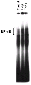
Activation of NF-κB in human OA chondrocytes. Nuclear extracts from human OA articular chondrocytes of 3 patients were subjected to EMSA with consensus NF-κB double stranded oligonucleotide probes. A representative example of one of the samples is shown. Note the clear increase in NF-κB DNA binding-activity in the cells treated with either IL-1β or TNF-α compared with control cells.
Figure 3
Figure 3
Protein (transcription factor)/DNA array. Representative images of TranSignal™ Protein/DNA Array I blots containing 54 different transcription factor DNA-binding sites obtained from control nuclear extracts or from nuclear extracts from one of the three nuclear extracts examined from human OA chondrocytes treated with TNF-α or IL-1β.The transcription factors that were down regulated in all 3 samples are shown in boxes. The DNAs were spotted in duplicate in two rows (top: undiluted; bottom: dilution 1/10). Biotinylated DNA was spotted for alignment along the right and bottom sides of the array.
Figure 4
Figure 4
EMSA showing a reduction in Smad3/4 DNA binding-activity in human OA chondrocytes treated with either IL-1β or TNF-α compared with control cells .
Figure 5
Figure 5
Reduction of TGF-β induced Smad3/4 DNA-binding activity by IL-β. A. Nuclear extracts from bovine articular chondrocytes were examined by EMSA with a probe containing a consensus site for NF-κB, showing a marked increase in the NF-κB DNA binding-activity in the cells treated with IL-1β alone and in the cells pretreated with IL-1β followed by TGF-β stimulation compared with control cells. Note that TGF-β alone had only minimal effect. NP: no nuclear protein. M: mutant probe. B. The same nuclear extracts were subjected to EMSA with a probe containing a consensus site for Smad3/4. The EMSA shows that treatment with TGF-β caused induction in Smad3/4 DNA binding activity whereas pretreatment with IL-1β reduced the TGF-β induced Smad3/4 DNA-binding activity.
Figure 6
Figure 6
Reduction of TGF-β induced Smad3/4 phosphorylation by IL-β. Human OA and bovine chondroyctes were treated with either TGF-β for 30 min, or pretreated before TGF-β treatment with IL-1β for 30 min, or treated with IL-1β alone for 60 min. Nuclear extracts were then prepared and subjected to Western analysis using anti phospho-Smad2/3 antibodies. Western analysis shows that the levels of phospho-Smad2/3 are significantly reduced in IL-1β pretreated chondrocytes vs TGF-β treatment alone. β-actin protein levels are shown as a control for protein loading and transfer. The data shown are representative of 2 independent experiments.
Figure 7
Figure 7
Effect of IL-1β on Smad7 mRNA or Smad7 protein levels in TGF-β treated cells A. Smad7 mRNA levels in human OA chondrocytes and bovine articular chondrocytes examined by real time-PCR. Note that Smad7 mRNA levels were increased by TGF-β treatment and pretreatment with IL-1β followed by TGF-β treatment partially reversed the Smad7 mRNA levels. COL2A1, aggrecan, and SOX-9 mRNA levels increased with TGF-β treatment, however, pretreatment with IL-1β abolished or significantly reduced this TGF-β-induced increase. GAPDH mRNA levels were used as internal controls. Three independent experiments were carried out. Student’s t analysis was performed to asess the statistical differences between groups .Values of p < 0.05 were considered significant. B. Human OA and bovine articular chondrocytes were treated with either TGF-β for 30 min or pre-treated with IL-1β for 30 min, or treated with IL-1β alone for 60 min. Whole cell lysates were then prepared and subjected to Western analysis using anti Smad7 antibodies. Western analysis shows that the Smad7 protein levels were only mildly affected by TGF-β or by IL-1β β-actin protein levels are shown as a control for protein loading and transfer.
Figure 7
Figure 7
Effect of IL-1β on Smad7 mRNA or Smad7 protein levels in TGF-β treated cells A. Smad7 mRNA levels in human OA chondrocytes and bovine articular chondrocytes examined by real time-PCR. Note that Smad7 mRNA levels were increased by TGF-β treatment and pretreatment with IL-1β followed by TGF-β treatment partially reversed the Smad7 mRNA levels. COL2A1, aggrecan, and SOX-9 mRNA levels increased with TGF-β treatment, however, pretreatment with IL-1β abolished or significantly reduced this TGF-β-induced increase. GAPDH mRNA levels were used as internal controls. Three independent experiments were carried out. Student’s t analysis was performed to asess the statistical differences between groups .Values of p < 0.05 were considered significant. B. Human OA and bovine articular chondrocytes were treated with either TGF-β for 30 min or pre-treated with IL-1β for 30 min, or treated with IL-1β alone for 60 min. Whole cell lysates were then prepared and subjected to Western analysis using anti Smad7 antibodies. Western analysis shows that the Smad7 protein levels were only mildly affected by TGF-β or by IL-1β β-actin protein levels are shown as a control for protein loading and transfer.
Figure 8
Figure 8
A. Diagram showing the potential sites of modulation of the TGF-β signaling pathway by IL-1β or TNF-α in articular chondrocytes. Modulation may occur through mechanisms other than NF-κB/Smad7 activation including: 1 and 2. Enhancement of ubiquitination and/or proteosome-mediated degradation of Smad proteins and type I TGF-β receptors. Smad proteins can be targeted for ubiquitination at different points in the TGF-β signaling pathway; in the cytoplasm by Smurf E3 ligases, or in the nucleus by SCF class of E3 ubiquitin ligase complexes. Type I TGF-β receptors can be ubiquitinated by Smurf E3 ligases interacting with I-Smads. Further degradation by the 26S proteosome follows the ubiquitination process (–20,30,31). 3. Interfering the interactions between Smad-activated TGF-β target genes and other transcription factors, co-activators and co-repressors. Transcription factors that interact with Smads include the JNK-dependent activated AP-1 (c-Jun) (27,28), as well as, co-repressors such as cSki/SnoN, c-Myc, Evi1, ATF3, TGIF, SNIP1, SIP1 and Tob (15,16). 4. Inhibitory effect upon Smad 3 phosphorylation. B. Postulated mechanisms responsible for inhibition of Smad pshosphorylation. TGF-β activated kinase 1 (TAK1) can directly inhibit Smad3 phosphorylation or can activate the JNK pathway leading to the formation of a cJun-Smad3 complex not compatible with Smad3-DNA interaction (48,53).

Similar articles

Cited by

References

    1. Grimaud E, Heymann D, Redini F. Recent advances in TGF-β effects on chondrocyte metabolism. Potential therapeutic roles of TGF-β in cartilage disorders. Cytokine Growth Factor Rev. 2002;13:241– 257. - PubMed
    1. Johnstone B, Hering TM, Caplan AI, Goldberg VM, Yoo JU. In vitro chondrogenesis of bone marrow-derived mesenchymal progenitor cells. Exp Cell Res. 1998;238:265– 272. - PubMed
    1. Tuli R, Tuli S, Nandi S, Huang X, Manner PA, Hozack WJ, Danielson KG, Hall DJ, Tuan RS. TGF-β 1-mediated chondrogenesis of human mesenchymal progenitor cells involves N-cadherin and MAP kinase and Wnt signaling crosstalk. J Biol Chem. 2003;278:41227– 41236. - PubMed
    1. Ballock RT, Heydemann A, Wakefield LM, Flanders KC, Roberts AB, Sporn MB. TGF-β 1 prevents hypertrophy of epiphyseal chondrocytes: regulation of gene expression for cartilage matrix proteins and metalloproteases. Dev Biol. 1993;158:414– 429. - PubMed
    1. Bohme K, Winterhalter KH, Bruckner P. Terminal differentiation of chondrocytes in culture is a spontaneous process and is arrested by transforming growth factor-β 2 and basic fibroblast growth factor in synergy. Exp Cell Res. 1995;216:191–198. - PubMed

Publication types

MeSH terms