
A mammalian microRNA expression atlas based on small RNA library sequencing

Pablo Landgraf et al. Cell. .

Abstract

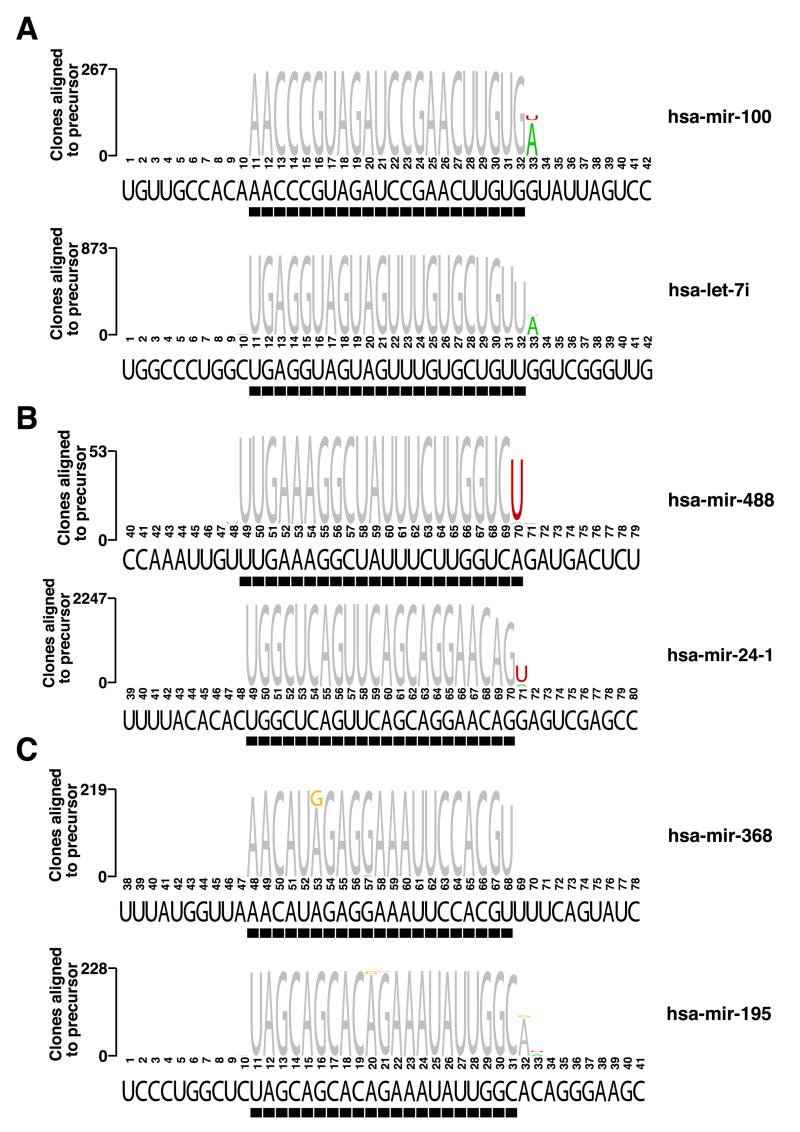
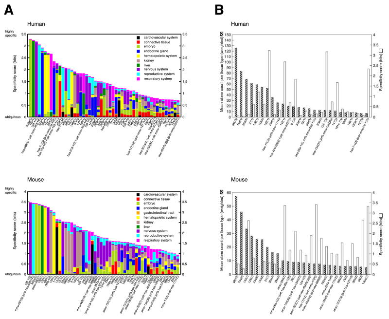
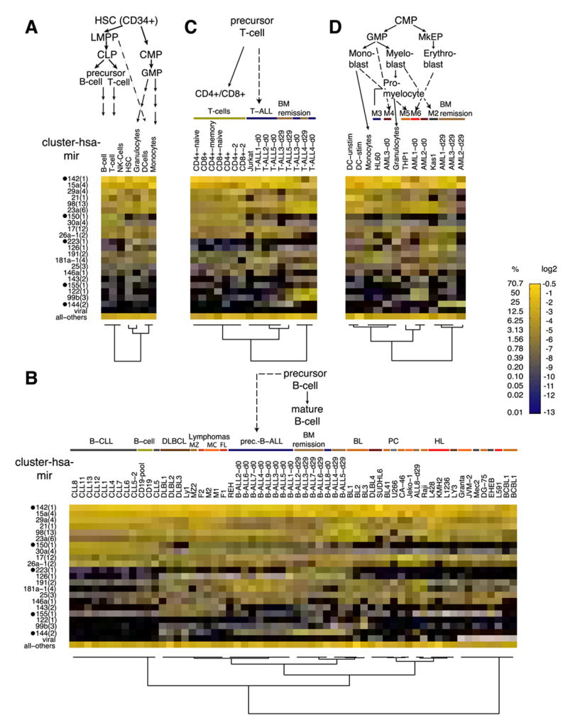
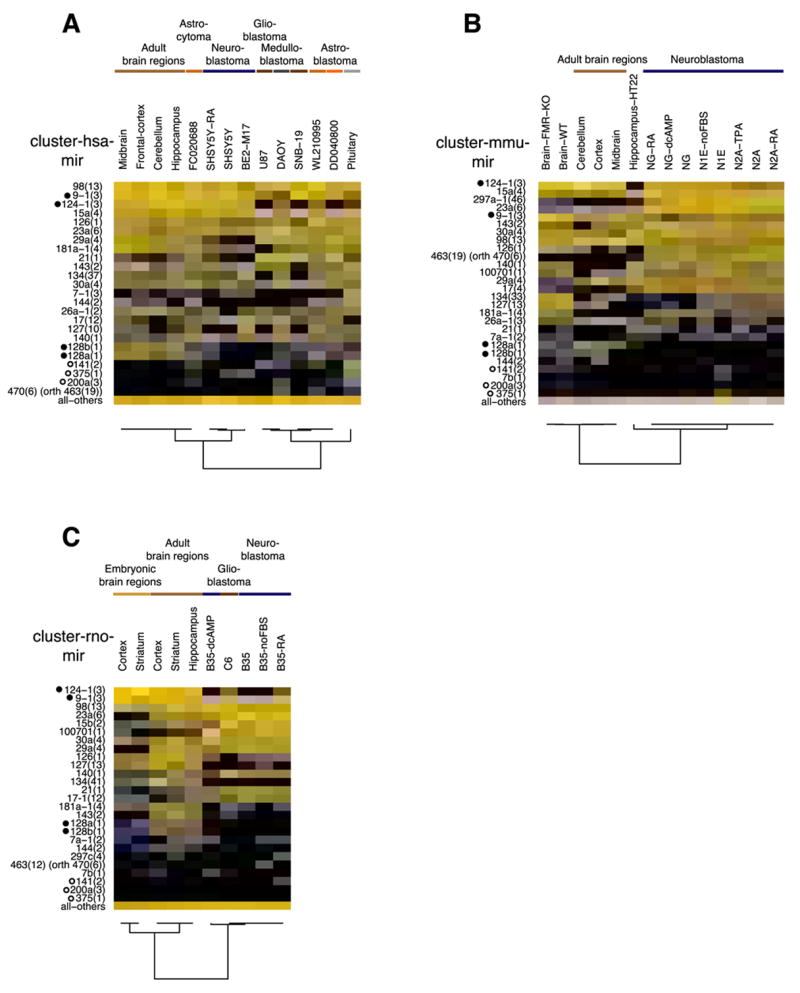
MicroRNAs (miRNAs) are small noncoding regulatory RNAs that reduce stability and/or translation of fully or partially sequence-complementary target mRNAs. In order to identify miRNAs and to assess their expression patterns, we sequenced over 250 small RNA libraries from 26 different organ systems and cell types of human and rodents that were enriched in neuronal as well as normal and malignant hematopoietic cells and tissues. We present expression profiles derived from clone count data and provide computational tools for their analysis. Unexpectedly, a relatively small set of miRNAs, many of which are ubiquitously expressed, account for most of the differences in miRNA profiles between cell lineages and tissues. This broad survey also provides detailed and accurate information about mature sequences, precursors, genome locations, maturation processes, inferred transcriptional units, and conservation patterns. We also propose a subclassification scheme for miRNAs for assisting future experimental and computational functional analyses.


Figures

Figure 1
Figure 1. Nucleotide 3′ additions and editing events in mature miRNA sequences
Examples for 3′ A additions (A), U additions (B), and A to G nucleotide exchanges due to possible deamination events (C) are given. Nucleotides of sequenced miRNAs that do not match their genomic sequence are colored. The black bar underneath each sequence logo indicates the predominantly cloned sequence. A list of all modifications and their occurrence in the different libraries is given in Tables S24 and S25.
Figure 2
Figure 2. Tissue specificity of miRNA expression
(A) The 51 most specific miRNA precursor clusters for human and mouse are depicted. The total height of each bar represents the information content reflecting tissue specificity, while the relative heights for each of the tissues are proportional to the clones of a miRNA precursor cluster in a given tissue type relative to all tissue types. The enrichment of miR-124 in the endocrine tissue in mouse is due to high expression of this miRNA in the MIN-6 beta-cell line, but miR-124 is not expressed in primary β-cells residing in mouse or human islets. (B) The 26 most expressed miRNA precursor clusters for human and mouse are depicted. The mean clone count for each tissue type (striped bars) and the tissue specificity (white bars) are indicated. The most abundant miRNA clusters are ubiquitously expressed and show little tissue specificity.
Figure 3
Figure 3. miRNA expression in hematopoietic lineage differentiation
miRNA precursor clusters with at least 5% relative cloning frequency in at least one of the samples are depicted individually in a color code. All other miRNAs and viral miRNAs were pooled and are displayed together. Undetected miRNAs are displayed in black. Hematopoietic-specific miRNA clusters are marked by black dots. B-, T- and dendritic cell (DC) samples were combined and their composite profiles are shown in panel (A) while panel B, C, and D, respectively, show the individual samples. The lineage is indicated above each figure. Different disease entities and normal cells are indicated with color bars. Patient-derived samples are labeled with the disease name and patient number, while for cell lines the respective names are given. The FAB classification is given for the AML samples and the progenitor cells are indicated. Abbreviations: LMPP, lymphoid primed multipotent progenitor; CLP, common lymphoid progenitor; CMP, common myeloid progenitor; GMP, granulocyte/macrophage progenitor; MkEP, megakaryocyte/erythrocyte progenitor; BM remission, bone marrow in remission; DLBCL, diffuse large B-cell lymphoma; MZ, marginal zone; MC, mantle cell; FL, follicular lymphoma; BL, Burkitt’s lymphoma; PC, plasmocytoma; HL, Hodgkin lymphoma.
Figure 4
Figure 4. miRNA expression in the nervous system
Expression profiles of miRNA precursor clusters with at least 5% relative expression in one of the samples as well as the neuronal-specific miRNA clusters (marked with black dot) are depicted individually for human (A), mouse (B) and rat (C) brain sections, brain tumor samples (FC0020688, WL210995, DD040800) and cell lines as indicated (color code of expression values see Figure 3). The open circles indicate miRNAs enriched in endocrine tissue including the pituitary gland. All panels show orthologous miRNA precursor clusters (if present). Abbreviations: dcAMP, dibutyryl cAMP; FBS, fetal bovine serum; TPA, trans-phorbol ester; RA, retinoic acid; orth, indicates that the hsa-mir-470 cluster is orthologous to the rodent mir-463 clusters.
Figure 5
Figure 5. miRNA expression in cell differentiation experiments
Cell lines were treated and differentiated into neurons (A) and osteoblasts (B). (C) A thyroid stimulating hormone (TSH)-dependent rat cell line FRTL5 was dedifferentiated by induction of a chimeric RAS protein. Expression profiles for orthologous miRNA precursor clusters with 5% or more relative cloning frequency in at least one sample are depicted in a color-coded manner (see Figure 3 for details). Abbreviations: dcAMP, dibutyryl cAMP; FBS, fetal bovine serum; TPA, trans-phorbol ester; RA, retinoic acid; USSC, human unrestricted somatic stem cell; orth, indicates that the hsa-mir-470 cluster is orthologous to the rodent mir-463 clusters.

Comment in

  • Mustering the micromanagers.
    Lim LP, Linsley PS. Lim LP, et al. Nat Biotechnol. 2007 Sep;25(9):996-7. doi: 10.1038/nbt0907-996. Nat Biotechnol. 2007. PMID: 17846629 No abstract available.

References

    1. Ambros V, Bartel B, Bartel DP, Burge CB, Carrington JC, Chen X, Dreyfuss G, Eddy SR, Griffiths-Jones S, Marshall M, et al. A uniform system for microRNA annotation. RNA. 2003;9:277–279. - PMC - PubMed
    1. Aravin A, Tuschl T. Identification and characterization of small RNAs involved in RNA silencing. FEBS Lett. 2005;579:5830–5840. - PubMed
    1. Bartel DP. MicroRNAs: genomics, biogenesis, mechanism, and function. Cell. 2004;116:281–297. - PubMed
    1. Baskerville S, Bartel DP. Microarray profiling of microRNAs reveals frequent coexpression with neighboring miRNAs and host genes. RNA. 2005;11:241–247. - PMC - PubMed
    1. Bentwich I, Avniel A, Karov Y, Aharonov R, Gilad S, Barad O, Barzilai A, Einat P, Einav U, Meiri E, et al. Identification of hundreds of conserved and nonconserved human microRNAs. Nat Genet. 2005;37:766–770. - PubMed

Publication types