. 2009 Jan;30(1):103-11.
doi: 10.1016/j.neurobiolaging.2007.05.014. Epub 2007 Jul 2.

Atrophy progression in semantic dementia with asymmetric temporal involvement: a tensor-based morphometry study

Affiliations

Atrophy progression in semantic dementia with asymmetric temporal involvement: a tensor-based morphometry study

S M Brambati et al. Neurobiol Aging. 2009 Jan.

Abstract

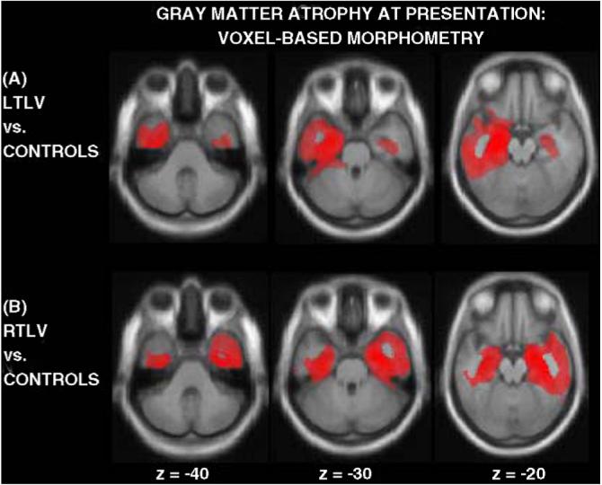
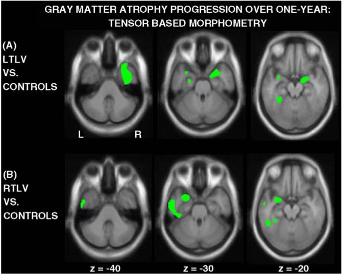
We performed a longitudinal anatomical study to map the progression of gray matter atrophy in anatomically defined predominantly left (LTLV) and right (RTLV) temporal lobe variants of semantic dementia (SD). T1-weighted MRI scans were obtained at presentation and one-year follow-up from 13 LTLV, 6 RTLV, and 25 control subjects. Tensor-based morphometry (TBM) in SPM2 was applied to derive a voxel-wise estimation of regional tissue loss over time from the deformation field required to warp the follow-up scan to the presentation scan in each subject. When compared to controls, both LTLV and RTLV showed significant progression of gray matter atrophy not only within the temporal lobe most affected at presentation, but also in the controlateral temporal regions (p<0.05 FWE corrected). In LTLV, significant progression of volume loss also involved the ventromedial frontal and the left anterior insular regions. These results identified the anatomic substrates of the previously reported clinical evolution of LTLV and RTLV into a unique 'merged' clinical syndrome characterized by semantic and behavioral deficits and bilateral temporal atrophy.


Figures

Fig. 1
Fig. 1
Example of main TBM pre-processing steps in a LTLV patient. (A) Presentation scan; (B) 1-year follow-up scan coregistered with the presentation scan; (C) Jacobian determinant map representing voxels of tissue expansion (voxel Jacobian value greater than 1) or contraction (voxel Jacobian value less than 1) from follow-up to presentation scan. Jacobian determinants were estimated by warping together the images A and B; (D) image representing gray matter volume changes between scans A and B. It was obtained by multiplying gray matter segment of the presentation scan with (Jacobian minus 1) values; (E) image D after normalization and smoothing process.
Fig. 2
Fig. 2
Whole-brain VBM analysis showing patterns of temporal gray matter atrophy in LTLV (A) and RTLV (B) patients compared with controls. Statistical maps are displayed on sections of the study-specific template used for normalization. The threshold for display is p < 0.05 corrected for multiple comparisons (SPM family-wise error—FWE). For display purpose, the result maps were multiplied by a binary inclusive mask derived by the GM a priori image.
Fig. 3
Fig. 3
Whole-brain TBM analysis showing patterns of GM atrophy progression in LTLV patients versus controls (A) and RTLV patients versus controls (B) over 1 year after diagnosis. Statistical maps are displayed on sections of the study-specific template used for normalization. The threshold for display is p < 0.05 corrected for multiple comparisons (SPM family-wise error—FWE).

Similar articles

Cited by

References

    1. Ashburner J, Andersson JL, Friston KJ. Image registration using a symmetric prior—in three dimensions. Hum. Brain Mapp. 2000;9(4):212–225. - PMC - PubMed
    1. Ashburner J, Friston KJ. Image segmentation. In: Frackowiack KJFRSJ, Frith C, Dolan R, Friston KJ, Price CJ, Zeki S, Ashburner J, Penny WD, editors. Human Brain Function. Academic Press; 2003.
    1. Ashburner J, Friston KJ. Nonlinear spatial normalization using basis functions. Hum. Brain Mapp. 1999;7(4):254–266. - PMC - PubMed
    1. Avants B, Grossman M, Gee JC. The correlation of cognitive decline with frontotemporal dementia induced annualized gray matter loss using diffeomorphic morphometry. Alzheimer Dis. Assoc. Disord. 2005;19(Suppl 1):S25–S28. - PubMed
    1. Brambati SM, Renda NC, Rankin KP, Rosen H, Seeley WW, Ashburner J, Miller BL, Gorno-Tempini ML. A tensor based morphometry study of longitudinal gray matter contraction in FTD. NeuroImage. 2007;35(3):998–1003. - PMC - PubMed

Publication types