Case Reports
. 2007 Jul 2:8:41.
doi: 10.1186/1471-2350-8-41.

Dominant inhibition of Fas ligand-mediated apoptosis due to a heterozygous mutation associated with autoimmune lymphoproliferative syndrome (ALPS) Type Ib

Affiliations
Case Reports

Dominant inhibition of Fas ligand-mediated apoptosis due to a heterozygous mutation associated with autoimmune lymphoproliferative syndrome (ALPS) Type Ib

Lilia L Bi et al. BMC Med Genet. .

Abstract

Background: Autoimmune lymphoproliferative syndrome (ALPS) is a disorder of lymphocyte homeostasis and immunological tolerance due primarily to genetic defects in Fas (CD95/APO-1; TNFRSF6), a cell surface receptor that regulates apoptosis and its signaling apparatus.

Methods: Fas ligand gene mutations from ALPS patients were identified through cDNA and genomic DNA sequencing. Molecular and biochemical assessment of these mutant Fas ligand proteins were carried out by expressing the mutant FasL cDNA in mammalian cells and analysis its effects on Fas-mediated programmed cell death.

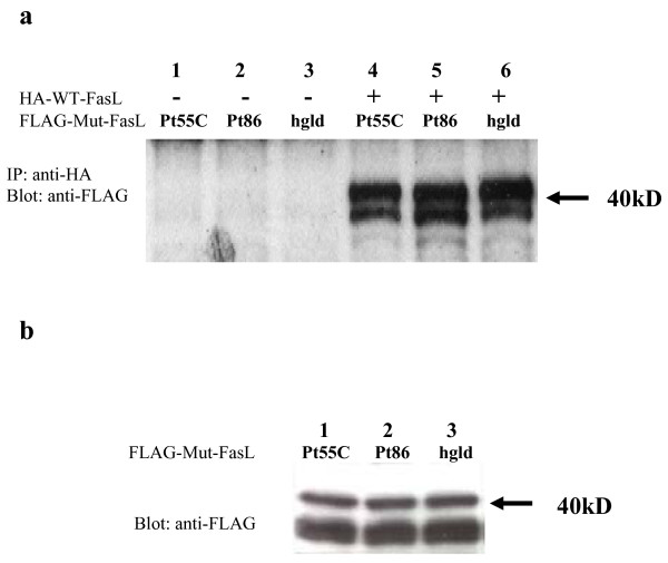
Results: We found an ALPS patient that harbored a heterozygous A530G mutation in the FasL gene that replaced Arg with Gly at position 156 in the protein's extracellular Fas-binding region. This produced a dominant-interfering FasL protein that bound to the wild-type FasL protein and prevented it from effectively inducing apoptosis.

Conclusion: Our data explain how a naturally occurring heterozygous human FasL mutation can dominantly interfere with normal FasL apoptotic function and lead to an ALPS phenotype, designated Type Ib.


Figures

Figure 1
Figure 1
Fas ligand mutations in the Pt 86, Pt 55C and the gld mouse. Sequencing of genomic DNA shows a heterozygous A530G mutation in Pt 86 and a A320G mutation in Pt55C depicted in panel (a) for double peaks of A and G from normal and mutant alleles amplified from genomic DNA. The family pedigree in panel (b) reveals the same mutation was found in his father, and paternal grandmother (labeled with m) but not in his healthy mother or two sisters. Black, the affected individual; gray, individuals with the mutation but not the full picture of ALPS. This mutation leads to an amino acid change of A156G, the heterozygous A320G mutation found in Pt 55C FasL genomic DNA leads to an amino acid change M86V in the transmembrane domain (TMD), and the gld mutation in mouse is shown at amino acid site 275, all compared to the wild type sequences at these sites in 5 species, as aligned in panel c.
Figure 2
Figure 2
(a) Molecular modeling of the extracellular, soluble portion of rat FasL. The soluble rat FasL trimer fragment was visualized based on the structure of TNF-α [33]. The Pt 86 FasL mutation is labeled in red and the hgld mutation is labeled in blue in side (left) and front (right) projections. The Pt 55C FasL mutation is not indicated in this model since it is located in the transmembrane domain, which is outside of the region in the model depicted. (b) A diagram of cloning strategy for epitope-tagging Fas-ligand protein.
Figure 3
Figure 3
Fas-mediated T-cell apoptosis in Family 86. The percentage of T cells killed by the treatment with an agonistic anti-Fas antibody is shown for the patient, members of his extended family, a normal control, and an ALPS Type Ia patient with a mutation in the Fas death domain. Percent cell loss is calculated as (1-number of cells with antibody stimulation/number of cells without stimulation) 100%. One representative of three repeated experiments is shown.
Figure 4
Figure 4
Cytotoxic effects of transfected FasL constructs on Jurkat cells. Cytotoxicity is reflected by the percentage of Fas-positive Jurkat cells killed by co-culture with transfected 293T cells. (a) Ratios of 1:1 or 3:1 were used for 293T cells (effectors) transfected with either of 4 constructs (WT; Pt 86, black; Pt 55C, open; and hgld, shaded) to Jurkat cells (targets). Averages of % cell death and error bars are shown as normalized to that of WT FasL-induced death. Data represent at least 3 experiments. (b) Cotransfection of the Pt 86 (R156G) mutant FasL with wild type FasL. 293T cells were transfected with a total of 0.4 or 0.2 μg of wild-type FasL only (open bars); with Pt 86 mutant mixed with wild-type FasL at ratio of 1:1 (black bars); and with Pt 86 mutant FasL only (shaded bars). The ratio of effector to target cells was 2:1 in this experiment. Means and error bars of the percentage Jurkat cells death are shown. Data represent two independent duplicate experiments. (c) Surface staining of FLAG-tagged FasL in 293T cells transfected with empty vector, hgld-, WT-, and Pt 86- FasL as indicated.
Figure 5
Figure 5
Cytotoxic effects of patient PBLs on Jurkat cells. The activated PBLs from normal controls (open), Pt 86 (FasL R156G, black), and father of Pt 86 (shaded) were used as the effector cells to mix with Jurkat (target) cells at the E/T ratios shown. Means and error bars of % target cell death are shown, data represent two independent experiments.
Figure 6
Figure 6
Physical association of FLAG-tagged mutant FasL with HA-tagged wild type FasL. (a) Lanes 1, 2 and 3 contain extracts from cells transfected with the FLAG-tagged mutant FasL constructs (A320G, A530G and hgld) alone and lanes 4, 5 and 6 are from cells co-transfected with one of FLAG-tagged mutant FasL constructs and the HA-tagged wild type FasL construct. An anti-HA antibody was used for the immunoprecipitation and an anti-FLAG antibody was used for Western blot analysis. The double bands appearing at about 40 kD represent two FasL isoforms with different glycosylation patterns. (b) Western blot analysis of transfected cell lysates. The FLAG-tagged FasL with the mutations (lanes 1–3) were expressed in 293T cells, and FasL expression was detected by Western blotting with an anti-FLAG antibody.

References

    1. Straus SE, Sneller M, Lenardo MJ, Puck JM, Strober W. An inherited disorder of lymphocyte apoptosis: the autoimmune lymphoproliferative syndrome. Ann Intern Med. 1999;130:591–601. - PubMed
    1. Oliveira JB, Bidère N, Niemela J, Zheng LX, Sakai K, Puro C, Danner R, Barb J, Munson P, Puck J, Dale J, Straus S, Fleisher TA, Lenardo MJ. NRAS mutation causes a human autoimmune lymphoproliferative syndrome. Proc Natl Acad Sci USA. 2007;104:8953–8958. doi: 10.1073/pnas.0702975104. - DOI - PMC - PubMed
    1. Rieux-Laucat F, Le Deist F, Hivroz C, Roberts IA, Debatin KM, Fischer A, de Villartay JP. Mutations in Fas associated with human lymphoproliferative syndrome and autoimmunity. Science. 2003;268:1347–1349. doi: 10.1126/science.7539157. - DOI - PubMed
    1. Fisher GH, Rosenberg FJ, Straus SE, Dale JK, Middleton LA, Lin AY, Strober W, Lenardo MJ, Puck JM. Dominant interfering Fas gene mutations impair apoptosis in a human autoimmune lymphoproliferative syndrome. Cell. 1995;81:935–946. doi: 10.1016/0092-8674(95)90013-6. - DOI - PubMed
    1. Puck JM, Straus SE, Le Deist F, Rieux-Laucat F, Fischer A. Inherited Disorders with Autoimmune and Defective Lymphocyte Regulation. In: Ochs H, Smith CIE, Puck JM, editor. Primary Immunodeficiency Diseases: A Molecular and Genetic Approach. New York: Oxford University Press;; 1999. pp. 339–352.

Publication types

MeSH terms

Substances