. 2007 Jul 2:2:18.
doi: 10.1186/1745-6150-2-18.

Small but versatile: the extraordinary functional and structural diversity of the beta-grasp fold

Affiliations

Small but versatile: the extraordinary functional and structural diversity of the beta-grasp fold

A Maxwell Burroughs et al. Biol Direct. .

Abstract

Background: The beta-grasp fold (beta-GF), prototyped by ubiquitin (UB), has been recruited for a strikingly diverse range of biochemical functions. These functions include providing a scaffold for different enzymatic active sites (e.g. NUDIX phosphohydrolases) and iron-sulfur clusters, RNA-soluble-ligand and co-factor-binding, sulfur transfer, adaptor functions in signaling, assembly of macromolecular complexes and post-translational protein modification. To understand the basis for the functional versatility of this small fold we undertook a comprehensive sequence-structure analysis of the fold and developed a natural classification for its members.

Results: As a result we were able to define the core distinguishing features of the fold and numerous elaborations, including several previously unrecognized variants. Systematic analysis of all known interactions of the fold showed that its manifold functional abilities arise primarily from the prominent beta-sheet, which provides an exposed surface for diverse interactions or additionally, by forming open barrel-like structures. We show that in the beta-GF both enzymatic activities and the binding of diverse co-factors (e.g. molybdopterin) have independently evolved on at least three occasions each, and iron-sulfur-cluster-binding on at least two independent occasions. Our analysis identified multiple previously unknown large monophyletic assemblages within the beta-GF, including one which unifies versions found in the fasciclin-1 superfamily, the ribosomal protein L25, the phosphoribosyl AMP cyclohydrolase (HisI) and glutamine synthetase. We also uncovered several new groups of beta-GF domains including a domain found in bacterial flagellar and fimbrial assembly components, and 5 new UB-like domains in the eukaryotes.

Conclusion: Evolutionary reconstruction indicates that the beta-GF had differentiated into at least 7 distinct lineages by the time of the last universal common ancestor of all extant organisms, encompassing much of the structural diversity observed in extant versions of the fold. The earliest beta-GF members were probably involved in RNA metabolism and subsequently radiated into various functional niches. Most of the structural diversification occurred in the prokaryotes, whereas the eukaryotic phase was mainly marked by a specific expansion of the ubiquitin-like beta-GF members. The eukaryotic UB superfamily diversified into at least 67 distinct families, of which at least 19-20 families were already present in the eukaryotic common ancestor, including several protein and one lipid conjugated forms. Another key aspect of the eukaryotic phase of evolution of the beta-GF was the dramatic increase in domain architectural complexity of proteins related to the expansion of UB-like domains in numerous adaptor roles.


Figures
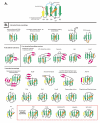
Figure 1
Figure 1
Topology diagrams of selected β-GF members. A generalized representative is shown in (A) with the key structural features found in certain lineages of the fold labeled, while (B) depicts idealized versions of specific lineages, the names of which are given above the diagrams. Strands are shown as arrows with the arrowhead at the C-terminal end. Strands belonging to the 4-stranded β-GF core are colored green, the additional strand found in the 5-stranded assemblage is colored yellow, strands forming a conserved insert within the β-GF scaffold are colored magenta, and other strands specific to a certain lineage are colored grey and outlined with a broken line. Helices are depicted as rectangles, with the core absolutely conserved helix colored orange and other helices specific to a certain lineage colored grey and outlined with a broken line. The diagrams are grouped and labeled in a manner consistent with the structural classes described in the text, with members of the eukaryotic UB-like superfamily nested within other members of the 5-stranded assemblage. The 2Fe-2S cluster of the ferredoxins is shown as four small ovals bound to cysteine residues represented by the letter "C".
Figure 2
Figure 2
Cartoon representations of distinct β-GF domains. Critical residues in MutT and HisI that are involved in enzyme catalysis are also shown.
Figure 3
Figure 3
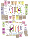
Reconstructed evolutionary history of β-grasp fold. Individual lineages are listed to the left of the figure grouped according to classifications given in the text, with their inferred evolutionary depth traced by solid horizontal lines across the relative temporal epochs representing major evolutionary transitional periods shown as vertical lines. The horizontal lines are color-coded according to their observed phyletic distributions, the key for this coloring scheme is given at the bottom of the figure. Dashed lines indicate uncertainty in terms of the origins of a lineage, while grey ellipses group lineages of relatively restricted phyletic distribution with more broadly distributed lineages, indicating that the former likely underwent rapid divergence from the latter. Major predicted structural/functional transitions of the fold are marked by green ellipses with a brief description given. Colored, labeled squares immediately to the left of the lineage names represent broad functional categories: E, enzymatic activity; LMB, ligand or metal-binding; CO, conjugated versions; AD, mediator of protein-protein interactions; RNA, RNA metabolism-related.
Figure 4
Figure 4
Reconstructed evolutionary history of eukaryotic ubiquitin superfamily. Similar to Figure 3, however, major evolutionary transitions are now shown as horizontal lines and the maximum depth to which these individual lineages can be traced is now shown with solid vertical lines. Functional categories are the same as described in Figure 3.
Figure 5
Figure 5
A) Architectural complexity plot of β-grasp domains found in eukaryotes and prokaryotes. The complexity quotient for a given species (y-axis) is plotted against the total number of β-grasp domain containing proteins in the same species. Names of species are given next to plot points. B) Domain architectures of β-grasp domains. Only a small sample of architectures is shown. These mainly represent novel or recently reported architectures that are described in the text. The TRS4 C-terminal domain, also found fused to certain E1-enzymes that lack the C-terminal UFD has a highly conserved ExxxH implying enzymatic function (see Additional file 1 for an alignment). Orange ellipses represent the conserved cysteine clusters observed in the NPL4-N family (see Additional file 1). A straight line with a small green box in the Ddi1 family architecture represents a possible cleavage site located between the domains. The proteins are not drawn to scale as only globular segments are show. Explanation of abbreviations/domain names: B3, DNA-binding domain; Auxin response, auxin-responsive transcription factor domain; OTU, OTU-like family of cysteine proteases; Znf, zinc-finger; Znf_LF, little finger family of zinc finger domains; R, Ring-finger domain; β-P, β-propeller domain; X, previously uncharacterized BofC C-terminal domain also found fused to a serine/threonine phosphatase in actinobacteria (see Additional file 1 for alignment). Organism abbreviations: Ehis, Entamoeba histolytica; Ath, Arabidopsis thaliana; Hsap, Homo sapiens; Rnor, Rattus norvegicus; Blic, Bacillus licheniformis; Mmaz, Methanosarcina mazei; Ddis, Dictyostelium discoideum; Lmaj, Leishmania major; Tcru, Trypanosoma cruzi; Pfal, Plasmodium falciparum; Tthe, Tetrahymena thermophila; Ncra, Neurospora crassa; Drer, Danio rerio; Cele, Caenorhabditis elegans; Dmel, Drosophila melonogaster; Scer, Saccharomyces cerevisiae; Tvag, Trichomonas vaginalis; Uma, Ustilago maydis; Spom, Schizosaccharomyces pombe; Cneo, Cryptococcus neoformans; Glam, Giardia lamblia; Cpar, Cryptosporidium parva; Tmar, Thermotoga maritima; Mpne, Mycoplasma pneumoniae; Ecol, Escherichia coli; Vcho, Vibrio cholerae; Hpyl, Helicobacter pylori; Nmen, Neisseria meningitides; Msp., Mesorhizobium sp.; Ctet, Clostridium tetani; Aaeo, Aquifex aeolicus; Tden, Treponema denticola; Drad, Deinococcus radiodurans; Mtub, Mycobacterium tuberculosis; Save, Streptomyces avermitilis; Bfra, Bacteroides fragilis; Ctep, Chlorobium tepidum; Nsp., Nostoc sp.; Ssp., Synecococcus sp.; Cpneu, Chlamydophila pneumoniae.
Figure 6
Figure 6
Diagram of relative location of β-grasp interacting partners. The strands and core helix of an idealized β-GF domain have been broken into interaction zones, and the names of representatives of the fold that interact using each of these zones is listed. The top view depicts the exposed face while the bottom view depicts the obscured face. Coloring of the boxes containing lists of specific β-GF domains interacting via a particular region correspond to coloring of structural elements (i.e. a particular strand or loop) involved in the interaction.

References

    1. Glickman MH, Ciechanover A. The ubiquitin-proteasome proteolytic pathway: destruction for the sake of construction. Physiol Rev. 2002;82:373–428. - PubMed
    1. Goldstein G, Scheid M, Hammerling U, Schlesinger DH, Niall HD, Boyse EA. Isolation of a polypeptide that has lymphocyte-differentiating properties and is probably represented universally in living cells. Proc Natl Acad Sci U S A. 1975;72:11–15. doi: 10.1073/pnas.72.1.11. - DOI - PMC - PubMed
    1. Wilkinson KD. The discovery of ubiquitin-dependent proteolysis. Proc Natl Acad Sci U S A. 2005;102:15280–15282. doi: 10.1073/pnas.0504842102. - DOI - PMC - PubMed
    1. Vijay-Kumar S, Bugg CE, Cook WJ. Structure of ubiquitin refined at 1.8 A resolution. J Mol Biol. 1987;194:531–544. doi: 10.1016/0022-2836(87)90679-6. - DOI - PubMed
    1. Vijay-Kumar S, Bugg CE, Wilkinson KD, Cook WJ. Three-dimensional structure of ubiquitin at 2.8 A resolution. Proc Natl Acad Sci U S A. 1985;82:3582–3585. doi: 10.1073/pnas.82.11.3582. - DOI - PMC - PubMed