. 2007 Jul 3:7:80.
doi: 10.1186/1471-213X-7-80.

Extra-embryonic endoderm cells derived from ES cells induced by GATA factors acquire the character of XEN cells

Affiliations

Extra-embryonic endoderm cells derived from ES cells induced by GATA factors acquire the character of XEN cells

Daisuke Shimosato et al. BMC Dev Biol. .

Abstract

Background: Three types of cell lines have been established from mouse blastocysts: embryonic stem (ES) cells, trophoblast stem (TS) cells, and extra-embryonic endoderm (XEN) cells, which have the potential to differentiate into their respective cognate lineages. ES cells can differentiate in vitro not only into somatic cell lineages but into extra-embryonic lineages, including trophectoderm and extra-embryonic endoderm (ExEn) as well. TS cells can be established from ES cells by the artificial repression of Oct3/4 or the upregulation of Cdx2 in the presence of FGF4 on feeder cells. The relationship between these embryo-derived XEN cells and ES cell-derived ExEn cell lines remains unclear, although we have previously reported that overexpression of Gata4 or Gata6 induces differentiation of mouse ES cells into extra-embryonic endoderm in vitro.

Results: A system in which GATA factors were conditionally activated revealed that the cells continue to proliferate while expressing a set of extra-embryonic endoderm markers, and, following injection into blastocysts, contribute only to the extra-embryonic endoderm lineage in vivo. Although the in vivo contribution is limited to cells of parietal endoderm lineage, Gata-induced extra-embryonic endoderm cells (gExEn) can be induced to differentiate into visceral endoderm-like cells in vitro by repression of Gata6. During early passage, the propagation of gExEn cells is dependent on the expression of the Gata6 transgene. These cells, however, lose this dependency following establishment of endogenous Gata6 expression.

Conclusion: We show here that Gata-induced extra-embryonic endoderm cells derived from ES cells mimic the character of XEN cells. These findings indicate that Gata transcription factors are sufficient for the derivation and propagation of XEN-like extra-embryonic endoderm cells from ES cells.


Figures
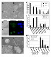
Figure 1
Figure 1
Induction of gExEn cells from ES cells by inducible Gata6 or Gata4 expression systems. (A-D) Morphology of differentiated SKG612 (A, B) and EBRTc-G6 (C, D) cells induced by withdrawal of Tc on gelatinized dishes (A, C) or under MEF culture conditions for 5 days (B, D). (E, F) Confocal microscopic image of ES cells with introduced pCAG-EGFP-G6GR 2 hr after the addition of 70% EtOH, without (E) and with (F) Dex. The left panels show phase-contrast images, the center panels show localization of EGFP-G6GR monitored by EGFP fluorescence, and the right panels show nuclei stained with Hoechst33342. (G-J) Morphology of 5G6GR11-2 ES cells with or without Dex. 5G6GR11-2 ES cells were grown in the presence (G) or absence (H) of LIF for 5 days, or were treated with 100 nM Dex for 5 days on gelatinized dishes (I, high-density conditions in inset) or on MEF (J). (K) Expression levels of endogenous Gata6 and Gata4 in ES-derived ExEn cells 4 days after activation of exogenous Gata on gelatinized dish cultures or PrE induced by withdrawal of LIF for 5 days. XEN2 and XEN9 cells; derived from embryo and MEF-dependent propagation. MEF; MEF alone. All values were normalized relative to the level of Gapdh and plotted relative to levels of expression in XEN2 cells. (L) Expression levels of endogenous Fgf3 in ES-derived ExEn cells 4 days after activation of exogenous GATA on gelatinized dish cultures, or PrE induced by withdrawal of LIF for 5 days. XEN2 and XEN9 cells; derived form embryo and MEF-dependent propagation. MEF; MEF alone. All values were normalized relative to the level of Gapdh and plotted relative to levels of expression in XEN2 cells. (M) GATA-dependent enhancer activity of the element containing GATA-binding site of pFgf3-luc in ES-derived ExEn cells. Reporter plasmids were transfected into 5G6GR11-2 and 5G4GR4-3 ES cells followed by culture with (exo-GATA ON) or without Dex (OFF), or EBRTc-G6 and SKG612 ES cells followed by culture with (OFF), or without Tc (exo-GATA ON). Relative expression levels of pFgf3-luc (filled; exo-GATA ON, hatched; OFF) are shown. All results were normalized relative to the luciferase activities of pCMV-RL and plotted relative to the luciferase activities of pGL3-luc in Dex-non-treated 5G6GR11-2, set at 1.0.
Figure 2
Figure 2
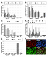
Marker gene expression of gExEn cells. (A-E) Q-PCR analysis of gene expression in Dex-treated gExEn cells after 10 passages. ExEn markers (A), PE markers (B and C), VE markers (D) and stem cell markers (E). Relative expression levels of the indicated marker genes in two independent clones of g6ExEn (2-3 and 11-2) and g4ExEn (1-1 and 4-3) and in 5G6GR ES cells, in the presence and absence of LIF for 5 days, are shown. All results were normalized relative to the level of expression Gapdh and plotted relative to expression levels in 5G6GR-derived PrE without LIF (A-D) or that in 5G6GR ES cells (E). (F) g4ExEn4-3 were stained with anti-GATA6 (red), anti-DAB2 (green), and nuclear staining by Hoechst33342 (blue). The lower right panel shows marginal images.
Figure 3
Figure 3
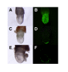
Contribution of gExEn cells to ExEn lineage in vivo. (A, B) 8.5 dpc chimeric embryos derived from 5G6GR-GFP ES cells. 5G6GR-GFP ES cells, kept in undifferentiated state without Dex, give rise to embryonic chimeras. The 8.5 dpc chimeric embryos with g6ExEn-GFP (C, D) derived from 5G6GR-GFP ES cells or g4ExEn-GFP cells (E, F) derived from 5G4GR-GFP ES cells, cultured in the presence of Dex for the activation of GATA-GR after several passages, contributed only to the distal parietal yolk sac.
Figure 4
Figure 4
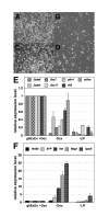
Effect of the extinction of exogenous GATA activity in gExEn cells. (A-D) Photomicrographs of g6ExEn11-2 (A, B) and g4ExEn4-3 (C, D) cells cultured with (A or C) or without (B or D) Dex for 4 days after 2 passages in the presence of Dex. (E, F) Q-PCR analysis of PE or VE marker gene expression in g6ExEn11-2 cells, with or without Dex. Withdrawal of Dex after 2 passages in the presence of Dex decreased expression of a set of PE marker genes (E), while expression of VE marker genes increased in parallel (F). All results were normalized relative to expression of Gapdh and plotted relative to the expression level in Dex-treated g6ExEn11-2 cells.
Figure 5
Figure 5
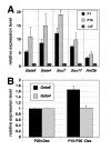
Expression of endogenous Gata4 and Gata6 in late-passage gExEn cells. (A) Marker gene expression in late passage: g6ExEn cells expressed endogenous Gata4 and Gata6 slightly higher than in early passage g6ExEn cells. All results were normalized relative to expression of Gapdh and plotted relative to the expression level in 5G6GR-derived PrE without LIF for 5 days. (B) After removal of Gata6-GR activity by withdrawal of Dex from passage10 to passage20 (P10-P20), the expression levels of endogenous Gata4 and Gata6 were maintained as same amount level in the presence of Dex culture condition (P20). All results were normalized relative to expression of Gapdh and plotted relative to the expression level in P20 g6ExEn cells.
Figure 6
Figure 6
Knock-down of Gata6 in late-passage gExEn cells. (A-C) Efficient transfection of g6ExEn cells by EGFP expression vector. After drug selection, almost all transfectants showed GFP expression microscopically (A: phase-contrast image, B: fluorescent image for EGFP), which was confirmed by FACS analysis for EGFP fluorescence (C). (D, E) Morphology of g6ExEn cell line, 1D3, transfected with the Gata6 silencing vector pSil-G6 (D) or empty vector (D) after 5 days transfection. (F) Expression level of Gata6 and Afp in Gata6-silenced 1D3 cells by Q-PCR at day 5. Results were normalized relative to expression of Gapdh and plotted relative to expression level in 1D3 cells transfected with pSil-H1puro.

Similar articles

Cited by

References

    1. Enders AC, Schlafke S. Comparative aspects of blastocyst-endometrial interactions at implantation. Ciba Found Symp. 1978:3–32. - PubMed
    1. Hogan BL, Cooper AR, Kurkinen M. Incorporation into Reichert's membrane of laminin-like extracellular proteins synthesized by parietal endoderm cells of the mouse embryo. Dev Biol. 1980;80:289–300. doi: 10.1016/0012-1606(80)90405-4. - DOI - PubMed
    1. Morrisey EE, Ip HS, Lu MM, Parmacek MS. GATA-6: a zinc finger transcription factor that is expressed in multiple cell lineages derived from lateral mesoderm. Dev Biol. 1996;177:309–322. doi: 10.1006/dbio.1996.0165. - DOI - PubMed
    1. Koutsourakis M, Langeveld A, Patient R, Beddington R, Grosveld F. The transcription factor GATA6 is essential for early extraembryonic development. Development. 1999;126:723–732. - PubMed
    1. Chazaud C, Yamanaka Y, Pawson T, Rossant J. Early lineage segregation between epiblast and primitive endoderm in mouse blastocysts through the Grb2-MAPK pathway. Dev Cell. 2006;10:615–624. doi: 10.1016/j.devcel.2006.02.020. - DOI - PubMed

Publication types

MeSH terms

LinkOut - more resources