. 2007;35(14):4728-36.
doi: 10.1093/nar/gkm507. Epub 2007 Jul 1.

The crystal structure of the Thermus aquaticus DnaB helicase monomer

Affiliations

The crystal structure of the Thermus aquaticus DnaB helicase monomer

Scott Bailey et al. Nucleic Acids Res. 2007.

Abstract

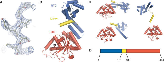
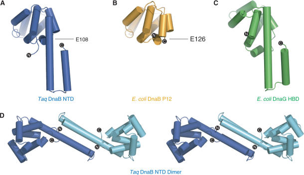
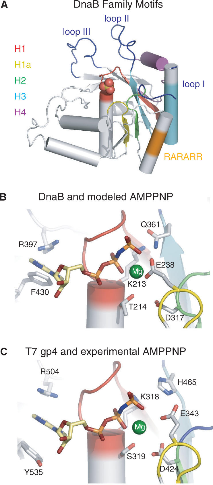
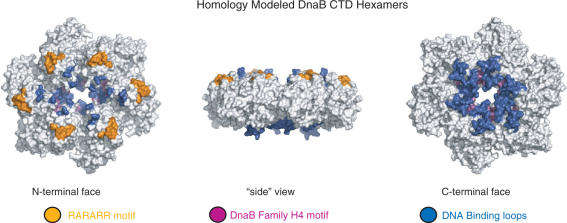
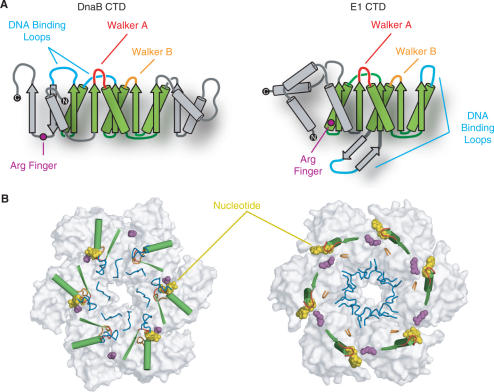
The ring-shaped hexameric DnaB helicase unwinds duplex DNA at the replication fork of eubacteria. We have solved the crystal structure of the full-length Thermus aquaticus DnaB monomer, or possibly dimer, at 2.9 A resolution. DnaB is a highly flexible two domain protein. The C-terminal domain exhibits a RecA-like core fold and contains all the conserved sequence motifs that are characteristic of the DnaB helicase family. The N-terminal domain contains an additional helical hairpin that makes it larger than previously appreciated. Several DnaB mutations that modulate its interaction with primase are found in this hairpin. The similarity in the fold of the DnaB N-terminal domain with that of the C-terminal helicase-binding domain (HBD) of the DnaG primase also includes this hairpin. Comparison of hexameric homology models of DnaB with the structure of the papillomavirus E1 helicase suggests the two helicases may function through different mechanisms despite their sharing a common ancestor.


Figures

Figure 1.
Figure 1.
(A) Portion of a sigma-A weighted 2FoFc electron density omit map calculated at 2.9 Å resolution. The diffraction amplitudes were sharpened by a temperature factor of 50. (B) Ribbon representation of the backbone structure of the DnaB monomer. The N-terminal domain (NTD), C-terminal domain (CTD) and linker region are colored blue, red and yellow, respectively. (C) Similar ribbon representations of the three additional copies of the DnaB monomer found in the asymmetric unit. Each of the monomer structures in panels B and C were orientated by pairwise superposition of their respective CTDs. (D) A schematic diagram of the domain structure of the DnaB helicase. Residues found at the domain boundaries are labeled.
Figure 2.
Figure 2.
Ribbon representations of (A) Taq NTD, (B) E. coli P12 fragment and (C) E. coli helicase-binding domain (HBD) of primase. The Taq NTD is colored as in Figure 2. The E. coli P12 fragment and HBD are colored orange and green, respectively. Escherichia coli P12 and HBD were orientated by superposition of their structures on that of the Taq NTD. The C-terminal residue of the E. coli P12 fragment and the equivalent residue in the intact Taq NTD are labeled. (D) Stereo view of a ribbon representation of the Taq NTD dimer observed in the crystal lattice.
Figure 3.
Figure 3.
(A) Ribbon representation of the Taq CTD colored according to the conserved DnaB family motifs: H1 red, H1a yellow, H2 green, H4 cyan and H4 purple. The RARARR basic region (see text) and the three proposed DNA-binding loops are colored orange and blue, respectively. (B) DnaB nucleotide-binding site with the homology modeled AMPPNP (stick model) and magnesium ion (green sphere). Side chains of key residues are displayed. Secondary structure elements are colored as in panel A. (C) T7 gp4 helicase nucleotide-binding pocket (PDB code: 1E0J). Representations and coloring are the same as panel B.
Figure 4.
Figure 4.
Three perpendicular views of a surface representation of the Taq DnaB CTD hexamer that has been constructed by homology modeling using the T7 gp4 helicase structure (PDB code: 1E0J). The loops implicated in DNA binding are colored blue, motif H4 magenta and the RARARR sequence orange.
Figure 5.
Figure 5.
Comparison of the ternary and quaternary structures of DnaB and the E1 helicase. (A) Topology diagrams of the CTD of DnaB (left) and the E1 helicase (right). The core RecA fold is colored green. Unique structural elements within each domain are colored gray. The Walker A and Walker B motifs are colored red and orange, respectively. Arginine fingers are highlighted as magenta circles and DNA-binding loops are in blue. The N and C-termini are indicated as black circles with white text. (B) Arrangement of essential helicase elements within (left) the DnaB CTD hexamer modeled by superposition (see text) onto the T7 gp4 structure and right the E1 CTD assembly. Key elements are colored as in panel A. For clarity only the β-strand preceding and the α-helix following the Walker A motif are shown. Shown in white are the molecular surfaces of the two structures with the nucleotide (yellow) displayed in a space-filling representation.

References

    1. LeBowitz JM, McMacken R. The Escherichia coli dnaB protein is a DNA helicase. J. Biol. Chem. 1986;261:4738–4748. - PubMed
    1. Galletto R, Jezewska MJ, Bujalowski W. Unzipping mechanism of the double-stranded DNA unwinding by a hexameric helicase: quantitative analysis of the rate of the dsDNA unwinding, processivity and kinetic step-size of the Escherichia coli DnaB helicase using rapid quench-flow methods. J. Mol. Biol. 2004;343:83–99. - PubMed
    1. Bird LE, Pan H, Soultanas P, Wigley DB. Mapping protein-protein interactions within a stable complex of DNA primase and DnaB helicase from Bacillus stearothermophilus. Biochemistry. 2000;39:171–182. - PMC - PubMed
    1. Gorbalenya AE, Koonin EV. Helicases: amino acid sequence comparisons and structure-function relationships. Curr. Opin. Struct. Biol. 1993;3:419–429.
    1. Story RM, Weber IT, Steitz TA. The structure of the E. coli recA protein monomer and polymer. Nature. 1992;355:318–325. - PubMed

Publication types

Associated data