. 2007 Jul 9;204(7):1717-27.
doi: 10.1084/jem.20070255. Epub 2007 Jul 2.

Role for DNA repair factor XRCC4 in immunoglobulin class switch recombination

Affiliations

Role for DNA repair factor XRCC4 in immunoglobulin class switch recombination

Pauline Soulas-Sprauel et al. J Exp Med. .

Abstract

V(D)J recombination and immunoglobulin class switch recombination (CSR) are two somatic rearrangement mechanisms that proceed through the introduction of double-strand breaks (DSBs) in DNA. Although the DNA repair factor XRCC4 is essential for the resolution of DNA DSB during V(D)J recombination, its role in CSR has not been established. To bypass the embryonic lethality of XRCC4 deletion in mice, we developed a conditional XRCC4 knockout (KO) using LoxP-flanked XRCC4 cDNA lentiviral transgenesis. B lymphocyte restricted deletion of XRCC4 in these mice lead to an average two-fold reduction in CSR in vivo and in vitro. Our results connect XRCC4 and the nonhomologous end joining DNA repair pathway to CSR while reflecting the possible use of an alternative pathway in the repair of CSR DSB in the absence of XRCC4. In addition, this new conditional KO approach should be useful in studying other lethal mutations in mice.


Figures

Figure 1.
Figure 1.
Creation of X4T transgenic mice. (A) Schematic representation of the pTRIP-X4 lentiviral vector. Restriction sites: E, EcoNI; A, AvaII. Filled circle, central DNA flap; filled box, X4 Orf. UbiC, human Ubiquitin C promoter. (B) Schematic representation of pTRIP-X4 provirus integrated in the genome of the X4T transgenic mouse line selected for this study. *, the positions of these sites are specific to the integration site. (C) Southern blot analysis of tail DNA shows a single integration of the transgene in the genome of X4T mice. EcoNI–AvaII–digested DNA from X4T-positive and control littermate X4T-negative mice was hybridized with the 5′ flanking probe A, the internal probe B, and the 3′ flanking probe C as indicated in A. Refer to B for expected sizes of bands. (D) Analysis of X4T expression in transgenic mice. Western blot analysis of X4T (top) and endogenous X4 (bottom) expression in purified splenic mature B cells and thymocytes of X4T mice and control X4T-negative mice (all on a wild-type background) revealed by anti-X4 antibody.
Figure 2.
Figure 2.
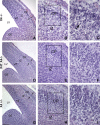
X4T reverts neuronal cell death of X4/ embryos. (A–I) Hematoxylin and eosin staining of littermate X4−/−, X4T X4−/−, and X4+/− brain sections. Coronal brain sections of E14.5 X4−/− (A–C) reveal a severe acellularity in the CP (small arrows) in the cerebral hemisphere. Higher magnifications show normal ventricular zone (VZ) of the mutant cortex, whereas numerous cells with pyknotic nuclei (dense hematoxylin stain, long arrow in C) are present in the intermediate zone (IZ) extending to the CP. In contrast, coronal brain sections of E14.5 X4T X4−/− mice (D–F) do not reveal significant difference in cortical structure with cerebral hemisphere of X4+/− brain (G–I) and an absence of cells with pyknotic nuclei. LV, lateral ventricle; GE, ganglionic eminence. Boxed areas in B, E, and H are enlarged in C, F, and I by image processing. Bars: (A, D, and G) 150 μm; (B, E, and H) 40 μm; (C, F, and I) 20 μm.
Figure 3.
Figure 3.
Conditional X4 deletion in mature B cells. (A) Schematic representation of the X4T locus before (X4T) and after (ΔX4T) Cre recombination and of a WT allele. Restriction sites: E, EcoNI; N, NdeI. (B) Analysis of X4T deletion by PCR using primers 1 and 2 located in A. Genomic DNA from total BM, purified splenic mature B cells, and thymocytes from X4T X4−/− mice expressing Cre (+) or not (−) were used in the PCR reaction. ΔX4T and WT fragments are indicated. Because of its large size, the X4T fragment is not detected in these PCR conditions. GAPDH-specific PCR was used as loading control. (C) Southern blot analysis of X4T deletion. DNA was prepared from liver, thymus, and from purified splenic mature B cells from X4T X4−/− (CD21-Cre-), ΔX4T X4−/− (CD21-Cre+), and nontransgenic (WT) mice, digested with EcoNI–NdeI, and hybridized with chromosome 16 integration site-specific probe D (refer to A for expected sizes of bands). Quantification of deletion is indicated below lanes 7, 8, and 9 as the percentage of the amount of ΔX4T compared with WT allele.
Figure 4.
Figure 4.
Ig production is impaired in conditional X4 KO mice. Sera from ΔX4T and control X4T mice were collected and total IgM, IgG1, IgG2b, IgG3, and IgA were determined by ELISA (microgram/milliliter). The results of statistical tests are indicated; * indicates a statistically significant difference (P < 0.05, two-tailed Mann-Whitney test).
Figure 5.
Figure 5.
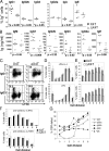
CSR defect in X4-deficient B lymphocytes in vitro. Surface expression and secretion of switched isotypes. Purified splenic mature B cells from ΔX4T and control X4T mice were labeled with CFSE and stimulated for 4 or 5 d with various polyclonal B cell activators for switching. (A) Percentage of total B cells expressing indicated isotypes of Ig, as determined by flow cytometry analysis, after 4 d of in vitro stimulation. (B) Secreted Ig (nanogram/milliliter) was analyzed in supernatants from splenic B cells cultures after 5-d in vitro stimulation. IgA was undetectable in supernatants. For A and B, the results of statistical tests are indicated; * indicates a statistically significant difference (P < 0.05, two-tailed Mann-Whitney test). (C–G) Analysis of CSR in the various subsets of proliferative B cells according to CFSE intensity. (C) Plots show IgG1 (top) and IgG3 (bottom) staining together with CFSE, on viable (PI) lymphoid cells. The percentages of switched cells are given. (D–F) Percentages of total (D and F) and IgG1-switched (E, top) or IgG3-switched (E, bottom) B cells that have undergone the indicated numbers of cell division, for ΔX4T (white) and X4T (gray) mice. (G) In each cell division, the decrease of CSR to IgG1, IgG2a, IgG2b, IgG3, and IgA, in B cells from ΔX4T compared with X4T mice is represented as a percentage of CSR defect.
Figure 6.
Figure 6.
Ig γ1 circle transcript production is impaired in X4-deficient B cells. Purified splenic mature B cells from ΔX4T and X4T mice were stimulated with LPS and IL-4 for 4 d, and μGT, γ1GT, γ1CT, and AID transcripts were quantified by real-time RT-PCR. Each point represents the level for the indicated transcript in ΔX4T mice (which has been normalized to CD19 transcripts) relative to X4T mice analyzed in the same experiment.
Figure 7.
Figure 7.
Production of episomal DNA carrying X4T upon Cre-mediated deletion. (A) Schematic representation of X4T locus before (X4T) and after (ΔX4T) Cre recombination, of episomal form of X4T (eX4T) and of X4 genomic locus (WT). Restriction sites: E, EcoNI; N, NdeI. (B) Southern blot analysis of X4T deletion and eX4T detection, using X4 third exon–specific probe E depicted in A. EcoNI–NdeI–digested DNA from total liver, thymus, or purified splenic mature B cells from X4T X4−/− (CD21-Cre-), ΔX4T X4−/− (CD21-Cre+) and non transgenic (WT) mice, was hybridized with probe E (refer to A for expected sizes of bands). (C) Detection of eX4T by PCR using primers 1 and 2 located in (A), on genomic DNA from the following: (left) purified splenic mature B cells, liver and thymocytes from ΔX4T mice; (right) purified splenic mature B cells from ΔX4T mice before (d0) and after (d4) 4 d of stimulation with LPS and IL-4. eX4T fragment is indicated. Artemis- (left) or GAPDH-specific (right) PCR was used as loading control. (D) Real-time quantitative RT-PCR analysis of X4 transcripts in purified splenic mature B cells from ΔX4T and X4T mice before (d0) and after stimulation with LPS and IL-4 for 4 d (d4). Bars represent the level of X4 transcript in ΔX4T B cells relative to X4T B cells. Results are means of two independent experiments.

References

    1. de Villartay, J.P., A. Fischer, and A. Durandy. 2003. The mechanisms of immune diversification and their disorders. Nat. Rev. Immunol. 3:962–972. - PubMed
    1. Revy, P., D. Buck, F. le Deist, and J.P. de Villartay. 2005. The repair of DNA damages/modifications during the maturation of the immune system: lessons from human primary immunodeficiency disorders and animal models. Adv. Immunol. 87:237–295. - PubMed
    1. Buck, D., L. Malivert, R. de Chasseval, A. Barraud, M.C. Fondaneche, O. Sanal, A. Plebani, J.L. Stephan, M. Hufnagel, F. le Deist, et al. 2006. Cernunnos, a novel nonhomologous end-joining factor, is mutated in human immunodeficiency with microcephaly. Cell. 124:287–299. - PubMed
    1. Callebaut, I., L. Malivert, A. Fischer, J.P. Mornon, P. Revy, and J.P. de Villartay. 2006. Cernunnos interacts with the XRCC4 x DNA-ligase IV complex and is homologous to the yeast nonhomologous end-joining factor Nej1. J. Biol. Chem. 281:13857–13860. - PubMed
    1. Ahnesorg, P., P. Smith, and S.P. Jackson. 2006. XLF interacts with the XRCC4-DNA ligase IV complex to promote DNA nonhomologous end-joining. Cell. 124:301–313. - PubMed

Publication types