. 2007 Jul 9;204(7):1553-8.
doi: 10.1084/jem.20070823. Epub 2007 Jul 2.

Cerebellar neurodegeneration in the absence of microRNAs

Affiliations

Cerebellar neurodegeneration in the absence of microRNAs

Anne Schaefer et al. J Exp Med. .

Abstract

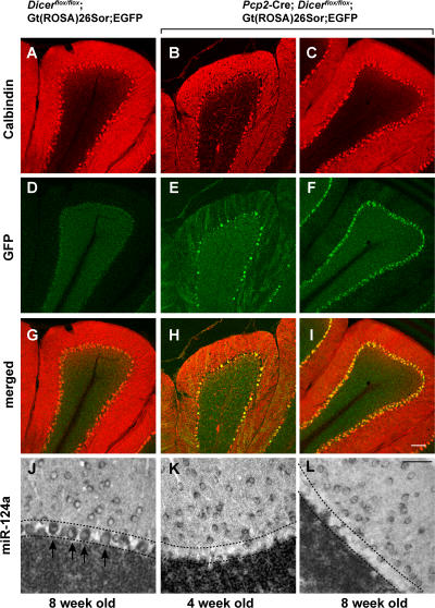
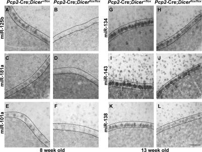
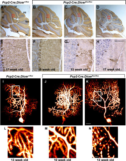
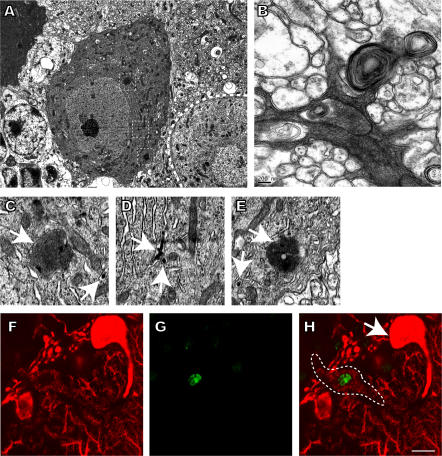
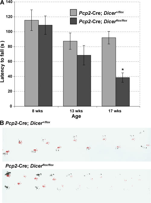
Genome-encoded microRNAs (miRNAs) are potent regulators of gene expression. The significance of miRNAs in various biological processes has been suggested by studies showing an important role of these small RNAs in regulation of cell differentiation. However, the role of miRNAs in regulation of differentiated cell physiology is not well established. Mature neurons express a large number of distinct miRNAs, but the role of miRNAs in postmitotic neurons has not been examined. Here, we provide evidence for an essential role of miRNAs in survival of differentiated neurons. We show that conditional Purkinje cell-specific ablation of the key miRNA-generating enzyme Dicer leads to Purkinje cell death. Deficiency in Dicer is associated with progressive loss of miRNAs, followed by cerebellar degeneration and development of ataxia. The progressive neurodegeneration in the absence of Dicer raises the possibility of an involvement of miRNAs in neurodegenerative disorders.


Figures

Figure 1.
Figure 1.
Dicer inactivation in postnatal Purkinje cells. 20-μm-thick cerebellar sections of 8-wk-old control Dicerflox/flox; Gt(ROSA)26Sor;EGFP (A, D, and G), and experimental Pcp2-Cre; Dicerflox/flox; Gt(ROSA)26Sor;EGFP mice at 4 (B, E, and H) and 8 (C, F, and I) wk of age are shown. To visualize Purkinje cells, the cerebella sections were stained with anti-calbindin antibody (red). Purkinje cells expressing Cre at levels inducing loxP recombination were visualized by expression of eGFP using an anti-GFP antibody (green). The expression of brain-specific miRNA miR-124a in 8-wk-old control (J) and Pcp2-Cre; Dicerflox/flox; Gt(ROSA)26Sor;EGFP mice at 4 (K) and 8 (L) wk of age was detected by in situ hybridization. Arrows indicate individual Purkinje cells, and the Purkinje cell layer is indicated by dashed lines. Bars: A–I, 100 μm; J–L, 50 μm.
Figure 2.
Figure 2.
Expression of miRNAs in Dicer-deficient Purkinje cells. 12-μm-thick cerebellar sections of 8 (A–F) or 13 (G–L) wk control Pcp2-Cre; Dicer+/flox and experimental Pcp2-Cre; Dicerflox/flox mice are shown. The expression of miR-125b (A and B), miR-181a (C and D), and miR-101a (E and F), representative of miRNAs that are below detection limit by 8 wk of age, and miR-134 (G and H), miR-143 (I and J), and miR-138 (K and L), representative for miRNAs that remain detectable at 13 wk of age, were detected by in situ hybridization with the corresponding specific probes. Bar: A–L, 50 μm.
Figure 3.
Figure 3.
Postnatal inactivation of Dicer leads to cerebellar degeneration. 12-μm-thick saggital cerebellar sections of 17-wk-old control Pcp2-Cre; Dicer+/flox mice (A and E) and 10- (B and F), 13- (C and G), and 17- (D and H) wk-old experimental Pcp2-Cre; Dicerflox/flox mice are shown. Purkinje cells were visualized by immunohistochemistry using an antibody to calbindin (brown). Sections are counterstained using Nissl stain (blue). Cerebellar lobules are indicated by roman numerals. The sections outlined by red rectangles in A–D are amplified in E–H. Morphological changes in Dicer-deficient Purkinje cells were visualized using two-photon images after intracellular injection of the calcium-sensing dye Fura 2. 13-wk-old control Pcp2-Cre; Dicer+flox (I and L) and 10- (J and M) and 13- (K and N) wk-old Pcp2-Cre; Dicerfloxflox mice are shown. The individual images were stacked and visualized using Imaris software. The sections outlined by white rectangles in I–K are shown in more detail in L–N. Bars: A–D, 1 mm; E–H, 50 μm; I–K, 20 μm; L–N, 5 μm.
Figure 4.
Figure 4.
Purkinje cell death in Pcp2-Cre; Dicerfloxflox mice. Electron microscopy images of degenerating Dicer-deficient Purkinje cells (A–E). Degenerating cells display condensation of the cytoplasm in cell bodies (A) and the dendritic compartment (B). Cytoplasmic condensation is associated with the occurrence of protein accumulations (A), autophagic vacuoles, and membrane whorls (B). Region of interests in A are marked by squares and shown in more detail (C–E). Membranes surrounding protein accumulations (C and E) and intracisternal inclusions (C–E) are indicated by arrows. The 12-μm-thick cerebellar section of a 13-wk-old Pcp2-Cre; Dicerflox/flox mouse is shown in F–H. Purkinje cells were visualized using anti-calbindin antibody (red), and apoptotic cells were detected by TUNEL staining (green). A TUNEL+ degenerating Purkinje cell (indicated by a dashed line) can be seen close to a TUNEL normal-appearing Purkinje cell (arrow). Bars: A, 5 μm; B, 0.2 μm; C–E, 1 μm; F–H, 10 μm.
Figure 5.
Figure 5.
Dicer deficiency in Purkinje cells causes ataxia. (A) Rotarod test on Pcp2-Cre; Dicerflox/flox mice compared with their littermate controls. Statistical analysis was performed using Anova single factor analysis (F = 5.976822099; P = 0.00024395; F crit = 2.41735603). Latency to fall from the rod was measured in control Pcp2-Cre; Dicer+/flox (gray) and mutant Pcp2-Cre; Dicerflox/flox (black) mice at 8 (control, n = 10; mutant, n = 8; P = 0.73494913), 13 (control, n = 10; mutant, n = 7; P = 0.28503378), and 17 (control, n = 9; mutant, n = 8; P = 0.00016573) wk of age. Results are expressed as means, and error bars represent ± SEM. p-values are from the unpaired two-tailed Student's t test. (B) Footprint analysis. Footprints of 17-wk-old male control and Pcp2-Cre; Dicerflox/flox mice are shown. The mutant animal exhibits a wide-based ataxic gait. Red, front paws; black, hind paws.

References

    1. Ambros, V. 2004. The functions of animal microRNAs. Nature. 431:350–355. - PubMed
    1. Bartel, D.P. 2004. MicroRNAs: genomics, biogenesis, mechanism, and function. Cell. 116:281–297. - PubMed
    1. He, L., and G.J. Hannon. 2004. MicroRNAs: small RNAs with a big role in gene regulation. Nat. Rev. Genet. 5:522–531. - PubMed
    1. Cobb, B.S., T.B. Nesterova, E. Thompson, A. Hertweck, E. O'Connor, J. Godwin, C.B. Wilson, N. Brockdorff, A.G. Fisher, S.T. Smale, and M. Merkenschlager. 2005. T cell lineage choice and differentiation in the absence of the RNase III enzyme Dicer. J. Exp. Med. 201:1367–1373. - PMC - PubMed
    1. Cobb, B.S., A. Hertweck, J. Smith, E. O'Connor, D. Graf, T. Cook, S.T. Smale, S. Sakaguchi, F.J. Livesey, A.G. Fisher, and M. Merkenschlager. 2006. A role for Dicer in immune regulation. J. Exp. Med. 203:2519–2527. - PMC - PubMed

Publication types