. 2007 Jul 10;104(28):11615-20.
doi: 10.1073/pnas.0704967104. Epub 2007 Jul 2.

Structural basis for conserved complement factor-like function in the antimalarial protein TEP1

Affiliations

Structural basis for conserved complement factor-like function in the antimalarial protein TEP1

Richard H G Baxter et al. Proc Natl Acad Sci U S A. .

Abstract

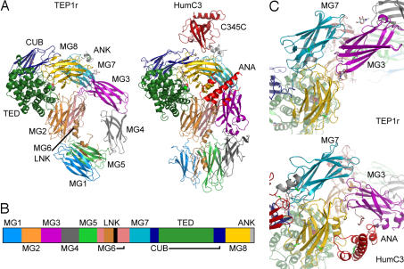
Thioester-containing proteins (TEPs) are a major component of the innate immune response of insects to invasion by bacteria and protozoa. TEPs form a distinct clade of a superfamily that includes the pan-protease inhibitors alpha(2)-macroglobulins and vertebrate complement factors. The essential feature of these proteins is a sequestered thioester bond that, after cleavage in a protease-sensitive region of the protein, is activated and covalently binds to its target. Recently, TEP1 from the malarial vector Anopheles gambiae was shown to mediate recognition and killing of ookinetes from the malarial parasite Plasmodium berghei, a model for the human malarial parasite Plasmodium falciparum. Here, we present the crystal structure of the TEP1 isoform TEP1r. Although the overall protein fold of TEP1r resembles that of complement factor C3, the TEP1r domains are repositioned to stabilize the inactive conformation of the molecule (containing an intact thioester) in the absence of the anaphylotoxin domain, a central component of complement factors. The structure of TEP1r provides a molecular basis for the differences between TEP1 alleles TEP1r and TEP1s, which correlate with resistance of A. gambiae to infection by P. berghei.


Conflict of interest statement

The authors declare no conflict of interest.

Figures

Fig. 1.
Fig. 1.
Overall structure of TEP1r. (A) Domain arrangements of TEP1r in comparison with human C3 (PDB ID 2A73). The mature protein commences with domain MG1 (medium blue) followed by MG2 (orange), MG3 (purple), MG4 (medium gray), MG5 (light green), MG6 (pink), LNK (light brown), MG7 (light blue), CUB (navy blue), TED (dark green), MG8 (yellow), and ANK (light gray). Additional domains in C3 are the ANA domain (red, between MG3 and MG8) and the C345C domain (dark red, top). (B) Sequence schematic of TEP1r, showing disposition of domains. (C) The relative position of MG3 to MG7-MG8 in TEP1r compared with human C3.
Fig. 2.
Fig. 2.
Stereo figure with refined density. 2FoFc density (cyan) contoured at 1σ for the MG3/MG8 interface.
Fig. 3.
Fig. 3.
The protease-sensitive region. (A) View of the cavity surrounding the protease-sensitive region of TEP1r. Positive FoFc density contoured at 2σ (green) for the end of the protease-sensitive region between domains MG3 and MG6. Dashed line represents line-of-sight path for residues 583–603. (B) Similar view for HumC3. (C) Coomassie-stained SDS/PAGE from limited proteolysis of purified TEP1r (160 kDa) shows two fragments identified as the N terminus (75 kDa) and the result of cleavage at position 601 (85 kDa) by N-terminal sequencing. (D) Silver-stained SDS/PAGE of dissolved crystals (>6 months old) displays autocatalytic cleavage at Gln 841 (105- and 55-kDa bands) upon heating in the absence of DTT, whereas unheated sample contains full-length TEP1r (160 kDa).
Fig. 4.
Fig. 4.
TEP1r and TEP1s substitutions. (A) Mapping of TEP1r/TEP1s substitutions onto TEP1r structure, peptide chain in gray, substitutions are pink spheres. The CUB, TED, and MG8 domains are colored according to Fig. 1 (B) alignment of TEP1r and TEP1s catalytic loop with HumC3, BovC3, and HumC4B. (C) The thioester (TE) chamber and surrounding aromatic residues. S1087 (unlabeled) is in the background between Y1286 and Y1287. (D) TED/MG8 interface. Residues K945 to E953 of catalytic loop and the pre-α4 loop (unlabeled) are to the left, MG8 loops interacting with the TED are to the right. (E) The β-hairpin motif between TED helices α9 and α10. In C–E, conserved residues have gray carbon atoms, and residues substituted in TEP1s have pink carbon atoms.

References

    1. Snow RW, Guerra CA, Noor AM, Myint HY, Hay SI. Nature. 2005;434:214–217. - PMC - PubMed
    1. Christophides GK. Cell Microbiol. 2005;7:325–333. - PubMed
    1. Cherry S, Silverman N. Nat Immunol. 2006;7:911–917. - PubMed
    1. Blandin S, Levashina EA. Mol Immunol. 2004;40:903–908. - PubMed
    1. Dodds AW. Immunobiology. 2002;205:340–354. - PubMed

Publication types

MeSH terms

Associated data