. 2007 Jul;117(7):1805-13.
doi: 10.1172/JCI30848.

Dysferlin-mediated membrane repair protects the heart from stress-induced left ventricular injury

Affiliations

Dysferlin-mediated membrane repair protects the heart from stress-induced left ventricular injury

Renzhi Han et al. J Clin Invest. 2007 Jul.

Abstract

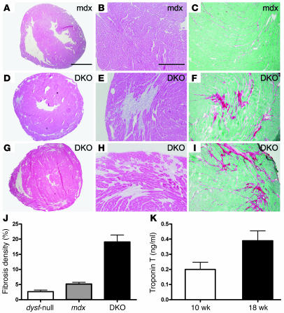
Dilated cardiomyopathy is a life-threatening syndrome that can arise from a myriad of causes, but predisposition toward this malady is inherited in many cases. A number of inherited forms of dilated cardiomyopathy arise from mutations in genes that encode proteins involved in linking the cytoskeleton to the extracellular matrix, and disruption of this link renders the cell membrane more susceptible to injury. Membrane repair is an important cellular mechanism that animal cells have developed to survive membrane disruption. We have previously shown that dysferlin deficiency leads to defective membrane resealing in skeletal muscle and muscle necrosis; however, the function of dysferlin in the heart remains to be determined. Here, we demonstrate that dysferlin is also involved in cardiomyocyte membrane repair and that dysferlin deficiency leads to cardiomyopathy. In particular, stress exercise disturbs left ventricular function in dysferlin-null mice and increases Evans blue dye uptake in dysferlin-deficient cardiomyocytes. Furthermore, a combined deficiency of dystrophin and dysferlin leads to early onset cardiomyopathy. Our results suggest that dysferlin-mediated membrane repair is important for maintaining membrane integrity of cardiomyocytes, particularly under conditions of mechanical stress. Thus, our study establishes what we believe is a novel mechanism underlying the cardiomyopathy that results from a defective membrane repair in the absence of dysferlin.


Figures

Figure 1
Figure 1. Expression and subcellular localization of dysferlin in cardiac muscle.
(A) Dysferlin transcripts were amplified by different primer sets (nos. 1–3) from the total RNA extracted from the WT mouse skeletal muscle (SK) and heart (HE). (B) Dysferlin (Dysf) proteins were detected among the SDS-extracted proteins of WT skeletal muscle and heart but not among those extracted from dysferlin-null skeletal muscle and heart. Caveolin-3 (Cav3) was detected and served as the loading control. (C) Subcellular membrane fractionation of heart homogenates from WT mice (see Methods for details) showed that dysferlin is localized in the plasma membrane (fractions 12 and 13; pellet 2) and in the intracellular vesicle fractions (fractions 2–11). α-DG, anti–α-dystroglycan; M, molecular weight marker.
Figure 2
Figure 2. Membrane damage-repair assay in cardiomyocytes in left ventricle slices from normal and dysferlin-null mice.
(A) Left panel: WT cardiomyocytes in the presence of physiological Ca2+ (WT + Ca); middle panel: WT cardiomyocytes in the absence of physiological Ca2+ (WT – Ca); right panel: dysferlin-null cardiomyocytes in the presence of physiological Ca2+ (dysf-null + Ca). Arrows indicate sites of laser damage. Scale bar: 50 μm. (B) Quantitative analysis of the membrane-repair assay of normal and dysferlin-null cardiomyocytes.
Figure 3
Figure 3. Mild cardiomyopathy in senescent dysferlin-null mice.
H&E staining of heart sections revealed necrosis and fibrosis in cardiac muscle of dysferlin-null mice (D, E, G, H) but not in WT mice (A and B). Sirius red staining (C, F, I) showed increased collagen deposits in aged dysferlin-null mice. Scale bars: 2 mm (AF); 500 μm (GI). H&E and Sirius red staining images were representatives of at least 4 mice in each group. (J) Quantitative analysis of total cardiac collagen deposits revealed about a 3-fold increase in aged dysferlin-null mice (n = 6 for each group; *P = 0.03, unpaired Student’s t test). (K) Serum troponin T was mildly elevated in the dysferlin-null mice at 30 (n = 5) and 70–90 weeks of age (n = 17) but not detected (ND) in age-matched WT mice (n = 10 for each age group).
Figure 4
Figure 4. Effect of stress exercise on echocardiography and cardiac uptake of EBD in dysferlin-null mice.
Stress exercise significantly decreased heart rate (A) and increased end-systolic volume (ESV) (B) in dysferlin-null mice. End-diastolic volume (EDV) (C) also tended to increase in response to stress exercise in dysferlin-null mice while ejection fraction (EF) (D) decreased in these animals. n = 6 for control and n = 9 for dysferlin-null mice. (E) H&E (bottom panel) and EBD uptake analysis (top panel) showed individual cardiac muscle fiber necrosis and EBD uptake (marked by asterisks) in heart sections of 24-week-old dysferlin-null mice following stress exercise. Scale bar: 100 μm.
Figure 5
Figure 5. Severe early-onset cardiomyopathy in DKO mice.
Histological examination revealed that the DKO mice presented large areas of necrosis and fibrosis at 11 (DF) and 16 (GI) weeks of age whereas mdx mice (AC) did not present any apparent fiber necrosis or fibrosis even by 16 weeks of age. Scale bars: 2 mm (A, D, and G); 500 μm (B, C, E, F, H, and I). (J) Quantitative analysis of total cardiac collagen deposits in dysferlin-null, mdx, and DKO mice at 16 weeks of age (n = 5 for each group; P < 0.01 for DKO versus either dysferlin-null or mdx mice). (K) The serum troponin T was dramatically elevated in the 10- (n = 5) and 18-week-old DKO mice (n = 5).
Figure 6
Figure 6. Large areas of acute necrosis and EBD uptake in the hearts of DKO mice following stress exercise.
Large areas of acute necrosis were detected in the ventricular walls of 9-week-old DKO mice following stress exercise (AD). Scale bars: 2 mm (A); 500 μm (BD). Hearts from DKO mice (G) had large areas of EBD uptake whereas those of dysferlin-null mice (E) showed only sporadic EBD uptake and those of mdx mice (F) only small focused areas of EBD uptake.

Comment in

Similar articles

Cited by

References

    1. Graham R.M., Owens W.A. Pathogenesis of inherited forms of dilated cardiomyopathy. N. Engl. J. Med. 1999;341:1759–1762. - PubMed
    1. Towbin J.A., Bowles N.E. Dilated cardiomyopathy: a tale of cytoskeletal proteins and beyond. J. Cardiovasc. Electrophysiol. 2006;17:919–926. - PubMed
    1. Muntoni F., et al. Brief report: deletion of the dystrophin muscle-promoter region associated with X-linked dilated cardiomyopathy. N. Engl. J. Med. 1993;329:921–925. - PubMed
    1. Franz W.M., et al. Association of nonsense mutation of dystrophin gene with disruption of sarcoglycan complex in X-linked dilated cardiomyopathy. Lancet. 2000;355:1781–1785. - PubMed
    1. Piccolo F., et al. Primary adhalinopathy: a common cause of autosomal recessive muscular dystrophy of variable severity. Nat. Genet. 1995;10:243–245. - PubMed

Publication types

MeSH terms