

Subcutaneous benign fibrous histiocytoma of the cheek. Case report and review of the literature
- PMID: 17608138
- PMCID: PMC2640009
Subcutaneous benign fibrous histiocytoma of the cheek. Case report and review of the literature
Abstract
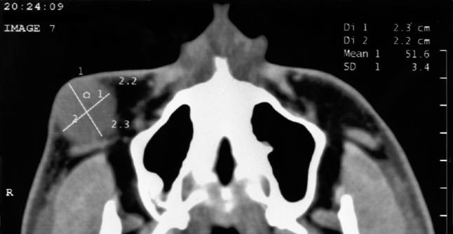
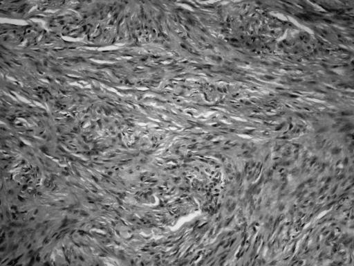
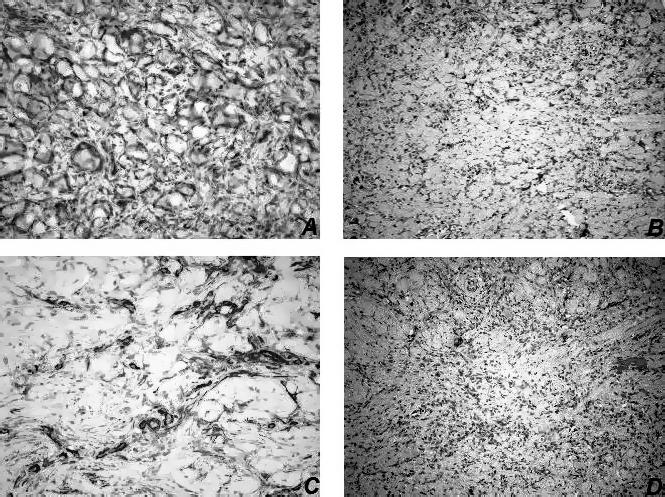
Fibrous histiocytoma is a benign tumour composed of a mixture of fibroblastic and histiocytic cells. Based on the location of this tumour, fibrous histiocytoma are usually divided into cutaneous types and those involving deep tissues. These lesions most often arise on the skin, but may rarely occur in soft deep tissues. The diagnosis of fibrous histiocytoma may be clinically difficult when the lesion is located in the deep tissues, and is frequently confirmed after local excision. The most important diagnostic distinction is the separation of this tumour from aggressive forms of fibrohistiocytic neoplasms, including dermatofibrosarcoma protuberans and malignant fibrous histiocytoma. A 19-year-old male presented with a painless swelling on the right cheek. Detailed clinical and laboratory examinations were performed. The lesion had been totally excised under local anaesthesia, and histopathology revealed a benign fibrous histiocytoma. The diagnosis, location, treatment and prognosis of fibrous histiocytoma are also discussed.
Il fibroma istiocitario è un tumore benigno composto da fibroblasti e istiociti. In base alla sede di questo tumore, i fibromi istiocitari sono divisi solitamente nei tipi cutanei ed in quelli che coinvolgono i tessuti profondi. Queste lesioni si presentano spesso sulla cute, mentre possono riscontrarsi raramente nei tessuti molli profondi. La diagnosi clinica del fibroma istiocitario può essere difficile quando la lesione è situata in profondità e frequentemente se ne ha conferma solo dopo l’asportazione. La distinzione diagnostica più importante è la differenziazione di questo tumore dalle forme aggressive del tumore fibroistiocitario, compreso il dermatofibrosarcoma protuberans e l’istiocitoma fibroso maligno. Caso clinico: un paziente maschio di 19 anni si presenta con tumefazione non dolorosa alla guancia destra. Vengono effettuati gli esami di laboratorio e clinici. La lesione viene completamente asportata in anestesia locale. L’esame istologico ha rivelato un istiocitoma fibroso benigno. Sono discussi la diagnosi, la sede, il trattamento e la prognosi del fibroma istiocitario.
Figures
References
-
- Bielamowicz S, Dauer MS, Chang B, Zimmerman MC. Non-cutaneous benign fibrous histiocytoma of the head and neck. Otolaryngol Head Neck 1995;113:140-6. - PubMed
-
- Batsakis JG. Fibrous lesions of the head and neck: Benign, malignant and indeterminate. In: Batsakis JG, editor. Tumours of the head and neck. 2nd edn. Baltimore, MD: Williams & Wilkins; 1979. p. 252-79.
-
- Sullivan BO, Audel N, Catton CN, Gullane PJ. Soft tissue and bone sarcomas of the head and neck. In: Harrison LB, Sessions RB, Hong WK, editors. Head and Neck Cancer. 2nd edn. Philadelphia, PA: Lippincott Williams & Wilkins; 2004. p. 786-823.
-
- Femiano F, Scully C, Laino G, Battista G. Benign fibrous histiocytoma of the cheek: CD 68-KP1 positivity. Oral Oncol 2001;37:673-5. - PubMed
-
- Fletcher CD. Benign fibrous histiocytoma of subcutaneous and deep soft tissue: a clinicopathologic analysis of 21 cases. Am J Surg Pathol 1990;14:801-9. - PubMed
Publication types
MeSH terms
LinkOut - more resources
Full Text Sources