. 2007 Jul;5(7):e179.
doi: 10.1371/journal.pbio.0050179. Epub 2007 Jul 3.

Rb-mediated neuronal differentiation through cell-cycle-independent regulation of E2f3a

Affiliations

Rb-mediated neuronal differentiation through cell-cycle-independent regulation of E2f3a

Danian Chen et al. PLoS Biol. 2007 Jul.

Abstract

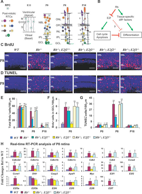
It has long been known that loss of the retinoblastoma protein (Rb) perturbs neural differentiation, but the underlying mechanism has never been solved. Rb absence impairs cell cycle exit and triggers death of some neurons, so differentiation defects may well be indirect. Indeed, we show that abnormalities in both differentiation and light-evoked electrophysiological responses in Rb-deficient retinal cells are rescued when ectopic division and apoptosis are blocked specifically by deleting E2f transcription factor (E2f) 1. However, comprehensive cell-type analysis of the rescued double-null retina exposed cell-cycle-independent differentiation defects specifically in starburst amacrine cells (SACs), cholinergic interneurons critical in direction selectivity and developmentally important rhythmic bursts. Typically, Rb is thought to block division by repressing E2fs, but to promote differentiation by potentiating tissue-specific factors. Remarkably, however, Rb promotes SAC differentiation by inhibiting E2f3 activity. Two E2f3 isoforms exist, and we find both in the developing retina, although intriguingly they show distinct subcellular distribution. E2f3b is thought to mediate Rb function in quiescent cells. However, in what is to our knowledge the first work to dissect E2f isoform function in vivo we show that Rb promotes SAC differentiation through E2f3a. These data reveal a mechanism through which Rb regulates neural differentiation directly, and, unexpectedly, it involves inhibition of E2f3a, not potentiation of tissue-specific factors.


Conflict of interest statement

Competing interests. The authors have declared that no competing interests exist.

Figures

Figure 1
Figure 1. E2f1, but Not E2f2 or E2f3, Loss Rescues Ectopic Division and Cell Death in the Rb KO Retina
(A) Retinal development. At E11 the retina is a NBL of dividing RPCs (white circle, green nuclei). RPC cell bodies oscillate along processes as they progress through the cell cycle. By P0 the NBL contains both RPCs and post-mitotic RTCs (coloured circles, red nuclei) and is separated from the GCL by the IPL. By P8 there are no RPCs, fewer RTCs, an OPL, and more differentiated rods (r) and cones (c) in the ONL; horizontal (h), bipolar (b), Müller (m), and amacrine (a) cells in the INL; and ganglion (g) and displaced amacrine cells in the GCL. Development is complete by ~P18. (B) Rb is thought to regulate cell cycle and apoptosis by repressing E2fs, but to promote differentiation by potentiating tissue-specific transcription factors. However, Rb loss could also perturb differentiation through the indirect effects of abnormal division or death, and/or through direct regulation of differentiation genes by E2fs. (C and D) Horizontal retinal sections of the indicated genotypes and ages were stained for nuclei (DAPI, blue), and (C) S-phase (anti-BrdU, red) or (D) apoptosis (TUNEL, red). Scale bars are 50 μm. (E–G) Quantification of (E) all BrdU+ cells, (F) ectopic BrdU+ cells in GCL at P0, and (G) total TUNEL+ cells. (H) Real-time RT-PCR analysis of E2fs and E2f target genes in P8 retinas of the indicated genotypes. Error bars represent SD of measurements from three animals, and asterisks indicate a significant difference between the WT and indicated genotypes (*, p <0.05; **; p <0.01; ANOVA and Tukey HSD test for [E–G] and Fisher test for [H]).
Figure 2
Figure 2. E2f1 Deletion Rescues Ganglion, Rod, and Bipolar Cells in the Rb KO Retina
(A) Horizontal retinal sections from mice of the indicated ages and genotypes were stained for nuclei (DAPI, blue) and markers that detect ganglion cells (Pou4f2, red), rods and cones (Sag [rod arrestin], green), and rod bipolar cells (Prkca, green, and Cabp5, red). Scale bars are 50 μm. (B) Quantification of Pou4f2+ ganglion cells. (C) Quantification of Prkca+ and Cabp5+ bipolar cells. (D) Thickness of the ONL, which represents the number of rods. Error bars represent SD of measurements from three animals, and asterisks indicate a significant difference between retinas of WT and the indicated genotypes, unless indicated otherwise by connecting lines (*, p < 0.05; **; p < 0.01; ANOVA and Tukey HSD test). (E and F) ERGs were recorded from the indicated genotypes under dark-adapted (scotopic) conditions, and (E) intensity series and (F) b-wave amplitudes as a function of the logarithm of the flash intensity were determined. (F) Further illustrates that the relative influence of the mutations on the photoreceptors (indicated by the saturated a-wave amplitude, right graph) was not substantially different from their effect on the b-wave response (dominated by the bipolars, left graph) at the same intensity of 10 cd·s/m2.
Figure 3
Figure 3. Differentiation Defects in Rb KO SACs
(A) P18 horizontal sections of WT, Rb KO, and Rb/E2f1 DKO retina were stained for nuclei (DAPI, blue), Calb2 (green), and Slc18a3 (red). (B) Confocal images of P30 horizontal sections of WT and Rb KO retina were stained for nuclei (DAPI, blue), Chat and Slc18a3 (both red), and Camk2a (green). In the Rb KO section, the red stain is Chat only, as Slc18a3 is missing (see [A]). (C) Quantification of dense Calb2+ cell bodies in the INL, total Slc18a3+ cell bodies, and Camk2a+ cell bodies in the INL. Error bars represent SD of measurements from three animals, and asterisks indicate significant differences between retinas of WT and the indicated genotypes (*, p <0.05; **; p <0.01; ANOVA and Tukey HSD test). Scale bars are 50 μm in (A) and 20 μm in (B).
Figure 4
Figure 4. E2f3 Loss Rescues Differentiation of Rb KO SACs
(A) Horizontal retinal sections of the indicated ages and genotypes were stained for nuclei (DAPI, blue), mitosis marker PH3 (green), and Slc18a3 (red), which marks SAC soma at early stages and processes from ~P5 onwards. Arrows show mitotic PH3+ nuclei in Rb KO, Rb/E2f2 DKO, and Rb/E2f3 DKO retinas. E2f1 loss rescues the ectopic mitosis and cell death defects, but not the SAC defect. E2f2 loss has no effect. E2f3 loss does not rescue the ectopic mitosis and cell loss defects, but rescues the SAC defect. Inactivating E2f1 and E2f3 together rescues the ectopic mitosis, cell death, and SAC defects. (B) The fraction of Camk2a+ cells that are Chat + and Slc18a3+ in the P30 retina. (C) Horizontal P5 retinal sections were stained for nuclei (DAPI, blue), Slc18a3 (green), and Isl1 (red). Arrows show double-labelled Isl1+/Slc18a3+ cells in the inner INL. (D) Horizontal P0 retinal sections of the indicated genotypes were stained for nuclei (DAPI, blue), cell division marker Mki67 (green), and Isl1 (red). Arrows show double-labelled dividing SACs. (E) The fraction of Isl1+ cells in the inner NBL (INBL) of P0 retinas that are dividing (Mki67+). Error bars represent SD of measurements from three animals, and asterisks indicate significant differences between retinas of WT and the indicated genotypes (*, p <0.05; **; p <0.01; ANOVA and Tukey HSD test). Scale bars in (A), (C), and (D) are 50 μm.
Figure 5
Figure 5. E2f3 and Rb Expression in SACs
(A) Left panels: horizontal P0, P8, and P18 retinal sections of the indicated genotypes were stained for E2f3 (red) and DAPI (blue). The arrow indicates the junction between the E2f3 null peripheral and WT central P0 retina. Note the absence of E2f3 protein in the peripheral E2f3 KO RPCs at P0 and in peripheral inner retinal neurons at P18. Far right panel: P18 retinal sections of the indicated genotypes were stained for Rb (red) and DAPI (blue). Note the absence of Rb protein in the peripheral Rb KO inner retinal neurons. (B) WT P18 retinal sections were stained for nuclei (DAPI, blue), E2f3 (red) or Rb (red), and Chat plus Slc18a3 (green). Arrows indicate double-labelled soma. Note that the IPL processes are also double-labelled. Scale bars are 50 μm.
Figure 6
Figure 6. Subcellular Distribution of E2f3 Isoforms and Other Cell Cycle Proteins in the Developing Retina
Nuclear and cytoplasmic extracts from an equivalent number of retinal cells from mice of the indicated genotypes and ages were analyzed by Western blotting to detect the indicated proteins. Lysate from E2f3a−/− mice was used as a control to confirm the location of E2f3a protein. C, cytoplasmic extracts; N, nuclear extracts.
Figure 7
Figure 7. The E2f3a Isoform Drives the Differentiation Defect in Rb KO SACs
(A) Schematic diagrams of the mouse WT, E2f3a−/−, and the Cre-recombined floxed E2f3 loci (indicated here as E2f3−/− for simplicity). E2f3a−/− mice lack most of E2f3 exon 1a and part of intron 1a (red dotted box). Arrows indicate PCR primers. Genotyping of an E2f3a+/− mouse is shown on the right. (B) RT-PCR detection of E2f3a and E2f3b mRNA in the retina. The sequences of primers are 1aF (5′-GCCTCTACACCACGCCACAAG-3′), 1bF (5′-CGGAAATGCCCTTACAGC-3′), and 4R (5′-CTCAGTCACTTCTTTGGACAG-3′). WT retina expresses both E2f3a and E2f3b mRNA. As expected, E2f3a−/− retina lacks E2f3a mRNA and still expresses E2f3b mRNA. E2f3−/− retina lacks full-length E2f3a and E2f3b mRNAs, and instead expresses a truncated mRNA lacking exon 3. (C) Real-time RT-PCR analysis of E2f genes in P8 retinas of the indicated genotypes. Error bars represent SD of measurements from three animals, and asterisks indicate a significant difference between WT and the indicated genotypes (*, p <0.05; **; p <0.01; ANOVA and Tukey HSD test). (D) Rescue of Rb KO SACs by E2f3a deletion. Horizontal retinal sections of the indicated genotypes and ages were stained for nuclei (DAPI, blue), M-phase (PH3, green), and the SAC marker Slc18a3 (red). E2f3a deletion does not suppress ectopic division, but rescues the SAC defect. Scale bars are 50 μm. M, molecular size marker.
Figure 8
Figure 8. Rb Regulates Distinct Processes through E2f1 and E2f3a
Red text and arrows indicate Rb-dependent events. Black text and arrows indicate events for which there is no direct evidence of Rb involvement. Rb does not appear to temper RPC expansion and is not required for differentiation of RPCs into RTCs, but is essential to couple RTC birth to terminal mitosis, thus locking them out of cycle. Rb performs this function by inhibiting E2f1. Rb is also required for SAC differentiation, and in this case, acts by inhibiting E2f3a. There is no direct evidence that Rb is required for terminal differentiation of other cell types. Colour codes and abbreviations as in Figure 1A.

Similar articles

Cited by

References

    1. Dyer MA, Bremner R. The search for the retinoblastoma cell of origin. Nat Rev Cancer. 2005;5:91–101. - PubMed
    1. Chen D, Livne-Bar I, Vanderluit JL, Slack RS, Agochiya M, et al. Cell-specific effects of RB or RB/p107 loss on retinal development implicate an intrinsically death-resistant cell-of-origin in retinoblastoma. Cancer Cell. 2004;5:539–551. - PubMed
    1. MacPherson D, Sage J, Kim T, Ho D, McLaughlin ME, et al. Cell type-specific effects of Rb deletion in the murine retina. Genes Dev. 2004;18:1681–1694. - PMC - PubMed
    1. Stevaux O, Dyson NJ. A revised picture of the E2F transcriptional network and RB function. Curr Opin Cell Biol. 2002;14:684–691. - PubMed
    1. Wu L, Timmers C, Maiti B, Saavedra HI, Sang L, et al. The E2F1–3 transcription factors are essential for cellular proliferation. Nature. 2001;414:457–462. - PubMed

Publication types

MeSH terms

LinkOut - more resources