

Lower cardiac output is associated with greater white matter hyperintensities in older adults with cardiovascular disease
- PMID: 17608877
- PMCID: PMC2721459
- DOI: 10.1111/j.1532-5415.2007.01226.x
Lower cardiac output is associated with greater white matter hyperintensities in older adults with cardiovascular disease
Abstract
Objectives: To preliminarily examine the association between cardiac output, a measure of systemic blood flow, and structural brain magnetic resonance imaging indices of white matter hyperintensities (WMHs).
Design: Cross-sectional.
Setting: University medical setting.
Participants: Thirty-six older adults without dementia with prevalent cardiovascular disease (aged 56-85).
Measurements: Cardiac output, WMHs.
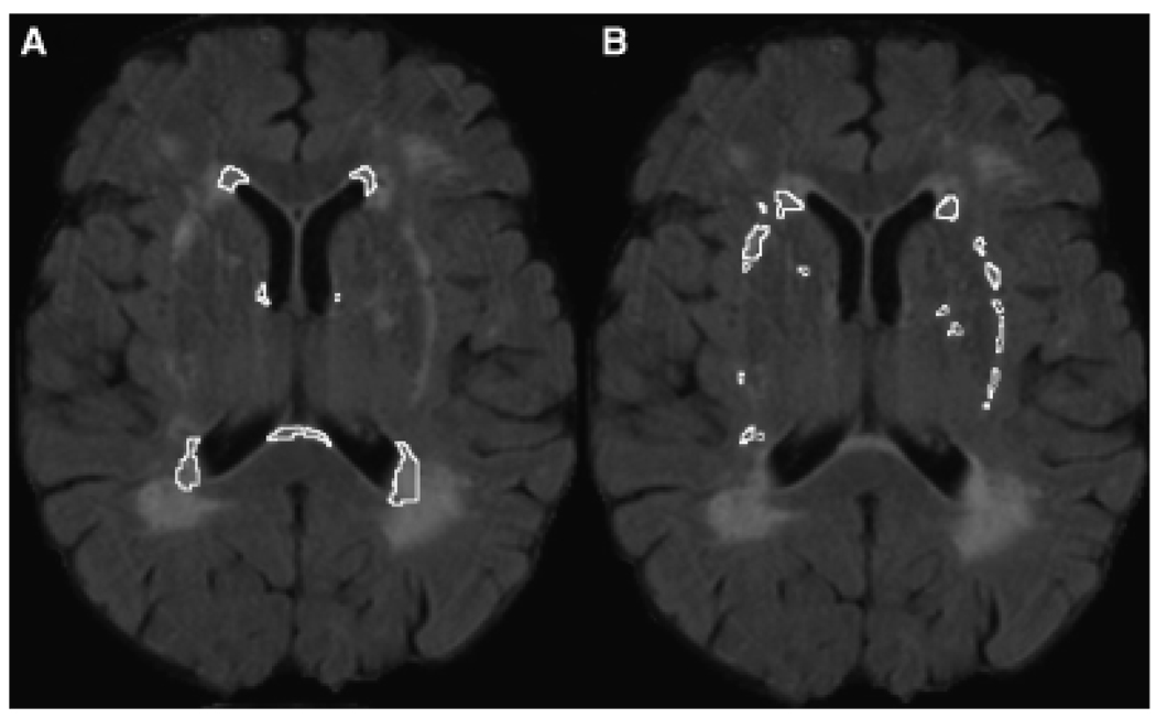
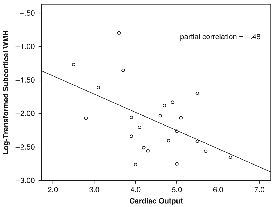
Results: Partial correlations, adjusting for age and history of hypertension, yielded an inverse relationship between WMHs adjacent to subcortical nuclei and cardiac output (correlation coefficient=-0.48, P=.03); as cardiac output decreased, WMHs increased significantly. No significant associations were found between cardiac output and total WMHs or periventricular WMHs.
Conclusion: These preliminary data suggest that systemic blood flow, measured according to cardiac output, is inversely associated with WMHs adjacent to the subcortical nuclei. Cerebrovascular degeneration and the chronicity of hypoperfusion may exacerbate the susceptibility of white matter integrity to alterations in blood flow in older adults.
Figures
References
-
- Gunning-Dixon FM, Raz N. The cognitive correlates of white matter abnormalities in normal aging: A quantitative review. Neuropsychology. 2000;14:224–232. - PubMed
-
- Kertesz A, Black SE, Tokar G, et al. Periventricular and subcortical hyperintensities on magnetic resonance imaging. ‘Rims, caps, and unidentified bright objects’. Arch Neurol. 1988;45:404–408. - PubMed
-
- de Leeuw FE, de Groot JC, Oudkerk M, et al. Atrial fibrillation and the risk of cerebral white matter lesions. Neurology. 2000;54:1795–1801. - PubMed
-
- de Leeuw FE, De Groot JC, Oudkerk M, et al. Aortic atherosclerosis at middle age predicts cerebral white matter lesions in the elderly. Stroke. 2000;31:425–429. - PubMed
-
- Lindgren A, Roijer A, Rudling O, et al. Cerebral lesions on magnetic resonance imaging, heart disease, and vascular risk factors in subjects without stroke. A population-based study. Stroke. 1994;25:929–934. - PubMed
Publication types
MeSH terms
Grants and funding
- P30-AG013846/AG/NIA NIH HHS/United States
- K23-MH065857/MH/NIMH NIH HHS/United States
- K23-MH073416/MH/NIMH NIH HHS/United States
- F32-HL74568/HL/NHLBI NIH HHS/United States
- K23 MH065857/MH/NIMH NIH HHS/United States
- R01 AG017975/AG/NIA NIH HHS/United States
- K23 AG030962/AG/NIA NIH HHS/United States
- K12-HD043444/HD/NICHD NIH HHS/United States
- F32-AG022773/AG/NIA NIH HHS/United States
- F32 HL074568/HL/NHLBI NIH HHS/United States
- P30 AG013846/AG/NIA NIH HHS/United States
- K12 HD043444/HD/NICHD NIH HHS/United States
- F32-AG024708/AG/NIA NIH HHS/United States
- F32 AG024708/AG/NIA NIH HHS/United States
- K23 MH073416/MH/NIMH NIH HHS/United States
- F32 AG022773/AG/NIA NIH HHS/United States
- R01-AG017975/AG/NIA NIH HHS/United States
LinkOut - more resources
Full Text Sources
Other Literature Sources
Medical
